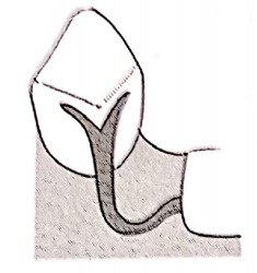
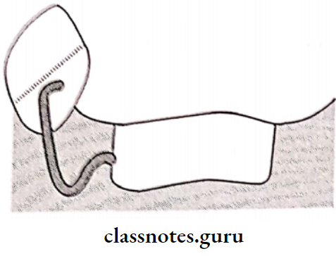
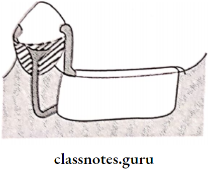
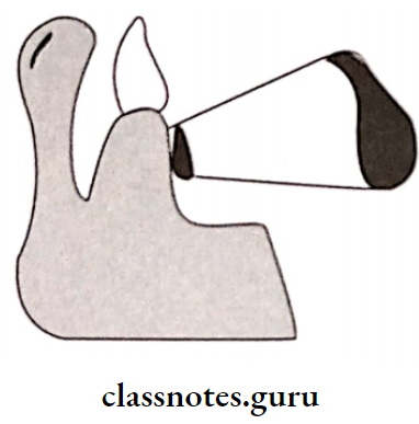
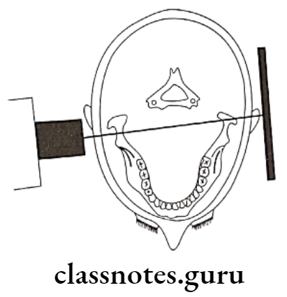
Epidemiology Of Gingival And Periodontal Disease

Epidemiology Of Gingival And Periodontal Diseases Short Essays

Question 1. Plaque Index.
Answer:

  • Selected tooth – entire dentition

1. Selected teeth

  • Surfaces: Dissocial, facial, mesiofacial and lingual
  • Site: Cervical third of the tooth

Epidemiology Of Gingival And Periodontal Diseases Plaque Index

Read And Learn More: Periodontics Question and Answers

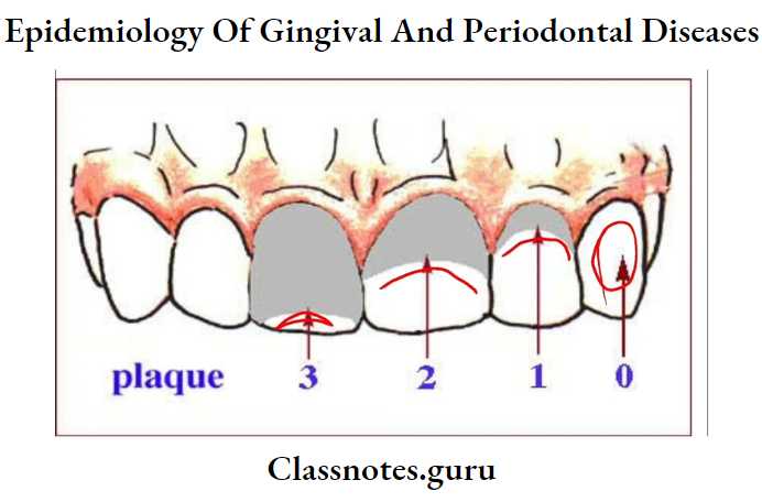
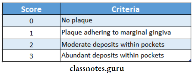
Plaque Index Scoring:

Epidemiology Of Gingival And Periodontal Diseases Plaque index

Plaque Index Calculation:

  • Per tooth-Score/4
  • Per person – Score per tooth/No. Of teeth

Question 2. OHI-S by Greene and Vermilion.
Answer:

  • The oral hygiene index-simplified was described by John C. Greene and Jack R. Vermilion in 1964
  • Tooth Examined:
  • 16/17, 11, 26/27
  • 46/47,31 36/37
  • Surfaces: Facial of 16/17, 11, 26/27, 36/37 Lingual of 31

OHI-S Sites:

  • D1-S – Incisal third to gingival third
  • C1-S Distal gingival crevice subgingivally from distal to medial contact

OHI-S Scoring: For D1-S:

  • 0-No debris
  • 1-Debris covering 1/3rd of tooth surface
  • 2-Debris covering more than 1/3rd and less than 2/3rd of tooth surface
  • 3-Debris covering more than 2/3rd of tooth surface

For CI-S:

  • O-No calculus
  • 1-Supragingival calculus covering 1/3rd of tooth surface
  • 2-Supragingival calculus covering more than 1/3rd but less than 2/3rd of tooth surface
  • 3-Supragingival calculus covering more than 2/3rd of the tooth surface, heavy bands of subgingival
    calculus also present

OHI-S Calculation:

  • OH1 – S = D1-S+C1-S

OHI-S Interpretation:

  • Good – 0.0 to 1.2
  • Fair – 1.3 to 3.0
  • Poor – 3.1 to 6.0

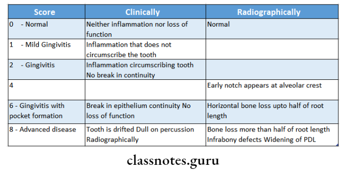
Question 3. Russel’s Periodontal Index.
Answer:

By Russell Al.

Teeth examined:

All

Surfaces:

Epidemiology Of Gingival And Periodontal Diseases Clinically and Radiographically

Interpretation:

  • 0-0.2 – Normal
  • 0.3-0.9 – Simple Gingivitis
  • 0.7-1.9 – Beginning of destructive disease
  • 1.6-5.0 – Established destructive disease
  • 3.8-8.0 – Terminal disease

Question 4. Gingival Index – by Loe and Stillness.
Answer:

  • Teeth Examined: All or selected

Gingival Index Surfaces:

  • Distal facial papillae
  • Facial margin
  • Mesial facial papillae
  • Lingual margin

Gingival Index Scoring:

  • 0-No inflammation
  • 1-Mild inflammation, no bleeding on probing
  • 2-Moderate inflammation, and bleeding on probing
  • 3-Severe inflammation

Gingival Index Calculation:

  • Per tooth-Score/4
  • Per person\( -\frac{\text { TotalScore }}{\text { Total teeth examined }}\)

Gingival Index Interpretation:

  • 0.1-1.0 – Mild gingivitis
  • 1.1-2.0 – Moderate gingivitis
  • 2.1-3.0 – Sever gingivitis

Question 5. CPITN.
Answer:

CPITN Objectives:

  • Mild gingivitis Moderate gingivitis Severe gingivitis
  • To survey and evaluate periodontal treatment needs, Identify actual and potential problems posed by periodontal diseases both in the community and in the individual

Teeth examined: Ten specified index teeth are

  • 17-16  11  26-27
  • 47-46  31  36-37

CPITN Scoring:

  • Code X – When only one tooth or no teeth are present in the sextant
  • Code 0 – Healthy periodontics
  • Code 1 – Bleeding on probing
  • Code 2 – Calculus present
  • Code 3 – Pocket of 4-5 mm
  • Code 4 – Pocket of more than 6 mm

CPITN Treatment Needs:

  • TN-0 -No treatment
  • TN-1 -Improvement of personal oral hygiene
  • TN-2 -Professional scaling
  • TN-3 Complex treatment involving deep scaling, root planning, and complex procedures

Question 6. Incidence and prevalence.
Answer:

Incidence:

  • It is defined as the number of new cases of a specific disease occurring in a defined population during a specified period of time

Incidence Uses:

  • Provide a clue for the etiology and pathogenesis of the disease
  • Study the distribution of the study
  • Control the disease

Incidence Types:

  1. Episode incidence
    • It is the rate of occurrence of new episodes of a dis-case arising in the population
  2. Cumulative incidence
    • Similar to incidence but the time interval is ex-presses as a fixed period

Prevalence:

  • The total number of all individuals who have an attribute or disease at a particular time divided by the population at risk of having the attribute or disease at this point or midway through the period

Prevalence Uses:

  • Estimate the magnitude of disease or health problems in the community
  • Identify the potential high-risk population
  • Useful in administrative and planning purposes like assessing manpower needs in health services

Prevalence Types:

1. Point prevalence:

  • It is the number of all current cases of a specific disease at one point in time with a defined population

2. Period prevalence:

  • It is defined as the total number of existing cases of a specific disease during a defined period of time expressed in relation to a defined population

Question 7. Describe the possible causes as to why the incidence, and prevalence of periodontal diseases are very high in India.
Answer:

  • The prevalence of periodontal disease in India is high due to:
  • Low socioeconomic group
  • Poor oral hygiene practice
  • Greater prevalence in mentally retarded children due to
    • Lack of awareness of oral hygiene
    • Nutritional deficiency
    • Malocclusion
    • Oral health habits like bruxism, tongue thrusting, mouth breathing
    • Low power of concentration
    • Low neuromuscular coordination
  • Vegetarian diet
  • Hereditary
  • Presence of habits like smoking and betel nut chewing
  • Evaluate the efficacy of preventive and therapeutic
  • Malnutrition
  • measures
  • Presence of systemic disease

Question 8. Define index  uses and ideal requirements of an index.
Answer:

Index Definition:

  • Numerical values describing the relative status of the population on a graduated scale with definite upper and lower limits designated to permit and facilitate comparisons with other populations that are classified by the same criteria and methods are referred to as index

Index Uses:

  1. In the case of individual patients
    • Provides individual assessment
    • Reveals the degree of effectiveness
    • Motivates the patient
    • Evaluates the progress of treatment
  2. In the case of research studies
    • Determines the baseline data before the introduction of experimental factors
    • Measures effectiveness of specific agents for pre-version control and treatment of oral conditions
    • Measures effectiveness of devices for personal care
  3. In community health
    • Provides baseline data
    • Assesses the needs of a community
    • Evaluates the results

Index Ideal Requirements:

  • Clarity simplicity
  • The examiner should remember the rules of the index clearly
  • The index should be simple & easy to apply
  • The criteria should be objective
  • Validity
  • The index should measure what it is intended to measure
  • It should correspond to the clinical stages of the disease under study
  • Reliability
  • The index should measure consistently at different times & under a variety of conditions
  • Quantifiability
  • The index should be amenable to statistical analysis so that the status can be expressed by a number
  • Sensitivity
  • The index should be able to detect small shifts in either direction
  • Acceptability
  • The use of an index should not be painful or demeaning to the subject

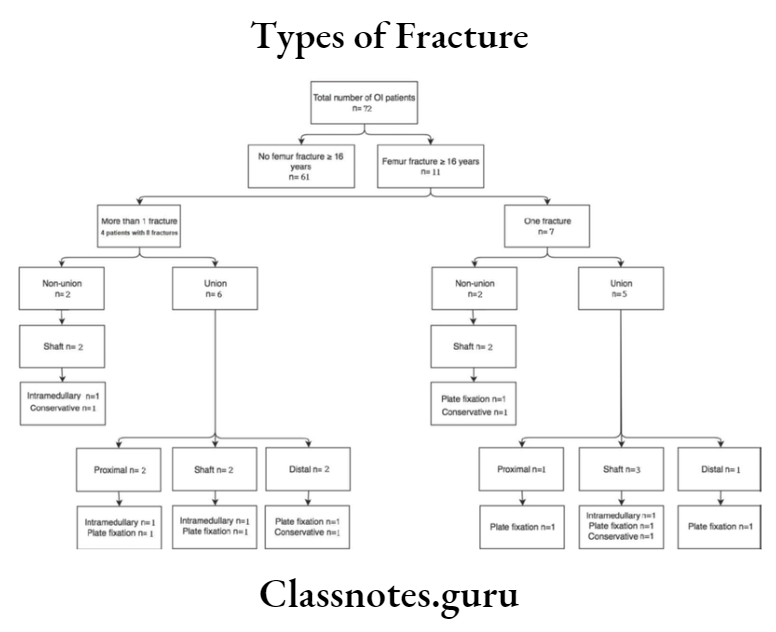
Types of Fracture Short Essays

Fractures General Principles Short Essays

Question 1. Compound fracture

Answer:

Compound Fracture

  • Compound fracture or open fracture involves wounds that communicate with the exterior and gets contaminated

Compound Fracture Classification:

  • Type 1
    • The wound is smaller than 1 cm
    • It is clean wound
  • Type 2
    • The wound is longer than 1 cm
    • It is clean and without any soft tissue damage
  • Type 3
    • The wound is longer than 1 cm with extensive soft tissue damage

Compound Fracture Management:

  • Aims
    • To prevent infection
    • To allow tire fracture to heal
    • To restore function
  • Treatment
    • Control of hemorrhage
    • Antibiotic administration
      • Cefazolin or clindamycin is preferred for type 1 and type 2
      • Metronidazole is preferred for type 3
    • Tetanus vaccination
    • Irrigation and debridement of the wound
    • Incision and drainage in case of type 3

Read And Learn More: General Surgery Question and Answers

Question 2. Pathological fractures

Answer:

Pathological Fractures

  • Pathological fracture is one that occurs due to underlying diseases like
  1. Tumors:
    • Giant cell tumour
    • Bone cysts
  2. Infections:
    • Acute osteomyelitis
  3. Metabolic bone diseases
    1. Hyperparathyroidism
    2. Osteoporosis
    3. Paget’s disease

Pathological Fractures Common Sites Involved:

  • Vertebral fractures
  • Fractures of the neck of femur
  • Colles fracture of the wrist

Pathological Fractures Diagnosis:

  • Laboratory investigation
    • To rule out the systemic diseases present
    • ESR estimation
    • Total blood count
  • A comminuted bone scan is done
  • Biopsy

Types of fractures short essay

Question 3. Fracture healing

Answer:

Stages Of Fracture Healing:

  • Stage of inflammation
    • Occurs soon after the fracture
    • Trauma to the blood vessels of the periosteum, endosteum, bone marrow, and Haversian system occurs
    • As a result hematoma formation occurs
    • This causes hypoxia and necrosis of the fragment ends
    • There is acute inflammatory reaction with edema at the site
    • Pleuripotent cells produce osteoblast, fibroblast, and chondroblasts
    • Granulation tissue is formed
    • Hematoma gets organized
  • Soft callus formation
    • There is formation of subperiosteal fibrous tissue with fibrocartilagenous and cartilagenous components
    • This is called callus
    • It is soft at this stage
  • Hard callus formation
    • The endosteal and periosteal blood supply improves
    • The callus gets converted into woven bone
    • This immature bone is called hard callus
  • Stage of remodeling
    • There is a continuous process of deposition and resorption of bone
    • The immature bone gets converted into mature lamellar bone

Question 4. Nonunion

Answer:

Nonunion

Lack of bony fusion of fractured ends

Nonunion Etiology:

  • Inadequate fixation
  • Infection of the fracture
  • Lack of adequate blood supply
  • Excessive periosteal stripping
  • Pathological fractures

Nonunion Features:

  • Pain
  • Difficulty in occlusion
  • Difficulty in mastication
  • Abnormality mobility of fractured fragments

Nonunion Management:

  • Expose the site
  • Graft the space
  • Stabilize the fractured ends
  • Fixation
  • Immobilization

Short note on bone fractures

Question 5. Dislocation and subluxation

Answer:

Dislocation And Subluxation

  • Dislocation refers to the condition in which the condyle is placed anterior to the articular eminence with collapse of the articular space
  • Subluxation is the partial dislocation

Dislocation And Subluxation Clinical Features

  • Pain
  • Inability to close the mouth
  • Tense masticatory muscles
  • Difficulty in speech
  • Excessive salivation
  • Open bite
  • Protruding chin
  • Deviation of the lower jaw

Dislocation And Subluxation Management:

  • Reassure the patient
  • Sedative drugs
  • Pressure and massage the area
  • Manipulation
  • The operator grasps the patient’s mandible
  • The thumb is placed over the occlusal surfaces of the lower molars
  • Fingertips are placed below the chin
  • Downward pressure is placed over posteriors
  • This overcomes spasms of muscles
  • Backward pressure is applied which pushes the entire mandible posteriorly
  • Immobilization is done

Classification of bone fractures

Question 6. General Management Of Patient With Head Injury

Answer:

General Management Of Patient With Head Injury

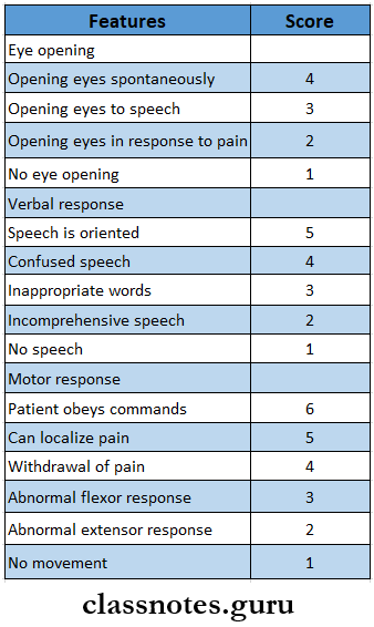
  • Management of the head injury depends on Glasgow Coma Scale
    • Less than 8 score- indicate severe injury
    • Score 9-12- moderate injury
    • Score 13-15- mild injury
  • Measures includes
    • Examination of the wound
    • Continued ventilation
    • Intensive care unit management of intracranial pressure
    • Oxygenation
    • Frequent neurological examination
    • CT scan

Fractures General Principles General Management Of Patient With Head Injury

Types Of Fracture Long Essays

Fractures General Principles Long Essays

Question 1. Lefort’s classification of fractures of the maxilla

Answer:

Lefort 1

Maxilla Clinical Features:

  • Oedema of lower part of face
  • Ecchymosis in buccal vestibule
  • Bilateral epitaxis
  • Mobility of upper teeth
  • Disturbed occlusion
  • Pain
  • Upward displacement of fragment- telescopic fracture H ‘Cracked cup’ sound on percussion of upper teeth
  • ‘Guerin sign- ecchymosis in the greater palatine region

Maxilla Management:

  1. Reduction
    • Reduction of the impacted fragment with the help of disimpaction forceps (Rowe’s and William’s forceps)
    • Placement of Rowe’s forceps:
      • A straight blade is placed into the nostrils
      • A curved blade is placed over the palate
    • Placement of William’s forceps
      • Placed over the buccal aspect
      • Displaces maxilla in mesiodistal direction
  2. Fixation:
    • Zygomatic suspension fixation is done
    • Holes are drilled over the zygomatic arch
    • Pass the wire through it
    • Bring it up to the arches
    • Twisted over are arch bars
  3. Inter Maxillary Fixation
    • IMF done for 3-4 weeks

Types of bone fractures long essay

Lefort 2 Clinical Features:

  • Cross edema of the middle third of the face.
  • Ballooning of face
  • Black eye
  • Lengthening of face
  • Bilateral subconjunctival hemorrhage
  • Depressed nasal bridge.
  • Anterior open bite
  • Bilateral epistaxis
  • Loss of occlusion
  • Difficulty in mastication and speech
  • Airway obstruction
  • CSF leak
  • Paraesthesia of cheek
  • Step deformity

Read And Learn More: General Surgery Question and Answers

Lefort 2 Management:

    • Reduction – reduction of the fragments through disimpaction forceps.
    • Fixation Zygomatic suspension fixation is done,
    • Inter – maxillary fixation
      • It is done for 3-4 weeks.

E:\Flow Charts\General Surgery\Types of Fracture Fractures General issues.png

Lefort’s 3 Clinical Features:

  • Ballooning of face
  • Panda facies
  • Racoon eyes
  • Bilateral subconjunctival hemorrhage
  • Lengthening of face
  • Separation of sutures
  • ‘Dish face’ deformity
  • Enophthalmus
  • Diplopia
  • Deviation of the nasal bridge
  • Epitaxis
  • CSF rhinorrhoea

Lefort’s 3 Management:

Bilateral frontomalar suspension

Application of arch bars

Intraosseous wiring

Classification of fractures in orthopedics

Question 2. Discuss the management of maxillofacial injuries

Answer:

Management Of Maxillofacial Injuries:

1. Primary assessment

  • Check for airway
  • Bilateral anterior mandibular fractures have the risk of the tongue falling back, check for it
  • Orotracheal intubation is carried out
  • Hemorrhage is controlled
  • Anterior and posterior nasal packing is used

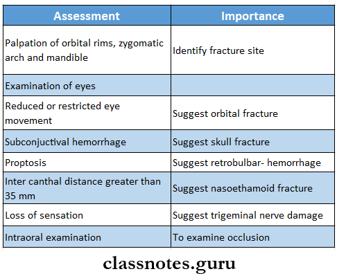
2. Secondary assessment

Fractures General Principles Secondary Assessment

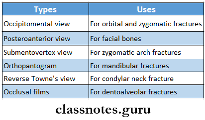
3. Radiography

Fractures General Principles Radiology

Principles Of Management:

  1. Reduction
    • Restoration of fractured fragments to their original position
    • Brought by
      • Closed reduction
      • Open reduction
  2. Fixation
    • Fractured fragments are fixed
    • This prevents displacement of the fragments
    • Consists of
      • Direct fixation
      • Indirect fixation
  3. Immobilization
    • The fixation device is retained in position till a bony union is obtained
    • Immobilization depends on the type of fracture and bone involved.

Question 3. Classify fractures of the face and discuss the management of each type of fracture

Answer:

Definition: Fracture is defined as a sudden break in the continuity of bone and it may be complete/incomplete

Fracture Classification:

  1. Lefort’s classification
    • Lefort 1
    • Lefort 2
    • Lefort 3
  2. Erich’s classification
    • Horizontal fracture
    • Pyramidal fracture
    • Transverse fracture
  3. Depending on the zygomatic bone
    • Sub zygomatic
    • Supra zygomatic
  4. Depending on level
    • Low level
    • Mid-level
    • High level

Fracture Management:

  • Open reduction
    • In it, the fractured fragments are surgically exposed and visualized
    • Indications
      • Dislocation of the condyle into the middle cranial fossa
      • Dislocation of condyle into the external auditory canal
      • Lateral extracapsular displacement
      • Inability to obtain the desired occlusion
      • Bilateral subcondylar fractures in edentulous
      • Bilateral subcondylar fractures associated with comminuted fractures
      • Consists of
        • Exposure of the site
        • Detachment of the bone from all muscle attachments
        • Reinserting
        • Fixation of the segment
  • Closed reduction
    • In it, the fractured fragments are not openly visualized for anatomical alignment
    • Consists of
      • Manipulation of joint
      • Intermaxillary fixation for 10 days
      • Mobilization of the jaw
    • Indications
      • Fractures of the condylar neck that are not displaced
      • Fractures of the condyle in children
      • Intracapsular fractures
  • Fixation
    • The anatomically aligned fragments are then held in place by devices to fix it in that position
    • It is divided into
      • Nonrigid
      • Semi-rigid
      • Rigid
  • Immobilization
    • The fragments are retained without any movement for at least 4-6 weeks
    • It enables callus formation and healing of fragments

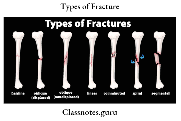
Types of Fracture Types of fracture

Essay on types of fractures

Question 4. Classify fractures. Describe the treatment of fractured mandible and clinical features.

Answer:

Fractured Mandible General Classification:

  1. Simple/ closed
    • Doesn’t communicate with the exterior
  2. Compound
    • It communicates with the exterior
  3. Comminuted
    • Bone is crushed into pieces
  4. Complex
    • Involvement of vital structures
  5. Impacted
    • One fragment is driven into other
  6. Greenstick
    • Fracture of one fragment and bending of other
  7. Pathological
    • Superimposition of disease

Management Of Fractured Mandible:

  • Closed Reduction And Indirect Fixation:

1. Wiring:

  1. Essig’s wiring
    • Used to stabilize dentoalveolar structures
    • Steps:
      • Move the luxated teeth back to the position
      • Adapt wire to the teeth
      • Pass the wire’s one end buccally and the other lingually
      • Join both ends
      • Pass small wires interdentally and fix it
      • Twist it, cut it, and adjust it interdentally
  2. Gilmer’s wiring
    • The pre-stretched wire is passed around the individual tooth
    • Both ends are brought together and twisted
    • Repeat for each tooth
    • Repeat for both the arches
    • Final twisting of mandibular and maxillary wires
    • Twist cut it, and adapt interdentally
  3. Risdon’s wiring
    • Pass the wire around both the 2nd molar
    • Both ends are twisted together
    • Repeat for each tooth
    • Both the base wires are bought to the midline
    • Twisted together
    • Cut it
    • Adapt it to the neck of the teeth
  4. Eyelet wiring
    • Prepare loops in the center of wire
    • Two tails of the wire are passed interdentally
    • One end is passed around the distal tooth from lingually to buccally
    • Another end is passed around the mesial tooth lingually to buccally
    • Twist both ends
    • Cut it short
  5. Multiloop wiring
    • Adapt solder wire around the buccal surface of the tooth
    • Adapt wire buccally from the last molar to the midline
    • Pass the other end distal to the 2nd molar over the lingual side
    • Pass interdentally bring it to the buccal side by passing under the wire
    • Now pass it from buccal to lingual
    • Round it around the tooth
    • Repeat the same procedure

2. Arch Bar Fixation:

  • Arch Bar Fixation is a method of indirect fixation used in the management of mandibular fractures
    • Open Reduction And Direct Fixation:
      • Transosseous wiring or osteosynthesis
      • Plating using compression plates
      • Lag screw fixation
      • Titanium or stainless steel mesh fixation

Fractured Mandible Clinical Features:

  • Change in the contour of the face
  • Lacerations
  • Ecchymosis of the floor of the mouth
  • Occlusal disturbances
  • Step deformity of the mandible
  • Pain and tenderness rismus
  • Deviated mouth opening
  • Anesthesia and paraesthesia of the lower lip and chin

Long answer on bone fractures and types

Question 5. Clinical signs, symptoms, and general principles of treatment of fractures.

Answer:

Treatment Of Fractures Clinical Features:

  • Pain at or near the site of fracture
  • Tenderness or discomfort on gentle pressure over the area
  • Swelling
  • Loss of sensation
  • The injured part cannot move normally
  • The contracting muscles may cause the broken ends of the bone to override
  • Irregularity of the bone
  • Crepitus may be heard or felt
  • Unnatural movement at the site of fracture

Principles Of Fracture Management:

  1. Reduction
    • Restoration of fractured fragments to their original position
    • Brought by
      • Closed reduction
      • Open reduction
  2. Fixation
    • Fractured fragments are fixed
    • This prevents displacement of the fragments
    • Consists of
      • Direct fixation
      • Indirect fixation
  3. Immobilization
    • The fixation device is retained in position till a bony union is obtained.
    • It depends on the type of fracture and bone involved.

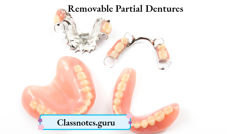
Removable Partial Dentures

Question 1. Removable Partial Dentures Internal attachments.
Answer:

Definition Of Removable Partial Dentures Internal Attachments:

“A retainer, used in removable partial denture construction, consisting of a metal receptacle and a closely fitting part:

  • The former is usually contained with the normal or expanded contours of
  • The crown of the abutment tooth and the latter is attached to a pontic or the denture framework”.

Types of Removable Partial Dentures Internal attachments:

Read And Learn More: Prosthodontics Question And Answers

  • Ney-Chayes attachment
  • Stern Goldsmith attachment
  • Baker attachment

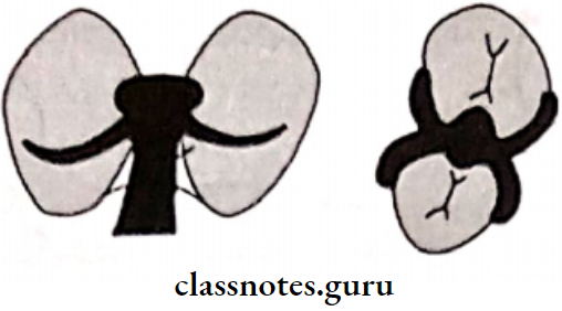
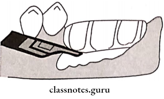
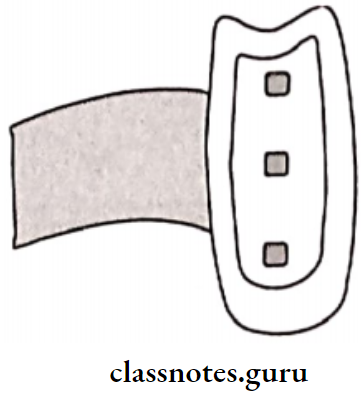
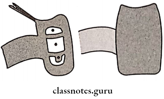
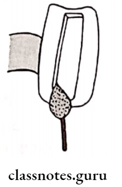
Removable Partial Dentures MAtrix And Patrix Of A Intracoronal Retainer

Removable Partial Dentures Notes

Advantages Of Removable Partial Dentures Internal attachments:

  • It eliminates the visible retentive component
  • It eliminates the visible vertical support
  • It provides horizontal stabilization
  • It stimulates the underlying tissues

Disadvantages Of Removable Partial Dentures Internal Attachments:

  • It requires the preparation of abutments
  • It requires tedious lab procedures
  • It leads to tooth wear
  • It is least effective in teeth with small crowns It is difficult to place

Removable Partial Dentures

Contraindications Of Internal Attachments::

  • Teeth with large pulp
  • Cost efficient

RPD Prosthodontics Guide

Question 2. Canine rests.
Answer:

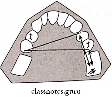
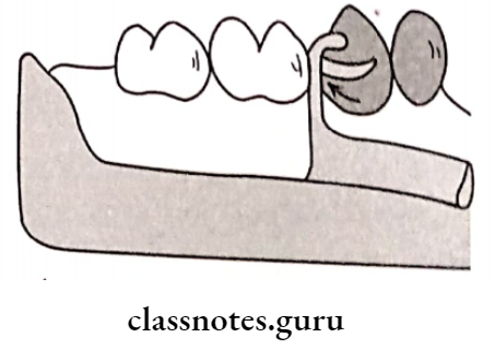
Canine Rests is a form of indirect retainer

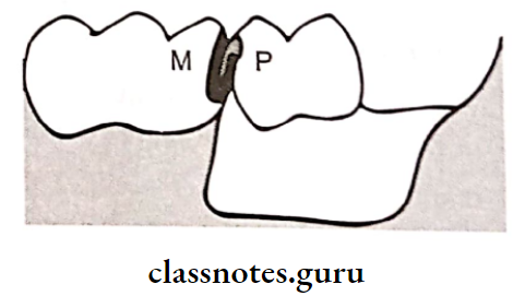
  1. Canine Rests Indication: If the mesial marginal ridge of the first premolar is close to the fulcrum line
  2. Canine Rests Modification: The minor connector can be placed anterior to the canine
  3. Canine Rests Advantage: It increases the efficacy of the indirect retainer

Question 3. Purposes of a surveyor.
Answer:

  • To survey the diagnostic and primary casts
  • For tripoding the cast
  • To transfer the tripod marks to another cast
  • To contour crowns and cast restorations
  • To perform mouth preparation directly on the cast to determine the outcome of treatment
  • To survey the master cast
  • To survey ceramic veneers before final glazing

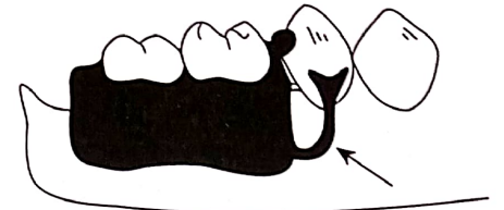
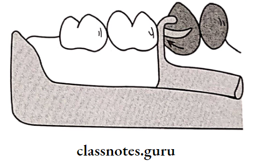
Question 4. Gingivally approaching clasps
Answer:

Gingivally Approaching Clasps is infrabulge clasp

Gingivally Approaching Clasps Advantages:

  • Easy to seat
  • Esthetic
  • Does not increases the occlusal load
  • No decalcification of teeth

Gingivally Approaching Clasps Disadvantages:

  • Gingivally Approaching Clasps tends to collect food debris
  • Gingivally Approaching Clasps needs additional stabilizing units

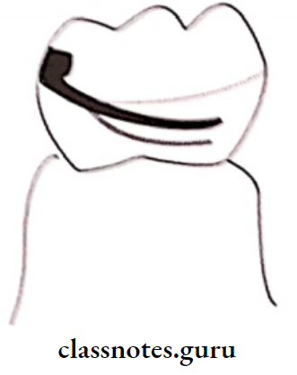
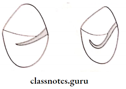
Question 5. Survey Lines

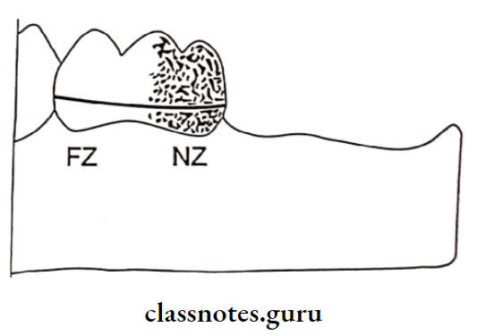
Survey Line: “A line drawn on a tooth or teeth of a cast by means of a surveyor for the purpose of determining the positions of the various parts of a clasp or clasps”.

  1. High Survey Line:
    • This line passes from the occlusal third in the near zone and to the occlusal third in the far zone
    • If the survey line lies higher in position, the undercut will be deep
      In this wrought wire clasp with more flexibility is used It is usually common in inclined teeth and in teeth with greater occlusal diameter than its cementoenamel junction
  2. Medium Survey Line:
    • It passes from the occlusal third in the near zone to the middle third in the far zone
    • In it Aker’s clasp is used
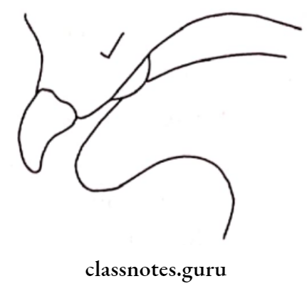
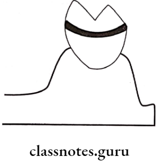
  3. Low Survey Line:
    • This survey line is closer to the cervical third of the tooth in both the near and far zone
    • A modified T clasp is used for teeth with low survey line

Removable Partial Denture Components

CLASP Used: A modified T – Clasp is used

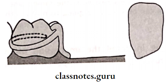
Removable Partial Dentures A Low Suevey Line Arising The Gingival Third The Near Zone

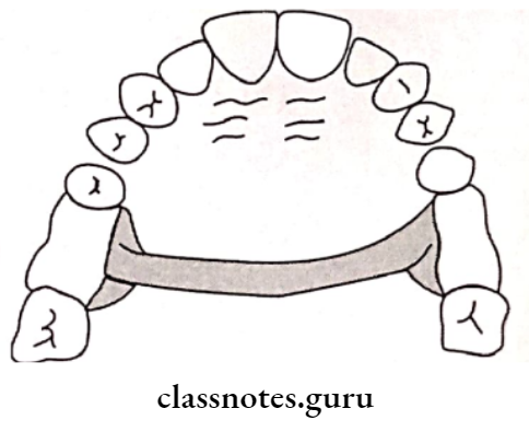
Question 6. U-shaped major connector.
Answer:

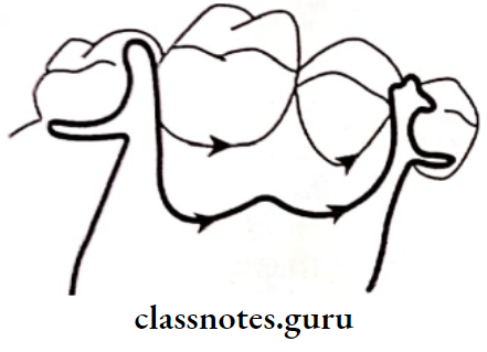
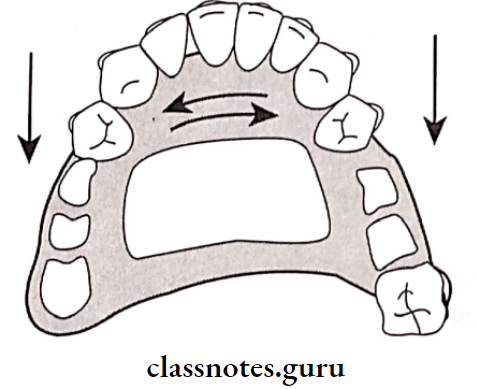
  • U-shaped Major Connector has a thin metal band running along lingual surface of posterior teeth
  • Anteriorly it covers cingula of the teeth

U-shaped Major Connector Indications:

  • Used in Kennedy’s class 4
  • Used in the presence of tori
  • Used in excessive over-bite

U-shaped Major Connector Advantages:

  • Reasonably strong
  • Has moderate indirect retention and support

U-shaped Major Connector Disadvantages:

  • When vertical forces are applied on either one or both ends it tends to straighten
  • Greater bulk is required to avoid flexing of the major , connector
  • Increased thickness may cause patient discomfort

Question 7. Indications for removable partial dentures (RPD).
Answer:

  • Length Of Edentulous Span – RPD are preferred for longer edentulous arches as it helps to distribute forces around the ridge evenly
  • Age – RPD are preferred in young patients due to presence of large dental pulps
  • Abutment – When there is no tooth posterior to the edentulous space to act as abutment, RPD is preferred
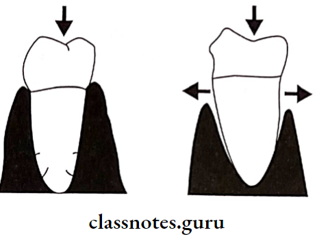
  • Periodontal Support – Tooth with weakened periodontal support are preferred for RPD as it requires less support from the abutment

Removable Partial Dentures Sound Tooth Will Distribuet The Forces And A Periodontally Weak Tooth

  • Cross- Arch Stabilization –  To stabilize the remaining teeth against lateral and anteroposterior forces, RPD is indicated

Removable Partial Dentures The RPD FrameWork Helps To Provide Stability

  • Excessive Bone Loss –  In case of ridge resorption, RPD can be fabricated as it can provide the required support and esthetics
  • Immediate Teeth Replacement – RPD is preferred as relining can be done
  • Esthetic Requirement – If required
  • Emotional Problems –  RPD is indicated as it requires shorter appointments
  • Patient Desires –
    • If the patient wants RPD due to
    • Avoidance of operative procedure
    • Economic reasons
    • Severe loss of tissue
    • Teeth with short clinical crown
    • When more than two posterior teeth or four anterior teeth are missing
    • If the canine and two of its adjacent teeth are missing
    • When there is no distal abutment
    • Presence of multiple edentulous spaces
    • In case of tilted abutment

Removable Partial Denture Designs

Question 8. Factors determining path of insertion.
Answer:

  • Aesthetics: The cast should be tilted so that the height of contour is shifted to a lower level This conceal the clasp arm as well as alters the path of insertion
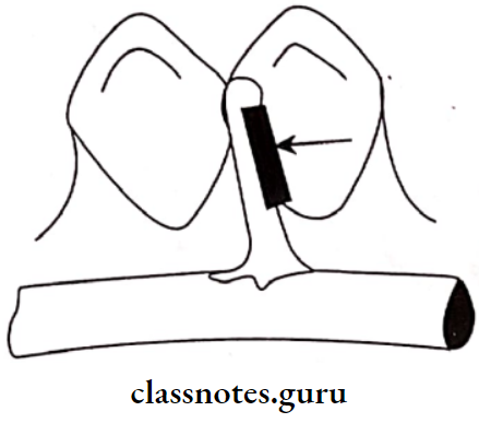
  • Guiding Planes: Two or more vertically parallel surfaces of abutment teeth so oriented as to direct the path of placement and removal of removable partial denture”
    • The path of insertion will always be parallel to the guide plane
    • The proximal plates on the partial denture will contact the guide planes during insertion

Removable Partial Dentures Since The Proximal Plates Of The Denture Forced

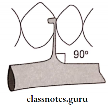
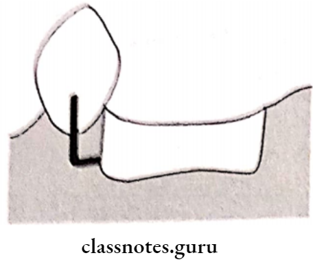
  • Location of the vertical minor connector
  • This minor connector will be parallel to the guide plane on the abutment which in turn determines the path of insertion

Removable Partial Dentures Vertical Minor Connector Parallel

  • Interference:
    • Certain areas can cause interference to the insertion
    • If these cannot be surgically removed, the path of insertion should be altered
    • Interference Examples:
      1. In Mandible:
        • Lingual tori
        • Lingually placed teeth
        • Bony exostoses
      2. In Maxilla:
        • Tori palatinus
        • Buccaly tipped teeth
        • Bony exostoses
  •  Retentive Undercuts:
    • Favourable undercuts should be evaluated to ob- tain good retention
    • The cast can be tilted until the height of contour lies between the gingival and middle third of the crown to obtain a good undercut
    • This tilting of the cast will alter the path of insertion

Removable Partial Dentures High Of Countour Located At The Middle Third Of The Tooth

RPD Procedure Explained

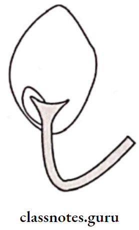
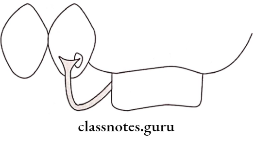
  •  Point Of Origin Of The Approach Arm Of A Bar Clasp:
    • The Approach arm forms a loop that produces terference during insertion
    • If the approach arm cannot be modified the path of insertion is to be altered
    • When the path of insertuion is altered the resulting bar clasp will not provide retention rather it will provide resistance to removal

Removable Partial Dentures Approac Arm of A Roach Or Bar Clasp

  • Denture Base:
    • Shape and extent of it determines the path of insertion
    • If the denture base extends anteriorly on both sides, it embraces abutment limiting multiple paths of insertion

Removable Partial Dentures The Proximal Plate Of The Direct Retainer

Question 9. Single Palatal Bar.
Answer:

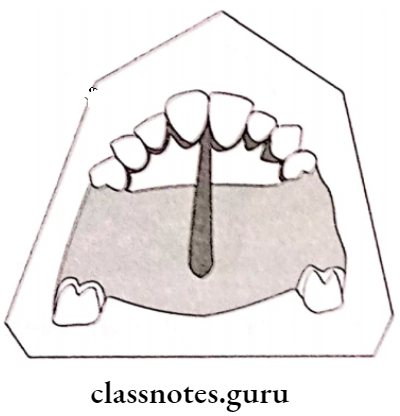
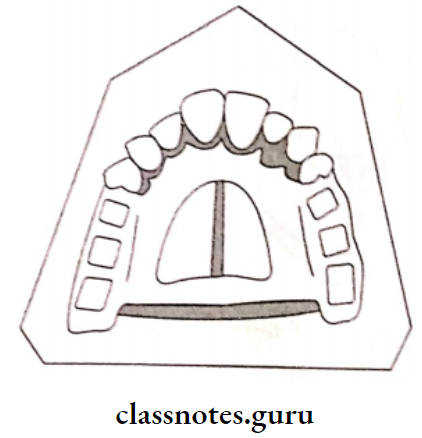
Single Palatal Bar is a bar running oval cross-section.

  • Single Palatal Bar Design:
    • It has a narrow half across the palate
    • It is thickest at the centre
  • Single Palatal Bar Fabrication:
    • Mark the extent of the bar on a cast
    • The wax pattern is fabricated and adapted over the area
  • Single Palatal Bar Indication:
    • For interim partial dentures
  • Single Palatal Bar Disadvantages:
    • Poor bony support
    • Cannot be used anterior to the premolar
    • It has poor vertical support
    • It can be used only in Kennedy’s Class 3 cases

Question 10. Factors determining clasp retention/flexibility.
Answer:

  • Depth Of The Undercut: The deeper the undercut the greater is the retention
  • Buccolingual Width Of The Undercut:
  • Clasp alloys are selected based on it
    • For 0.010″ undercut-cast chrome alloy
    • For 0.015″ undercut – Gold alloy
    • For 0.020″ undercut – Wrought wire
  • The Distance Between The Survey Line And The Tip Of The Retentive Clasp:

Removable Partial Dentures The Distance Between The Height Of Contour

  • The Mesiodistal Length Of The Clasp Arm Below The Height Of Contour:
    • Longer clasp arms offer more flexibility
    • Increased flexibility decreases the magnitude of the horizontal stresses acting on the abutment

Removable Partial Dentures For The same Tooth Looping The Retentive Arm

  • Taper Of The Clasp Arm:
    • The clasp arm should taper uniformly from its origin to the tip

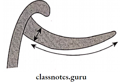
Removable Partial Dentures The RetentiveArm Should Taper

  • Cross-Sectional Form:
    • A half-round clasp arm is flexible only in one plane
    • While round clasp is flexible in all planes
    • A cast retentive clasp arm is used mainly in tooth-supported partial dentures because they need to flex only during placement and removal of the denture

Removable Partial Dentures Half Round Clasp

Question 11. Occlusal Rest Seat Preparation.
Answer:

The location and extent of the rest seat is determined during the surveying of a diagnostic cast

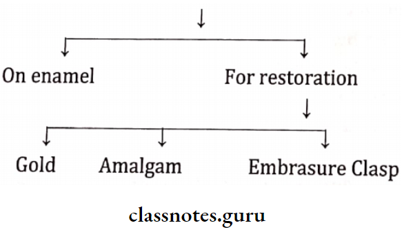
Removable Partial Dentures Occlusal Rest Seat Preparation

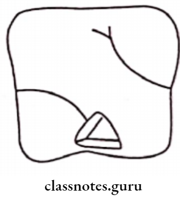
1. On Enamel:

  • A depth orientation groove is drawn along the desired outline
  • Next, the enamel is removed between these grooves using the same bur
  • The design is then verified with that marked on the primary cast
  • Occlusal clearance is checked with the help of a beading/utility
  • Rounding of internal line angles is carried out
  • Any unsupported enamel if remains is removed
  • Finally, the polishing of the rest seat is carried out

Removable Partial Dentures Occlusal Rest Seat On Enamel

2. Gold Restoration:

  • Tooth reduction/preparation is carried out for gold restoration
  • Depression is made on the tooth surface at the place of the rest seat
  • Initially, the rest seat is prepared on a wax pattern using a No. 4 round bur in slow-speed handpiece or with a discoid or cleoid excavator
  • It is polished using a finishing bur
  • Finally, the wax pattern is cast

Removable Partial Dentures Additional Tooth Reduction

  1. On Amalgam Restoration:
    • Rest seat preparation over amalgam restoration is usually avoided as it would weaken the restoration
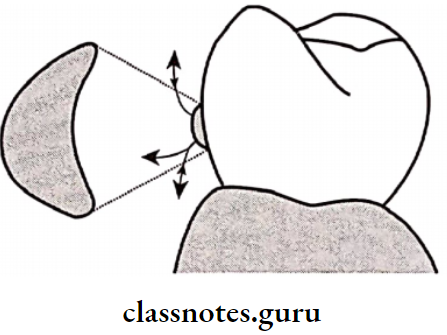
  2.  For Embrasure Clasp:
    1. Two occlusal reset seats are to be prepared over the involved teeth
    2. Both the marginal ridges are reduced equally, but the contact point should not be removed
    3. The outline must be verified
    4. Finally, finishing and polishing is done using No. 4 round steel bur

Removable Partial Dentures Preparation For And Embrasure Clasp

Question 12. Methods for obtaining functional support for distal extension base.
Answer:

Support for distal extension base is obtained by following methods

  • Functional Impression Technique:
    • The tissues get compressed during function leading to the vertical displacement of the denture
    • This type of tissue ward movement of the denture will produce rotation of the prosthesis around its terminal abutment axis
    • This is prevented if the tissues are recorded in a compressed form
    • The denture fabricated from it will seat and compress the tissues even during rest and there will be no additional tissue ward movement during function

Removable Partial Dentures The Sides Of The Cast Trimmed Parallel To The Surface

Removable Partial Dentures The Anterior Portion Of the Base Of The Maxillary Cast

  • Providing Minimal Occlusal Contact
    • The occlusal load can affect the support of the denture
    • The occlusal table of the artificial teeth should be narrowed to reduce the occlusal load
  • Identification Of Stress-Bearing Area
    • The distal extension base derives support from the stress-bearing area
    • These areas are identified in the maxillary and mandibular arch
    • Maxillary:
      • Buccal slopes of the ridge
      • Hard palate Crest of the ridge
    • Mandibular:
      • Buccal shelf area
      • Slopes of the ridge
  • Minimizing The Movement Of The Denture Base:
    • The denture base should be designed such that the forces acting on the edentulous ridge can be minimized
    • Minor connectors are added to avoid the rotation of the denture
  •  Increasing The Tissue Coverage Of The Denture Base:
    • Increasing the tissue coverage leads to a wider distribution of the occlusal load
    • Thus, it provides support

Question 13. Distal extension denture base.
Answer:

Distal extension denture bases are fabricated in case of Kennedy’s class 1 and class 2 cases

  • Support for such bases is necessary as the occlusal load may rotate the denture
  • Support for a distal extension denture is obtained from both the teeth and the tissues
  • Among it the teeth are less compressible and intrude little to the occlusal load compared to the supporting soft tissues.

Factors Affecting Distal Extension Denture Base

  • Quality of soft tissues
  • Quality of underlying bone Tissue coverage of denture base
  • Amount of forces acting on denture Stress bearing area
  • Fit of the denture
  • Type of Impression

Question 14. Methods of establishing occlusal relationship in the removable partial denture.
(or)
Occlusal relation in removable partial denture.
Answer:

  1. Noncontact Relation:
    1. Protrusion
    2. Lateral movement
    3. Rest position and freeway space
  2. Contact Relation:
    1. Protrusive
    2. Retrusion
    3. Lateral
    4. Canine guided
    5. Group function
    6. Intercuspal

Removable Partial Denture Methods:

  • Direct Apposition Of Cast:
    • Cast are fabricated
    • These casts are articulated
    • Occlusal is established over the cast on the articulator itself
  • Interocclusal Record On Remaining Posterior:
    • The upper and lower trial dentures are inserted into patient’s mouth
    • Wax or impression paste is loaded onto the occlusal surface of teeth in the mandibular occlusal rim
    • The patient is asked to slowly retrude the mandible and close on the wax till tooth contact occurs
    • The trial dentures are removed and the wax is allowed to cool
    • Both the maxillary and mandibular trial dentures are placed on their articulated casts.
  •  Occlusal Relation With Record Base And Rims:
    • Rims made from wax or modeling plastic are fabricated over record base
    • These are inserted into the patient’s mouth and various mandibular movements are recorded
  •  Entirely With Rims:
    • The occlusal relation is recorded with the help of extraoral tracing
    • The record bases attached to the recording devices are inserted in the patient’s mouth
    • The pointer is coated with precipitated chalk and denatured alcohol
    • The patient is asked to perform protrusive, lateral, and anteroposterior movements

Removable Partial Dentures Recording An Extra oral Arrow Point Tracing

  •  Registration Of Occlusal Pathways:
    • The record base is fabricated using metal/resin
    • A layer of sticky wax is placed over it
    • Next, the occlusal rim by hard inlay wax is fabricated It is inserted into the patient’s mouth
    • Mandibular movements are recorded

Question 15. Direct v/s Indirect Retention.
Answer:

Direct Retention:

  • Retention obtained in a removable partial denture by the use of clasps or attachments that resist removal from the abutment teeth.
  • Component of a removable partial denture used to re- tain and prevent dislodgement consisting of a clasp assembly called direct retainer

Removable Partial Dentures Direct Retention

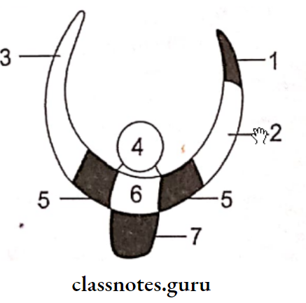
  1. Retentive terminal
  2. Retentive clasp arm
  3. Reciprocal arm
  4. Occlusal rest
  5. shoulder
  6. Body
  7. Minor Connector

Direct Retention is the ability of the component to prevent the distion and provide indirect retention placement of the denture

Indirect Retention:

  • It is the effect achieved by one or more indirect retainers of removable partial denture that reduce the tendency for a denture base to move in an occlusal direction or rotate about the fulcrum line.
  • The component of a removable partial denture that assists the direct retainer in preventing displacement of the distal extension denture base by functioning through lever action on the opposite side of the ful- crum line called indirect retainer

Removable Partial Dentures Direct Retainers

Indirect Retention is the ability of the component to retain dentures in place

Question 16. Immediate partial denture.
Answer:

“A complete removable partial denture constructed for insertion immediately following the removal of natural teeth”

Types Of Immediate Partial Denture:

  1. Temporary Immediate Partial denture
    • Indicated when the permanent immediate partial denture is likely to become ill-fitting due to more than normal bone resorption
  2. Permanent Immediate Partial denture
    • Indicated when bone resorption is expected to be less and denture prognosis is good

Advantages Of Immediate Partial Denture:

  • Immediate Partial Denture is more esthetics
  • Prevents supra-eruption and drifting of the opposing and adjacent teeth
  • Immediate Partial Denture acts like a splint over the surgical site thus controlling hemorrhage and swelling

Question 17. Swing Lock.
Answer:

  • Described by Dr. Joe J.Simmons
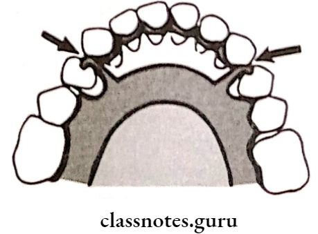
  • The swing lock has a labial bar in addition to lingual major connector
  • The labial bar extends labially all along the arch
  • The swing lock is attached to the remaining parts of the denture by a hinge on one side and a lock on the other

Removable Partial Dentures The Swing Lock

Swing Lock Method Of Use:

  • The swing lock is unlocked during insertion and locked after insertion
  • As the labial bar moves around a hinge joint these dentures are called “Swing lock dentures”

Swing Lock Function:

  • To support periodontally weak teeth

Swing Lock Indications:

  • If the functional abutment teeth are missing
  • In case of abutment teeth that has questionable prognosis
  • Presence of few remaining teeth
  • Presence of unfavorable tooth contours
  • When the position of the remaining teeth does not facilitates conventional design
  • Presence of unfavorable soft tissue contours
  • For retention and stability of a maxillofacial prosthesis.

Swing Lock Contraindications:

  • Poor oral hygiene
  • Shallow vestibule
  • High frenal attachment

Swing Lock Advantages:

  • Inexpensive
  • Provide stability and retention
  • It is possible to add additional teeth to it

Swing Lock Disadvantages:

  • Poor esthetics
  • Leads to lingual tipping of the teeth

Swing Lock Fabrication:

Metal selection chrome alloy

Surveying of cast

Designing of various components

Making the impression

Fabrication of framework

Trying of the framework

Jaw relation and teeth arrangement

Insertion of the prosthesis

Removable Partial Dentures Waxed Up Swing Lock prosthesis Ready For Processing

Removable Partial Dentures Short Answers

Removable Partial Dentures

Question 1. Contraindications for removable partial dentures
Answer:

  • Patients with macroglossia tending to push the denture away
  • Cannot be used in mentally retarded patients
  • This should be avoided in patients with poor oral hygiene

Read And Learn More: Prosthodontics Question And Answers

Question 2. Linguoplate major connector
Answer:

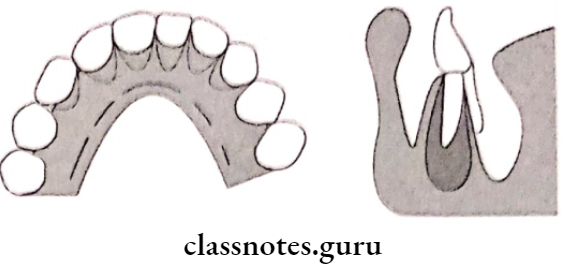
  • The superior border extends up to the cingulum
  • Linguoplate is scalloped in between the teeth
  • In the presence of large embrasures, it is made to dip down step back design
  • Anteriorly, it should be supported by the rest

Linguoplate Major Connector Advantages:

  • Rigid, stable
  • Provide indirect retention

Linguoplate Major Connector Disadvantages:

  • Food accumulation
  • Decalcification of teeth
  • Soft tissue irritation

Removable Partial Dentures Notes

Question 3. Ring clasp.
Answer:

Ring Clasp is example of a cast circumferential clasp

  • Ring Clasp Indication:
    • Distal extension denture
  • Ring Clasp Contraindication:
    • Soft tissue undercut
    • Buccinator’s attachment, if present close to the  lower molar
  • Ring Clasp Disadvantages:
    • Alter food flow
    • Increased tooth surface coverage
    • Cannot retain its physical qualities
    • Difficult to repair or adjust

Removable Partial Dentures Short Answers

Question 4. Objectives of surveying
Answer:

  • To achieve good retention and bracing by designing the rigid and flexible components of a removable prosthesis
  • Aids in marking the survey lines by determining the height of the contour of hard and soft tissue areas above the undercut
  • Decides the path of insertion
  • Helps in deciding the areas into which the prosthesis should not extend by determining the undesirable undercuts

Question 5. Cingulum rest
Answer:

  • Also known as lingual rest
  • Cingulum Rest is placed on the lingual surface of a tooth
  • Cingulum Rest is specially placed over maxillary canine

Question 6. Enameloplasty/ Dimpling.
Answer:

  • Enameloplasty is defined as the intentional alteration of the occlusal surface of the teeth to change their form
  • Enameloplasty is done to produce a retentive undercut
  • Enameloplasty is a gentle depression created on the enamel surface of the abutment teeth to provide a retentive undercut It is done when abutment teeth do not provide any surface undercut
  • Enameloplasty can also be done to modify the existing undercut

Question 7. Limitations of Kennedy’s classification.
Answer:

  • Did not explain the length of the edentulous span or the number of missing teeth only provide number of modification spaces.
  • Not enough consideration about the condition of teeth and the remaining supporting structures.
  • No distinction between modification spaces which occur in the anterior segment to those of the posterior segment

RPD Prosthodontics Guide

Question 8. Uses of surveyor.
Answer:

  • Surveying the diagnostic and primary casts
  • Tripoding the cast
  • Transferring the tripod marks to another cast
  • Contouring wax pattern
  • Contouring crowns and cast restoration Placing internal attachments and rests
  • Performing mouth preparation on casts
  • Surveying master cast
  • Surveying ceramic veneer

Question 9. Cingulum bar
Answer:

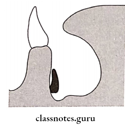
  • The Cingulum Bar is located on or slightly above the cingula of the anterior teeth
  • Indications:
  • Large embrasures
  • Large diastema cases

Question 10. Merits of occlusal approaching clasp.
Answer:

Occlusally Approaching Clasp is easy to fabricate and repair

  • Occlusally Approaching Clasp leads to less food retention
  • Occlusally Approaching Clasp can be best applied in a tooth-supported partial denture
  • Provides excellent support and retention

Question 11. Define  Interim Removable Denture.
Answer:

“A transitional denture may become an interim denture when all of the natural teeth have been removed from the dental arch”.

Question 12. Support for RPD
Answer:

  • Support for RPD depends on
  • Quality of the residual ridge
  • Total occlusal load applied
  • Accuracy of the denture base
  • Accuracy and type of impression registration

Question 13. Height of Contour.
Answer:

Height of Contour Definition: A line encircling a tooth designating its greatest circumference at a selected position.

Height of Contour Significance:

  • Height of the Contour is used as a guideline that helps in the placement of the components of the clasp
  • The retentive clasp arm of the retentive arm is located above the height of contour while the retentive terminal lies below the height of the contour
  • The reciprocal arm is located above the height of contour

Removable Partial Denture Components

Question 14. Guiding Planes.
Answer:

Guiding Planes Definition: “Two or more vertically parallel surfaces of abutment teeth so oriented as to direct the path of placement and removal of removable partial denture”.

Types Of Guiding Planes:

  • Guide planes on abutment teeth supporting a tooth supported partial denture
  • Guide planes on abutment teeth that supports a secondary distal extension denture base
  • Guide planes prepared on lingual surfaces of abutment teeth
  • Guide planes on anterior abutments

Removable Partial Dentures Guide Planes For Tooth Supported Partial Denture

Removable Partial Dentures Guide Planes For A Distal Extension Denture Base

Removable Partial Dentures Canine And Incisors

Functions Of Guiding Planes:

  • Minimizes the wedging stresses on the abutments
  • Makes insertion and removal easier
  • Aids to stabilize the prosthesis against horizontal stresses
  • Aids to stabilize individual teeth
  • Guiding Planes improves oral hygiene
  • Guiding Planes contributes to indirect retention and frictional retention

Question 15.  Internal Rests.
Answer:

  • Internal Rests are large box shaped metallic extensions that function as intracoronary retainers
  • Internal Rests differ from internal attachments
  • Internal Rests are not pre-fabricated instead the rest seat is formed by the sound tooth structure

Removable Partial Dentures Internal Rest Seat

Question 16. Prothero’s cone theory.
Answer:

  • Prothero described cone theory in 1916 to explain the basis for clasp retention
  • He described the shape of the crown of premolars and molars to be equivalent to two cones sharing a common base
  • The upper cone resembles the occlusal half of the tooth and the lower cone resembles the cervical half of the tooth
  • A clasp tip that ends below the junction of the two cones will resist movement in the upward direction
  • The degree of resistance to deformation determines the amount of clasp retention

Removable Partial Denture Designs

Question 17. Combination clasp.
Answer:

A Combination Clasp is a combination of wrought wire and rigid-cast alloy

  1. Combination Clasp Indication: Maxillary canines and premolars
  2. Combination Clasp Contraindications: Undercut adjacent to edentulous space
  3. Combination Clasp Advantages: It has thin line contact It can flex in all planes
  4. Combination Clasp Disadvantages:
    • Tedious lab procedures
    • Easily breaks or distorts
    • Poor stability

Question 18. Half and Half clasp.
Answer:

  • Half And Half Clasp has a retentive arm arising from one direction and a reciprocal arm arising from another
  • Thus it needs two minor connector
    1. First attaches the occlusal rest and the retentive arm to the major connector
    2. Second, connects the reciprocal arm with or without an auxiliary rest
  • Half And Half Clasp Modification:  The reciprocal arm is converted into short bar to reduce the tooth coverage
  • Half And Half Clasp Advantage: It provides dual retention

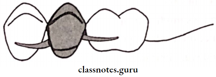
Removable Partial Dentures Occlusal View And Proximal View

Question 19. Fulcrum Line.
Answer:

  • Fulcrum Line Definition: “An imaginary line around which a partial denture tends to rotate”
  • Fulcrum Line Location: It is usually formed at the terminal abutment axis

Types of Fulcrum line:

  • Retentive Fulcrum Line: “An imaginary line connecting the retentive points of clasp arms, around which the denture tends to rotate when subjected to forces, such as the pull of sticky foods”.
  • Stabilizing Fulcrum Line: “An imaginary line, connecting occlusal rests, around which the denture tends to rotate under masticatory forces”.

Question 20. Displacing forces.
Answer:

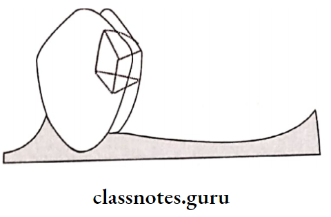
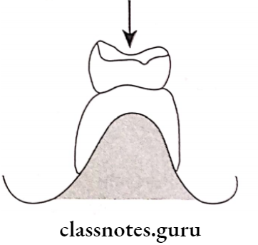
Removable Partial Dentures Displacing Forces

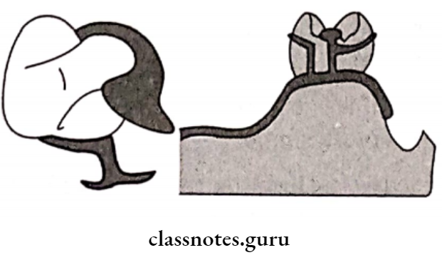
Removable Partial Dentures Tissue Supported Partial

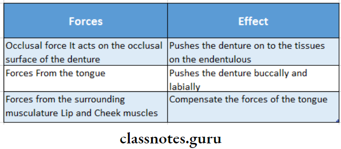
Removable Partial Dentures The Forces Of The Tongue And Cheek

Question 21. Group Function.
Answer:

Group Function Definition:

Group Function is multiple contract relations between the maxillary and mandibular teeth in lateral movements on the working side whereby simultaneous contact of several teeth acts as a group to distribute occlusal forces

Group Function Indications:

  • End-on-end bite
  • Anterior open bite
  • Class 2 and Class 3 molar relation

Group Function Types:

RPD Procedure Explained

  1. Group Function Partial group function:
    • In this occlusion, some of the posterior teeth are able to shear the load in excursion whereas others contact only in centric relation.
  2.  Anterior group function:
    • A most practical method of dis occluding
  3. Group Function Advantages:
    • Efficient and comfortable
    • Distributes wear over more teeth
    • Distributes stresses over more teeth
    • Distributes stresses to teeth farther from the condylar axis
  4. Group Function Limitations:
    • Not possible in all cases
    • Some arch relations do not permit it
    • Concave anterior guidance permits it whereas


Removable Partial Dentures Long Essays

Question 1. Define rests and rest seats. Discuss the various types of rests, their functions, and their design considerations.
Answer:

1. Rest: It is defined as “A rigid extension of a fixed or removable partial denture which contacts a remaining tooth or teeth to dissipate vertical or horizontal forces.”

Removable Partial Dentures Long Essay

2.  Rest Seat: The prepared recess in a tooth or restoration created to receive the occlusal, incisal, cingulum or lingual rest

  • Types Of Rest Seat:
    1. Based on the relation of rest to the direct retainer
      • Primary rest-placed along the clasp assembly
      • Secondary rest placed away from the clasp

Read And Learn More: Prosthodontics Question And Answers

    1. Based on the position of the rest
      • Occlusal rest placed on occlusal surface of posterior
      • Cingulum rest placed on lingual surface of the tooth
      • Incisal rest-placed on the incisal edge of the tooth
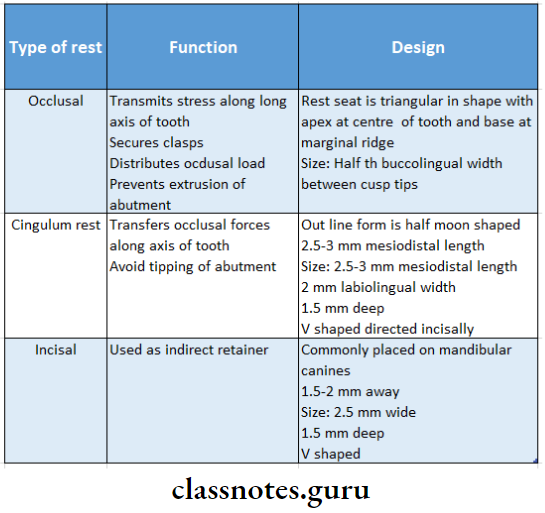
Removable Partial Dentures Type Of Rest And Function And Design

Question 2. Define indirect retainer. What are the functions of an indirect retainer?
Or
Write about various forms of indirect retainers in detail.
Or

Define indirect retainers and discuss in detail about various types of it.
Answer:

Indirect Retainers:

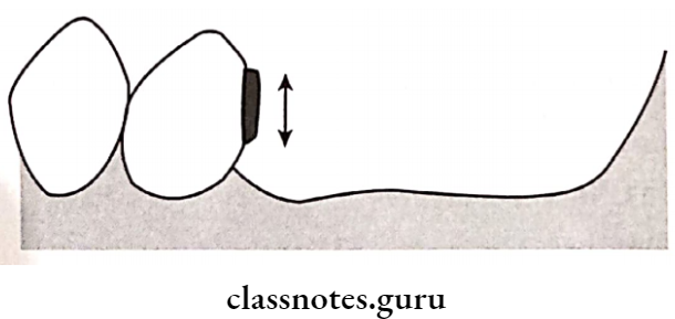
Indirect Retainers is defined as “a part of a removable partial denture which assists the direct retainers in preventing displacement of distal extension denture bases by functioning through lever action on the opposite side of the fulcrum line.
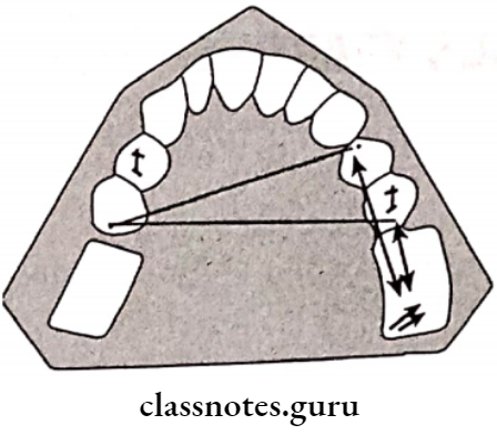
Removable Partial Dentures Moving The Stablizing Fulcrum Line

Functions Of Indirect Retainers:

  • Indirect Retainers counteract the lifting forces and stabilizes the denture It counteracts horizontal forces and provide stability and support to the denture
  • Indirect Retainers can splint and protect the anterior teeth
  • Indirect Retainers may act as an auxillary rest
  • The dislodgement of indirect retainer suggest the need of relining.

Types Of Indirect Retainers:

1. Auxillary Occlusal Rest:

  • Auxillary Occlusal Rest is located on the occlusal surfaces away from the distal extension base
  • Auxillary Occlusal Rest is placed perpendicular to the midpoint of the fulcrum line.
    • Location:
      • Bilaterally over 1st premolars in Kennedy’s class 1
      • Over 1st premolar of opposite side in Kennedy’s class 2

2. Canine Extension From The Occlusal Rest:

  • Occlusal Rest is a finger-like extension from a premolar rest is placed on the lingual slope of the adjacent canine
  • Occlusal Rest is used when the first premolar must also acts as a primary abutment
Removable Partial Dentures Long Essays

3. Canine Rest:

  • If the mesial marginal ridge of the first premolar is close to the fulcrum line, canine rest is used.

4. Continuous Bar Retainers And Lingual Plates:

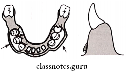
  • In Kennedy’s class I and class II, these retainers are placed just above the middle third of the anterior teeth to act as indirect retainers.
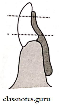
Removable Partial Dentures Continuous Bar Retainers And Linguoplates

5. Modification Areas:

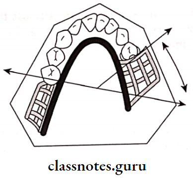
  • In class 2 modification I, the fulcrum line runs from the left second premolar to the right second molar
  • In such cases, the direct retainer resting on right first premolar act as an indirect retainer
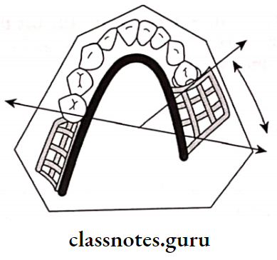
Removable Partial Dentures Direct Retainers

6. Rugae Support:

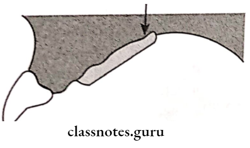
  • As the rugae area is firm and well placed it can be used for indirect retention for a palatal horseshoe major connector.
  • Horde shoe Connector lacks Posterior retention
  • In this case ruge acts as indirect retainer

Removable Partial Denture Prosthodontics Notes

7. Direct Indirect Retention:

The reciprocal arm of a direct retainer located anterior to the fulcrum line may act as an indirect retainer.
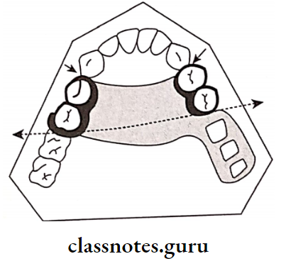
Removable Partial Dentures Reciprocal Arms Of Direct Retainers

8. Indirect Retention From Major Connector:

The major connector provides indirect rerention due to its rigidity
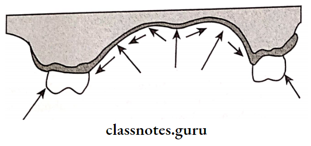
Removable Partial Dentures Rigid Major Connectors Resist

Question 3. Define direct retainers. Enumerate the requirements of direct retainers with a note on the circumferential clasp.
Answer:

Direct Retainers Definition:

Direct Retainers is defined as “A clasp or attachment placed on abutment tooth for the purpose of holding a removable denture in position

Direct Retainers Requirements:

  1. Passivity:
    • The quality or condition of inactivity or rest assumed by the teeth, tissues, and denture when a removable partial denture is in place but not under masticatory pressure
    • The retentive function should act only when dislodging forces are present
  2. Retention:
    • It is that quality inherent in the prosthesis that resists the force of gravity, the adhesiveness of foods, and the forces associated with the opening of the jaws
    • It is provided by the retentive arm of the clasp
  3. Reciprocation:
    • It is defined as the means by which one part of a prosthesis is made to counter the effect created by another part
    • It is provided by a rigid reciprocal arm
    • It resists the stresses generated by the retentive arm
  4. Encirclement:
    • It is the property of the clasp assembly to encompass more than 180 degrees of the abutment tooth either by continuous or broken contact to prevent dislodgement during the function
    • Each clasp much encircle more than 180 degree of the abutment tooth
    • There can be continuous contact or broken contact
  5. Stability:
    • It is defined as the quality of a denture to be firm, steady, or constant to resist displacement by functional stresses and not to be subject to change of position when forces are applied
    • Provided by shoulder and vertical arm of minor connector
  6. Support:
    • It is defined as holding up or serving as a foundation or prop for
    • It is resistance to the movement of the denture in a gingival direction provided by rest
    • Path of escapement should never coincide with the path of removal
    • Primary abutment should never exert tipping forces on the abutment
    • Place reciprocal elements at the height of the contour and retentive element below the height of contour

Circumferential Clasp/Maker’s Clasp

  • These clasps embrace more than half of the abutment tooth
  • Maker’s Clasp prevents the rotation of the denture
  • They approach the undercut from an occlusal direction

Maker’s Clasp Advantages:

  • Maker’s Clasp is easy to fabricate and repair
  • Maker’s Clasp leads to less food retention
  • Maker’s Clasp can be best applied in a tooth-supported partial denture
  • Provides excellent support and retention

Maker’s Clasp Disadvantages:

  • Maker’s Clasp covers a large tooth surface area
  • Alters buccolingual width of the crown
  • Interferes with the normal food flow
  • This leads to food accumulation and decalcification of the tooth structure
  • Deprives periodontal stimulation
  • Maker’s Clasp cannot be used for cases with an undercut away from edentulous space

Question 4. Classify partial edentulous areas according to Applegate-Kennedy’s classification and mention Applegate’s rules for Kennedy’s classification.
Answer:

Applegate Kennedy’s classification:

  • Proposed by Dr. Edward Kennedy in 1923
  • Kennedy classified partially edentulous arches and not the dentures
  • This classification is anatomical
  • In Kennedy’s classification, he included 4 classes Further, 2 additional classes were included by Apple-gate

Applegate Kennedy’s Classification Is As Follows:

  • Class 1:
    • Bilateral edentulous areas located posterior to the remaining natural teeth
    • There are two edentulous spaces located in the posterior region without any teeth posterior to it.
  • Class 2:
    • The unilateral edentulous area located posterior to the remaining natural teeth
    • There is a single edentulous space located in the posterior region without any teeth posterior to it.
  • Class 3:
    • Unilateral edentulous area with natural teeth anterior and posterior to it.
    • It indicates a single edentulous area that does not cross the midline of the arch
  • Class 4:
    • The single, bilateral edentulous area located anterior to the remaining natural teeth
    • It crosses the midline of the arch
    • Teeth are present only posterior to the edentulous arch
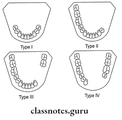
Removable Partial Dentures Teeth Are Preseny Only Posterior To The Edentulous Arch

  • Class 5:
    • The edentulous area bounded anterior and posteriorly by natural teeth
    • But the anterior abutment is not suitable for support
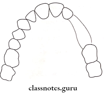
Removable Partial Dentures Kennedy Applegates Class 5 Partially Edentulous Condition

  • Class 6:
    • Edentulous area in which the teeth adjacent to the space are capable of total support of the required prosthesis
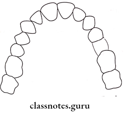
Removable Partial Dentures Kennedy Applegates Class 6 Partially Edentulous Condition

Applegate Kennedy’s Applegate’s Rules:

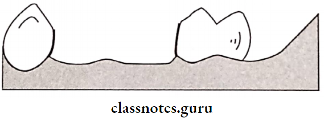
  1. Rule One: Classification should follow rather than preceed extractions that might alter the original classification
  2. Rule Two: If the third molar is missing and not to be replaced, it is not considered in the classification
  3. Rule Three: If the third molar is present and is used as abutment, it is considered in the classification
  4. Rule Four: If the second molar is missing and not to be replaced, it is not considered in the classification
  5. Rule Five: The most posterior edentulous area or areas always determine the classification
  6. Rule Six: Edentulous areas other than those, which determine the classification, are referred to as modification spaces and are designated by their number
  7. Rule Seven: The extent of the modification is not considered, only the number of additional edentulous spaces are considered
  8. Rule Eight: There can be no modification areas in class 4.

RPD Prosthodontics Long Questions

Question 5. Define surveyor. Mention its parts. Explain in detail step by step procedure in surveying and Functions.
Or
Define dental cast surveyor. Enumerate their function and Parts of surveyors.
Or
Describe the surveying procedure and Functions and Parts of surveyors.
Answer:

Definition Of Surveyor:

An instrument used in the construction of removable partial dentures to locate and delineate contours and relative positions of abutment teeth and associated structures

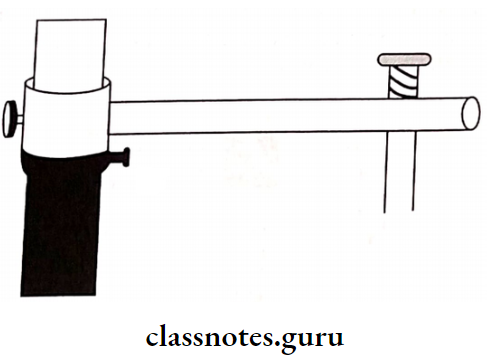
Parts Of Surveyor:

  • Surveying Platform:
    • The surveyor is metal plate parallel to the floor
    • The surveyor forms base of the surveyor
    • A Cast holder is placed over it
  • Cast Holder/Surveying Table:
    • Cast Holder has a base and table to place a cast
    • Cast Holder should not be tilted more than 10 degrees
  •  Vertical Arm:
    • The Vertical Arm arises vertically from the surveying platform
  • Horizontal Arm:
    • Vertical Arm extends from top of the vertical arm
    • Vertical Arm supports the surveying arm at its free end
    • Vertical Arm Variations:
      • Fixed- in Ney surveyor
      • Revolving- in Jelenko surveyor

Removable Partial Dentures Horizontal Arm

  • Surveying Arm:
    • Surveying Arm extends from free end of horizontal arm
    • Surveying Arm is parallel to the vertical arm
    • Surveying Arm can move upward and downward
    • Its lower end has mandrel for attachment of surveying tools
  • SurveyingTools:
    • Analyzing Rod:
      • Analyzing Rod is solid cylindrical metal rod
      • Analyzing Rod helps to analyze the location of height of contours, presence and absence of undercut
      • Analyzing Rod helps to determine path of insertion
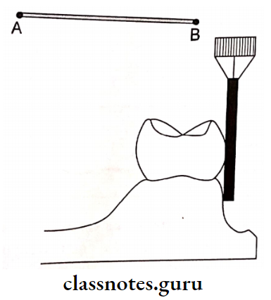
Removable Partial Dentures Using A Analyzing Rod To Analyze A Cast

    • Carbon Markers:
      • Carbon Markers resemble the lead points
      • Carbon Markers are used to draw height of contour of the object
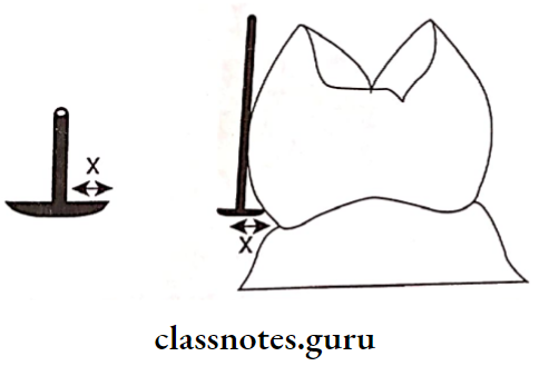
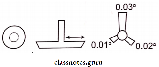
    • Undercut Gauges:
      • Undercut Gauges is used to measure the depth and location of the undercuts
      • Sizes:
        • According to Stewart-0.010″, 0.015″, 0.020″ According to McCracken- 0.010″, 0.020″, 0.030″
      • Shape:
        • Ney surveyor circular beaded
        • Jelenko- Fan shpaed bead

Removable Partial Dentures The Connector Area Unified

Removable Partial Dentures Net Under Cut Gauge

    • Wax Knife:
      • Used to trim the excess wax and to eliminate undesirable undercut
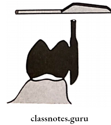
Removable Partial Dentures Surveying Wax Knife

Surveying Procedure:

  • The primary cast is mounted on the surveying table It is locked in the position with a zero-degree tilt with the help of clamps on the surveying table
  • Surveying is mounted such that the occlusal surfaces of the remaining teeth are parallel to the base
  • After this, the surveying arm is positioned in relation to the cast
  • Surveying is positioned as follows:
    • First, the horizontal arm is adjusted vertically such that the surveying arm can contact at least three different spaced-out points on the cast
    • Surveying is locked to the vertical arm with the help of a thumb screw Next, the cast is analyzed by using different analyzing tools

Components of Removable Partial Dentures Essay

Analyzing Tools:

  • Analyzing Rod:
    • Analyzing is the first surveying tool used
    • Analyzing is attached to the mandrel of the surveying arm
    • The cast is rotated against it to analyze the presence of undercuts whether favorable or unfavorable
  •  Carbon Markers:
    • These resemble the lead points
    • After analyzing the teeth with analysing rod, it is replaced by carbon markers
    • These are used to draw survey lines, which denote the height of the contour of the teeth
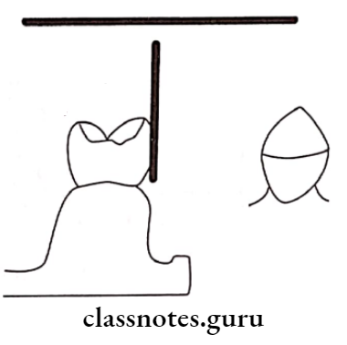
Removable Partial Dentures Marking Or Scribing The Height Contour A Carbon Marker

  • Undercut Gauges:
    • Undercut Gauges are used to measure the depth and location of the undercuts in three dimensions

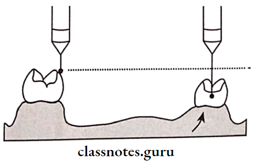
Tripoding:

  • Tripoding is done next to maintain the records of the primary cast in the master cast
  • A carbon marker is fixed to the mandrel of the surveying arm along with a locked horizontal arm
  • The surveying arm is moved freely
  • Due to this three points are marked that lie in the same plane
  • Additional reference points are marked
  • They are
    • A distal marginal ridge of 1st premolar Incisal edge of the lateral incisor
    • Lingual cusp tip of 1st premolar of opposite side
  • After marking the primary cast is removed and the master cast is placed on the surveying table
  • The master cast is adjusted in the surveying table such that the carbon marker contacts in the same manner as that with the primary cast
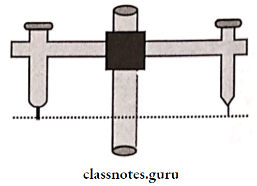
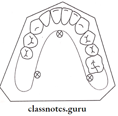
Removable Partial Dentures Once The Surveying Arm

Removable Partial Dentures Tripoding The Primary Cast

Removable Partial Dentures The Master Card

Functions of Surveyor:

  • Surveying the diagnostic and primary casts
  • Tripoding the cast
  • Transferring the tripod marks to another cast
  • Contouring wax pattern
  • Contouring crowns and cast restoration
  • Placing internal attachments and rests
  • Performing mouth preparation on casts
  • Surveying master cast
  • Surveying ceramic veneer

Uses Of Tripoding:

  • Helps in positioning the master cast
  • Helps to remount the diagnostic casts on the surveying table

Question 6. Define major connector. Mention different types and discuss criteria for the selection of major connectors.
Answer:

Definition of Major Connectors:

Major Connectors is defined as “A part of a removable partial denture which connects the components on one side of the arch to the components on the opposite side of the arch”

Types Of Major Connectors:

  1. Maxillary:
    • Single posterior palatal bar
    • Palatal strap
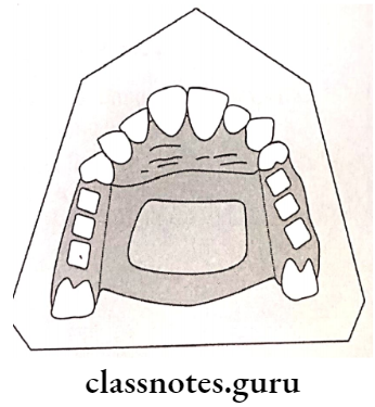
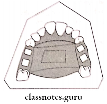
    • Palatal plate
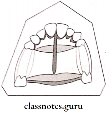
    • Antero posterior palatal bar
    • Horseshoe connector Closed horseshoe
    • Complete palate
  2. Mandibular:
    • Lingual bar Lingual plate
    • Kennedy bar
    • Sublingual bar
    • Cingulum bar
    • Labial bar

Criteria For Selection Of Major Connectors:

Major connector should fulfill the following criteria

  1.  Functional Requirement: It should distribute forces among all the teeth
  2. Anatomical Restraints: In case of mandibular connector less space is present, hence connector is designed accordingly
  3. Relief: Adequate amount of releif should be provided
  4. Hygiene: It should be enable to clean the connector
  5. Patient’s Compliance:
    • It should be accepted by the patient
    • It should be comfortable

Removable Partial Dentures Patients Compliance

Question 7. Define removable partial denture. How do you choose a direct retainer for RPD.
Answer:

Removable Partial Denture:

  • Any prosthesis that replaces some teeth in a partially dentate arch is called a removable partial denture
  • It can be removed from the mouth and replaced at will

Classification of RPDs in Prosthodontics

Selection Of Direct Retainer For RPD:

Type of direct retainer is selected according to the following condition
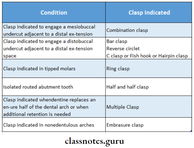
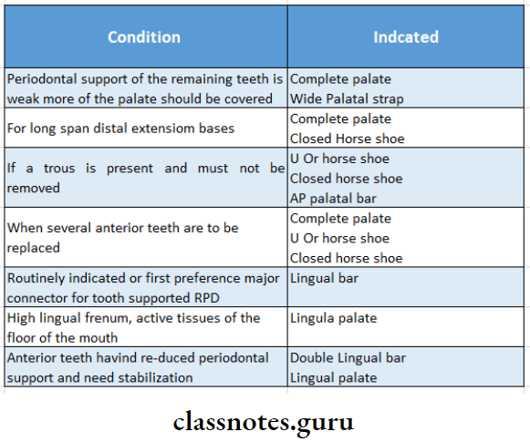
Removable Partial Dentures Selection Of Direct Retainer For RPD

Question 8. What is the direct retainer? Describe its parts
Answer:

Definition of Direct Retainer:

Direct Retainer is defined as “A clasp or attachment placed on an abutment tooth for the purpose of holding a removable denture in position

Parts of Direct Retainer:

1. Retentive Arm:

“A flexible segment of a removable partial denture which engages an undercut on an abutment and which is designed to retain the denture”

Retentive Arm Parts:

  1. Retentive Clasp Arm:
    • It is not flexible
    • It is located above the height of the contour
  2. Retentive Terminal:
    • It is flexible
    • It is located below the height of the contour
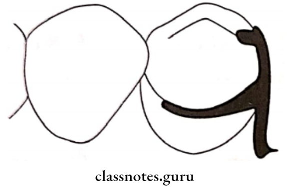
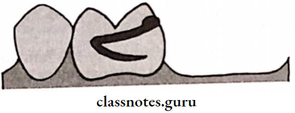
Removable Partial Dentures The Retentive Terminal

2. Reciprocal Arm:

  • “A clasp arm or other extension used on a removable partial denture to oppose the action of some other part or parts of the prosthesis”
  • Reciprocal Arm is located on the opposite side of retentive arm
  • Reciprocal Arm is placed above the height of the contour
    • Reciprocal Arm Functions:
      • Acts as an indirect retainer
      • Reciprocal Arm can resist the rocking of the denture base
      • Reciprocal Arm provides stability and reciprocation against the retentive arm
      • The denture is stabilized against horizontal movement
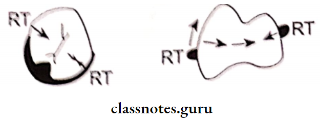
Removable Partial Dentures The Reciprocal Arm

3. Retentive Arm Shoulder:

  • The shoulder is part of the clasp that connects the body to the clasp terminals
  • The shoulder lies above the height of the contour and provides stabilization against horizontal displacement

4. Retentive Arm Body:

  • The Retentive Arm is part of the clasp that connects the rests and shoulders of the clasp to the minor connector.
  • Retentive Arm is rigidly above the height of the contour
  • Retentive Arm is designed such that it contacts the guide plane of the abutment during insertion and removal

5.Retentive Arm Rest:

  • Retentive Arm is rigid extension of the removable partial denture that contacts a remaining tooth or teeth to dissipate vertical or horizontal forces
  • Retentive Arm is the part of the clasp that lies on the occlusal or lingual or incisal edge or surfaces of the tooth
  • Retentive Arm resists tissue-ward movement of the clasp by acting like a vertical stop

6. Minor connector:

  • Minor Connector is connecting link between the major connector or base of a removable partial denture and other units of the prosthesis, such as clasps, indirect retainers and occlusal rests
  • Minor Connectror joins the clasp with the remaining part of the metal framework

Question 9. Discuss how to achieve stress equalization in Kennedy’s class 1 and 2 situations.
Answer:

Stress Equalization Or Stress Breaker:

This is used for stress equalization in Kennedy’s class I and II situation

Definition of Stress Breaker:

A device that relieves the abutment teeth of all or part of the occlusal forces

Significance of Stress Breaker:

  • In a tooth tissue-supported partial denture when an occlusal load is applied, the denture tends to rock due to the difference in the compressibility of the abutment teeth and the soft tissues
  • This can produce harmful effects on the abutment teeth In order to protect the abutment from such conditions, stress breakers are incorporated into a denture
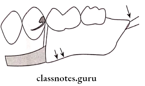
Removable Partial Dentures Stress Breakers

Advantages of Stress Breaker :

  • Stress Breaker preserves the alveolar support of the abutment
  • Stress Breaker helps to create balanced stress on the residual ridge and the abutment
  • Stress Breaker requires minimal direct retention
  • This avoids the frequent need for relining and rebasing Weak teeth are well splinted
  • Stress Breaker produces a massaging effect over soft tissues

Disadvantages of Stress Breaker:

  • Stress Breaker is expensive
  • Stress Breaker is difficult to repair
  • Stress Breaker gets easily distorted
  • Stress Breaker tends to food accumulation
  • Stress Breaker reduces the stability of the denture It reduces indirect retention

Types of Stress Breakers:

1. Stress Breakers Type 1:

  • Here a movable joint is placed between the direct retainer and a denture base
  • It decreases the amount of force acting on the abutment
  • A Movable joint is placed between the direct retainer and denture base
  • This joint may either be a Hinge or a ball and socket or a sleeve and cylinder
  • Example: DALBO

Removable Partial Denture Base Shows Independent Movement

2. Stress Breakers Type 2:

  • Stress Breakers has flexible connections between the direct retainer and the denture base
  • Stress Breakers can be a wrought wire connector divided or split major connector or a movable joint between two major connectors

Question 10. Classify direct retainers in the removable partial prosthesis. Explain about occlusal approaching clasps.
Answer:

Direct Retainer:

“A clasp or attachment applied to an abutment tooth for the purpose of holding a removable denture in position.”

Classification of Direct Retainer:

By Herman:

  1. Extracorporeal Direct Retainer: “A part of a removable partial denture which acts as a direct retainer for the denture by partially encircling or contacting an abutment tooth”
    • Extracorporeal Direct Retainer includes:
      • Suprabulge clasps
      • Infrabulge clasps
  2. Intracoronary Direct Retainer:
    1. In it, a part or the whole of the retentive components are located within the anatomic contour of the abutment teeth
      • Precision attachment
      • External attachment
      • Stud attachment
      • Bar attachment

Occlusally Approaching Clasps:

  • Circumferential Clasp/Aker’s clasp:
    • These clasps embrace more than half of the abutment tooth
    • Clasps prevents the rotation of the denture
    • They approach the undercut from an occlusal direction
  • Occlusally Approaching Clasps Advantages:
    • Clasps is easy to fabricate and repair
    • Clasps leads to less food retention
    • Clasps can be best applied in a tooth-supported partial denture
    • Provides excellent support and retention
  • Occlusally Approaching Clasps Disadvantages:
    • Clasps covers a large tooth surface area
    • Alters buccolingual width of the crown
    • Interferes with the normal food flow
    • Leads to food accumulation and decalcification of the tooth structure
    • Deprives periodontal stimulation
    • It cannot be used for cases with an undercut away from edentulous space
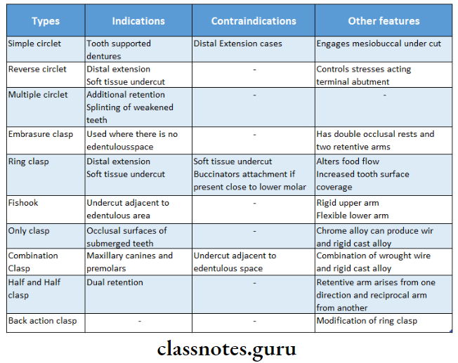
Removable Partial Dentures Types And Indications And Containdications And Other Features

Removable Partial Dentures Simple Circlet Clasp

Removable Partial Dentures Reverse Circlet Clasp

Removable Partial Dentures Multiple Circlet Clasp

Removable Partial Dentures Embrasure Clasp

Removable Partial Dentures Ring Clasp

Removable Partial Dentures Harpin Clasp

Removable Partial Dentures Onlay Clasp

Removable Partial Dentures Combination Clasp

Question 11. Give in detail the impression methods for providing adequate support in extension base removable partial denture.
Or
Physiologic impression in RPD
or
Functional relining methods
or
Fluid wax functional impression
or
Impression procedures for distal extension RPD
Answer:

  • Dual impression techniques are used for extension base removable partial dentures.
  • Such techniques record a part in the functional form and the remaining areas in the anatomical form

 Dual Impressions Are Classified As:

  • Physiological Or Functional Dual Impression:
    • McLean’s technique
    • Hindle’s modification
    • Functional relining method
    • Fluid wax method
  • Selective Pressure Technique:
    • McLean’s technique
    • A custom-made impression tray is fabricated over the edentulous areas of the primary cast
    • Occlusal rims are made on the custom tray
    • The tray is loaded with impression material
    • It is inserted into the patient’s mouth
    • Ask the patient to close on the rims
    • Due to this, the tissues under the tray are compressed and the impression is recorded in this relation
    • An alginate impression is made over the existing impression using a large stock tray
    • Finger pressure is applied on the stock tray
    • The impression is removed
    • This leads the alginate over the impression to carry the functional impression along with it
    • A cast is poured into the impression

Removable Partial Dentures Special Try Confined

Removable Partial Dentures the Patient Asked To Close On The Special Tray

Removable Partial Dentures the Patient Asked To Close On The Special Tray

Removable Partial Dentures Inverted Pick Up Impression

Removable Partial Dentures Pressure Applied Over The Stock Tray While

  • Hindle’s Modification:
    • A special tray with stoppers and occlusal rims is fabricated using primary cast
    • Hindle’s is loaded with impression material
    • Hindle’s is inserted in the patient’s mouth
    • A special stock tray with large holes loaded with alginate is used for over-impression
      • The finger is placed into the holes to apply pressure on the occlusal rim
      • Pressure is held till the alginate sets
      • This pressure pushes the tray against the edentulous ridge

Removable Partial Dentures Making The Pickup Impression Using Hindles

  • Functional Relining Method:
    • The single anatomic master impression is made
    • The master cast is made from it
    • It is then duplicated and a refractory cast is made • Soft metal spacer is adapted to the cast
    • A framework is fabricated using a refractory cast and tried in the patient’s mouth
    • Spacer is removed
    • A functional impression is made on the tissue surface of a framework using low-fusing modeling plastic
    • It is tempered and placed in the mouth
    • Sufficient pressure is applied
    • The modeling plastic at the borders of the framework is re-softened
    • Border molding is carried out
    • Modelling plastic is reduced by 1 mm
    • The final impression is made with zinc oxide eugenol impression paste.

Removable Partial Dentures Modeling Plastic

Removable Partial Dentures the Modeling Plastic Should Relived To Allow Space

  • Fluid Wax Functional Impression:
    • The metal framework is fabricated using a refractory cast
    • Fluid Wax is tried in the patient’s mouth
    • The framework is positioned on the master cast
    • The outline of the tray is drawn on the master cast
    • The cast is coated with separating media
    • A spacer is adapted over the crest of the edentulous ridge
    • The framework is placed over the spacer
    • Auto-polymerizing resin is adapted over the framework
  • Excess Material Is Trimmed:
    • Relief holes are made along the crest of the ridge
    • Impression wax is softened and painted over the tissue surface of the framework
    • It is seated in the patient’s mouth with half-open for at least 5 minutes
    • The framework is removed and the impression is examined for glossy and dull areas
    • Finally, place it for 12 minutes

Removable Partial Dentures Marking The Extent Of The Spacial Tray On The Cast

Removable Partial Dentures A Spacer Is Adapted Over The Required Area

Removable Partial Dentures After Adapting The Spacer

Removable Partial Dentures Excess Dough Material Is Trimmed Away

Removable Partial Dentures Relief Holes May Be provided In The Special Tray

Removable Partial Dentures Fluid Was painted In Layers Over The Special Tray

  •  Selective Pressure Technique:
    • The special tray is fabricated on the master cast
    • The tissue surface of the tray is trimmed
    • Selective Pressure is loaded with impression material
    • Selective Pressure is inserted in the patient’s mouth
    • Pressure is applied over the stress-bearing area.

Removable Partial Dentures the Special Tray Is Trimmed On The Tissue Surface

Question 12. Discuss various components of removable partial dentures and the functional role of each.
Answer:

Components Of Removable Partial Denture:

1. Removable Partial Denture Major Connector:

  • A removable Partial Denture is defined as “A part of a removable partial denture which connects the components on one side of the arch to the components on the opposite side of the arch”.
  • Removable Partial Denture is the largest and most important component of the removable partial denture
  • Removable Partial Denture forms the basic framework.

2. Removable Partial Denture Ideal Requirements:

  • Removable Partial Denture should be rigid enough to uniformly distribute occlusal forces
  • Removable Partial Denture should provide vertical support
  • Removable Partial Denture should protect soft tissues
  • Removable Partial Denture should provide indirect retention when needed It should be comfortable for the patient
  • Removable Partial Denture should not allow food accumulation
  • Removable Partial Denture should be self cleansable

3. Removable Partial Denture Classification:

  • Maxillary:
    • Single posterior palatal bar
    • Palatal strap
    • Palatal plate
    • Antero posterior palatal bar
    • Horseshoe connector
    • Closed horseshoe
    • Complete palate
  • Mandibular:
    • Lingual bar
    • Lingual plate
    • Kennedy bar
    • Sublingual bar
    • Cingulum bar
    • Labial bar

4. Removable Partial Denture Location:

  • Removable Partial Denture should be placed free of movable tissue
  • Removable Partial Denture should avoid impingement of gingival tissue
  • Bony and soft tissue prominence should be avoided
  • Relief should be provided
  • The border of the major connector should be 6 mm away from gingival margins in the maxillary arch and 3 mm in the case of the mandibular arch

Removable Partial Dentures 6 mm Intentional Relief Mandatory

Removable Partial Dentures 3 mm Intentional Relief And Cross Sectional View

Removable Partial Dentures Relief Provided Avoid Interference From Large Inoperate Tori

5. Removable Partial Denture Functions:

  • Removable Partial Denture connects all the components of RPD
  • Removable Partial Denture provide indirect retention

6. Removable Partial Denture Minor connector:

  • Removable Partial Denture is defined as “The connecting link between the major connector or base of a removable partial denture and other units of the prosthesis, such as clasps, indirect retainers, and occlusal rests”.

7. Removable Partial Denture Types:

  • The minor connector that connects the direct retainer to the major connector
  • The minor connector that connects auxillary rests to the major connector
  • The minor connector that connects the denture base to the major connector
  • The minor connector that extends as the approach arm of a bar clasp.

Removable Partial Denture Form And Location:

  • It should have sufficient bulk for rigidity
  • Its thickest portion should lie on lingual surface
  • It should taper in contact area to avoid sharp edges
  • It should not rest in convex area rather present within embrasure

1. Functions:

  • It connects the major connector to other parts
  • It transmits stresses evenly to all components
  • It transmits the force acting on the prosthesis to the edentulous ridge and the remaining natural teeth

Removable Partial Dentures Minor Connectors Support Clasp Assembelies

Removable Partial Dentures Minor Connectors Of Support Auxiliary Rest

Removable Partial Dentures Approach Arm Of A Bar Of Roach Clasp

Removable Partial Dentures Only Thr Transverse Struts

2. Rest:

  • “It is defined as “A rigid extension of a fixed or re- movable partial denture which contacts a remaining tooth or teeth to dissipate vertical or horizontal forces.”
  • Rests are metallic extensions in the denture frame. work that extend over the occlusal/lingual surface of the supporting teeth

Function Of Rest:

To transmit the occlusal forces acting on the denture along the long axis of the abutment teeth

Classification Of Rest:

  • Based On The Relation Of The Rest To The Direct Retainer
    • Primary rRest –  They are placed along with the clasp assembly
    • Secondary/Auxillary rest – These are the one placed for indirect retention

Removable Partial Dentures Primary And Auxiliary Rest

  • Based On The Location Of The Rest:

    • Occlusal Rest
    • Cingulum/ Lingual Rest
    • Incisal Rest

Removable Partial Dentures Triangular Occlusal Rest

Removable Partial Dentures Semilunar Cingulum Rest

Removable Partial Dentures V Shaped Incisal Rest

  • Based On The Shape And Structure Of The Rest:
    • Triangular
    • Boomerang shaped
    • Conservative circular

3. Direct Retainer:

  • Direct Retainer is defined as “A clasp or attachment placed on an abutment tooth for the purpose of holding a removable denture in position”.

The Function Of Direct Retainer:

  • Direct Retainer prevents the displacement of the denture
  • Direct Retainer provides retention through the retentive arm of the clasp
  • All components of the clasp except the retentive arm provide stability
  • Direct Retainer resist the movement of the denture against the path of insertion
  • Direct Retainer stabilizes the denture against horizontal movement
  • The encirclement of the clasp confines it to the tooth during function

Removable Partial Dentures Retention Is The Resistance To Movement Of The Denture Against

Removable Partial Dentures Encirclement Provided By The Retentive Arm

Classification of Direct Retainer:

  1. Intracoronary:
    • Precision attachment
    • Semiprecision attachment
  2. Extracoronal:
    • Attachment
    • Clasp assemblies
    • Suprabulge
    • Infrabulge

4. Indirect Retainers:

Indirect Retainer is defined as “a part of a removable partial denture which assists the direct retainers in preventing displacement of distal extension denture bases by functioning through lever action on the opposite side of the fulcrum”.’

Removable Partial Dentures Moving The Stablizing Fulcrum Line

Functions Of Indirect Retainers:

  • Indirect Retainers counteract the lifting forces and stabilize the denture
  • Indirect Retainers counteract horizontal forces and provide stability and support to the denture
  • Indirect Retainers can splint and protect the anterior teeth
  • Indirect Retainers may act as an auxiliary rest
  • The dislodgement of the indirect retainer suggests the need of relining.

Types  Of Indirect Retainers:

  • Auxillary occlusal rest
  • Canine extension
  • Canine rest
  • Continuous bar retainer Modification areas
  • Rugae support
  • Direct indirect retention
  • Indirect retention from major connectors

5. Denture Base:

Denture Base is defined as “that part of a complete or removable partial denture which rests upon the basal seat and to which teeth are attached”.

Requirements of Denture Base:

  • Accurate tissue adaptation Thermal conductivity
  • Sufficient strength
  • Cleanability
  • Cost-effective

Types of Denture Base:

  • Acrylic
  • Metal
  • Combination

Functions Denture Base:

  • Provide a framework for tooth adaptation
  • Restores the contour of the edentulous ridge

6. Artificial Teeth attached to the denture base by the following methods

  • Acrylic teeth fused with the denture base acrylic resin
    Porcelain or acrylic tube teeth are usually cemented to the metal base
    Resin teeth processed directly over the metal base
  • Metal teeth are cast along with the framework
  • Chemical bonding of acrylic with the metal base

Question 13. Enumerate various components of RPD and discuss in detail about the maxillary major connector.
Answer:

Components Of RPD:

  • Major connector
  • Minor connector
  • Direct retainer
  • Indirect retainer
  • Rest
  • Denture base
  • Artificial tooth

Maxillary Major Connector Requirements:

Beading should be given to the posterior margin of the maxillary major connector to provide a seal with the soft tissue in their margin

Removable Partial Dentures Beading provided At The Borders of A Maxillary Connector

  • It should be rigid enough to distribute occlusal forces
  • The border of the connector should be 6 mm away from the gingival margins
  • The borders should be parallel to the gingival margins
  • The borders should be rounded to avoid interference to the tongue

Removable Partial Dentures The Gingival Margin Of The Major Connector

Removable Partial Dentures the MArgins Of the Major Connector

  • The Anterior border should end in the  valley of the rugae and not on the crest

Removable Partial Dentures The Anterior Margin Of A Maxillaty Major Connector

Maxillary Major Connector Types Features and Indications:

  • Type 1: Single Posterior palatal bar
  • Single Posterior Palatal Bar Feature And Indication:  It is a bar running across the palate
    • Single Posterior Palatal Bar Used In:
      • Interim partial denture

Removable Partial Dentures Single Posterior Palatal Bar

  • Type 2: Palatal strap
  • Palatal Strap Feature And Indication: A thin Band of Metal plate runs across the palate
    • Palatal Strap Used in:
      • Unilateral distal extension partial denture

Removable Partial Dentures Palatal Strap

  • Type 3: Palatal plate
  • Palatal Plate Feature And Indication:  Broader than the palatal strap 

    • Palatal Plate Used in:
      • Class 1
      •  ‘V’ Or ‘U’ shaped palate
      • Strong abutment
      • More than six remaining anterior teeth

Removable Partial Dentures Single Broad Palatal Major Connector

  • Type 4: Anterior Posterior bar
  • Anterior Posterior Bar Feature And Indication: It is a combination of an anterior palatal strap and a posterior palatal bar
    • Anterior Posterior Bar Used in:
      • When anterior and posterior abutments are widely separated
      • Large inoperable tori

Removable Partial Dentures Anterior Posterior Doble Palatal Bar

Anterior Posterior bar Design Procedure:

  • Step 1-Outline the stress-bearing area
  • Step 2- Outline the non-stress-bearing area
  • Step 3 – Outline the extend of the connector
  • Step 4- Select the connector based on
    • Patient’s comfort
    • Rigidity required
    • Denture base
    • Amount of indirect retention required

Step 5-Connect All The Markings:

Removable Partial Dentures The Denture Base Area

Removable Partial Dentures The Relief Area

Removable Partial Dentures The Connector Area

Removable Partial Dentures Connecting Areas Unified
Question 14. Add a note on various types of clasp.
Answer:

Types Of Clasp:

1. Circumferential Clasp/Aker’s Clasp:

  • These clasps embrace more than half of the abutment tooth
  • Circumferential Clasp prevents the rotation of the denture
  • They approach the undercut from an occlusal direction

Circumferential Clasp Advantages:

  • Circumferential Clasp is easy to fabricate and repair
  • Circumferential Clasp leads to less food retention
  • Circumferential Clasp can be best applied in a tooth-supported partial denture
  • Provides excellent support and retention

Circumferential Clasp Disadvantages:

  • Circumferential Clasp covers a large tooth surface area
  • Alters buccolingual width of the crown Interferes with the normal food flow
  • This leads to food accumulation and decalcification of the tooth structure Deprives periodontal stimulation
  • Circumferential Clasp cannot be used for cases with an undercut away from edentulous space
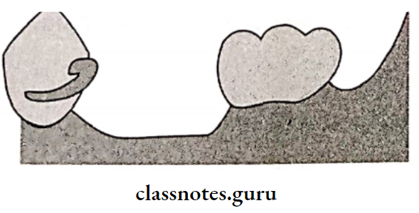
2. Bar Clasp:

  • Bar Clasp is an infra bulge clasp
  • Bar Clasp approaches the undercut gingivally
  • Bar Clasp has a push type of retention

Bar Clasp Parts:

  • Retentive arm
    • Gingival approach arm
    • Retentive terminal
  • Rest
  • Shoulder
  • Body
  • Proximal plate
  • Reciprocal arm

Removable Partial Dentures Retentive Finger Engages An Undercut

Bar Clasp Advantages:

  • Bar Clasp Is easy to Insert
  • It is more esthetic

Bar Clasp Disadvantages:

  • Bar Clasp tends to collect food debris
  • Bar Clasp has reduced bracing and stabilization
  • Bar Clasp needs additional stabilizing units

Types Of Claps:

  • T clasp
  • Modified T clasp
  • Y clasp
  • I clasp
  • Mirror view clasp
  • I bar

Removable Partial Dentures T Clasp


Removable Partial Dentures Modified T Clasp

Removable Partial Dentures Y Clasp

Removable Partial Dentures I Clasp

Removable Partial Dentures Mirror View clasp

Question 15. Discuss diagnosis and treatment planning for Kennedy’s Class 1 patient.
Answer:

Diagnosis:

1. Clinical Diagnosis:

  • Periodontal Health:
    • The periodontal condition of the existing teeth should be examined
    • Clinical signs of periodontal health should be evaluated
    • Oral hygiene is evaluated
    • Mobility of teeth, if present is measured
  • Occlusion of Existing teeth:
    • The teeth should have a good cusp-to-fossa relationship
    • Evaluate the alignment of teeth
    • Trauma from occlusion is evaluated
  • Conservative And Endodontic Status
    • The existing teeth should be examined to rule out the presence of carious lesions, vitality of the pulp
    • The teeth should be examined for cracks, chipped corners, and fractures
  • Radiological Examination
    • The bone loss should be evaluated
    • The structure of the basal bone in the denture-bearing area should be evaluated
    • Presence of periapical bone loss and furcation involvement should be examined
    • The presence of impacted teeth and submerged root stumps should be examined
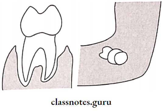
Removable Partial Dentures Severe Alveloar Bone Loss And Impacted Molar Tooth Bud

2. Derived Diagnosis:

Derived Diagnosis is obtained from a diagnostic cast that is obtained from a diagnostic impression.

Derived Diagnosis Preparation:

  • A stock tray of a suitable size should be selected Alginate is mixed in motion and loaded into the tray ray is inserted in the patient’s mouth
  • After the material gets set, it is removed by a single-stroke
  • Inspect for any void
  • Disinfect the impression
  • Pour the cast within 12 minutes
  • Trimming of the cast is carried out
  • Survey the cast

Derived Diagnosis Purpose:

  • To locate the favorable and unfavorable undercuts
  • To determine the need for pre-prosthetic mouth preparation
  • To determine the path of insertion of the denture

Derived Diagnosis Treatment Planning:

  • Treatment should be properly planned before starting the treatment
  • It can be divided into six different stages
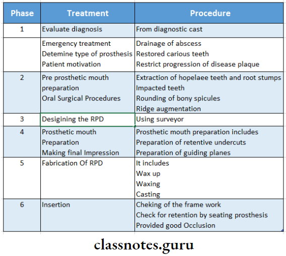
Removable Partial Dentures Treatment of Planning

Question 16. Types of mandibular major connectors.
Answer:

Mandibular Major connector:

1. Lingual Bar Design:

  • It is half pear-shaped
  • Its thickest portion is placed inferiorly
  • There must be 8 mm vertical clearance from the floor of the mouth
    The upper border should have 3 mm clearance from the marginal gingiva
  • The minimum height should be 5 mm
  • It should be as inferior as possible

Lingual Bar Design Advantages:

  • Lingual Bar Design is easy to fabricate
  • Lingual Bar Design has mild soft tissue contact

Lingual Bar Design Disadvantages:

  • Contraindicated in the presence of Tori
  • Contraindicated in short vestibule

Removable Partial Dentures Half Pear Shaped Cross Section

2. Lingual Plate Design:

  • The superior border extends up to the cingulum It is scalloped in between the teeth
  • In the presence of large embrasures, it is made to dip down-step back design
  • Anteriorly, it should be supported by rests

Lingual Plate Design Indications:

  • In the absence of posterior teeth
  • In the presence of periodontally weakened teeth
  • In the case of a short vestibule
  • In the case of resorbed ridge
  • In bilateral distal extension conditions
  • When additional teeth are to be added
  • In the case of retrognathic jaw

Removable Partial Dentures Lingual Major Connector

Lingual Plate Design Advantages:

  • Rigid, stable
  • Provide indirect retention
  • Provide the addition of teeth

Lingual Plate Design Disadvantages:

  • Food accumulation Decalcification of teeth
  • Soft tissue irritation

3. Double Lingual/Kennedy Bar Design:

  • The middle portion is cut off
  • The lower part is pear-shaped
  • The upper part is half oval 2-3 mm high and 1 mm thick
  • The upper bar should dip into the embrasure
  • Two bars are connected by a minor connector
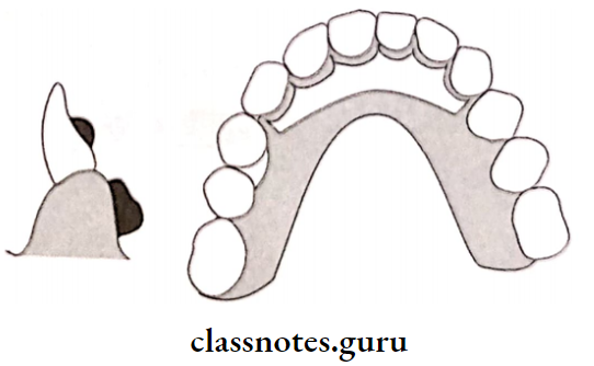
Removable Partial Dentures Doble Lingual Bar Occlusal And Cross Sectional View

Removable Partial Dentures The Vertical Major Connector Supporting

Double Lingual/Kennedy Bar Design Indications:

  • Large embrasures cases
  • Large diastema cases

Double Lingual/Kennedy Bar Design Advantages:

  • Provide indirect retention
  • Horizontal stabilization
  • Allow free flow of saliva

Double Lingual/Kennedy Bar Design Disadvantages:

  • Tongue interference
  • Food entrapment

4. Sublingual Bar:

  • Similar to the lingual bar
  • It is placed more inferiorly and posteriorly

Sublingual Bar Indications:

  • Short vestibular depth
  • Along with the linguistic plate
  • Presence of anterior lingual undercut

Removable Partial Dentures A Sub Lingual Bar Major Connector Placed Deep

Sublingual Bar Contraindications:

  • Lingual tori
  • High frenal attachments

5. Cingulum Bar:

  • The Cingulum Bar is located on or slightly above the cingula of the anterior teeth

Cingulum Bar Indications:

  • Large embrasures cases
  • Large diastema cases

6. Labial Bar:

  • Placed on the labial surface
  • Labial Bar is similar to the lingual bar, but broader and thicker

Labial Bar Indications:

  • Lingually inclined teeth
  • Presence of tori

Labial Bar Disadvantages:

  • Poor esthetics
  • Distort the lower lip
  • Patient discomfort

Removable Partial Dentures Labial Bar And Occlusal View And Labial Bar Cross Sectional View

Question 17. Classify clasps. Give the difference between gingivally and occlusally approaching clasps.
Answer:

1. Circumferential/Aker’s clasp/Occlusally Approaching:

“A clasp that encircles a tooth by more than 180°, including opposite angles, and which usually has total contact with the tooth with at least one terminal being in the infra bulge area”.

Sub-Types:

  • Simple circlet clasp
  • Reverse clasp
  • Multiple circle clasp
  • Embrasure clasp
  • Ring clasp
  • Fishhook or hairpin clasp
  • Onlay clasp
  • Combination clasp
  • Back action
  • Half and Half clasp
  • Grasso’s clasp

2. Vertical Projection/Bar/Roach Clasp:

“A clasp having arms which are bar-type extensions from major connectors or from within the denture base, the arms pass adjacent to the soft tissues and approach the point or area of contact on the tooth in a gingival-occlusal direction”

3. Continuous Clasp:

“A metal bar usually resting on the lingual surface of teeth to aid in their stabilization and to act as an indirect retainer”.
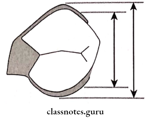
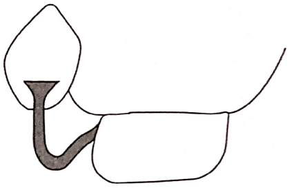
Removable Partial Dentures Notice That A Cast Circumferential Clasp

Removable Partial Dentures Approach Arm

Difference Between Occlusal Approaching And Gingival Approaching Clasp:
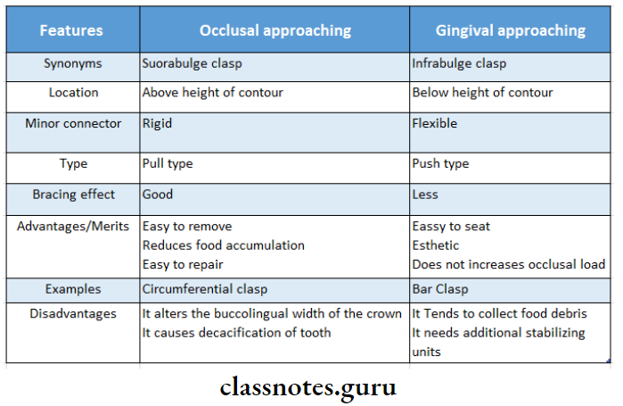
Removable Partial Dentures Difference Between Occlusally Approaching And Ginfival Approaching Clasp

Diseases Of The Arteries Veins And Lymphatic Question and Answers

Diseases Of The Arteries Veins And Lymphatic System Long Essays

Question 1. Describe the clinical features, diagnosis, and treatment of thromboangitis obliterans.

Answer:
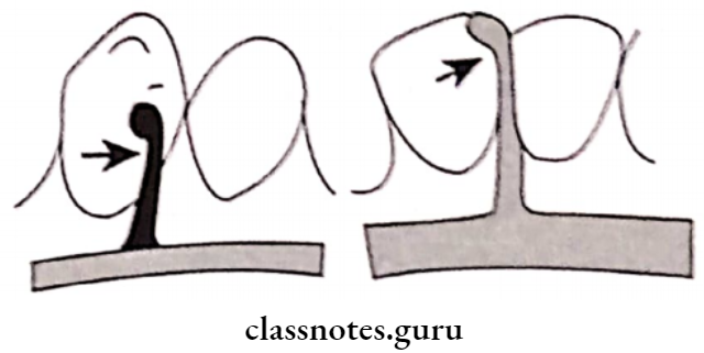
Thromboangitis Obliterans/Buerger’s Disease:
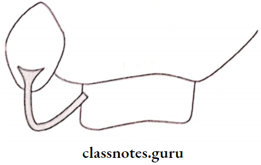
  • Thromboangitis Obliterans is the inflammatory reaction in the arterial wall with the involvement of the neighboring vein and nerve, terminating in thrombosis of the artery.

Thromboangitis Obliterans Clinical Features:

  • Age/sex – 20 – 40 years males.
  • Pain while walking at the arch of the foot
  • Pain increases when the muscle is exercised
  • Postural color changes appear followed by trophic changes.
  • Gradually ulceration and gangrene occur.
  • Gangrene starts from one digit and then involves the entire foot.
  • BP is normal in the normal limb but reduced in the diseased limb.

Thromboangitis Obliterans Diagnosis:

  • Arteriography.
    • Large arteries show abrupt areas of occlusion surrounded by extensive collateral circulation.
    • It gives a tree roof or ‘spider legs’ in appearance.
    • Peripheral arteries give a ‘corkscrew’ appearance.

Thromboangitis Obliterans Treatment:

  1. Conservative treatment.
    • Quit smoking.
    • Prostaglandin therapy to prevent platelet aggregation.
  2. Surgical treatment
    • Microvascular transplantation of free grafts
    • Amputation – to remove gangrenous area.

Read And Learn More: General Surgery Question and Answers

Question 2. Define gangrene. Describe the types, clinical features, and management of wet gangrene.

Answer:

Gangrene: Gangrene is the death of a portion of the body with putrefaction.

Gangrene Types:

  1. Dry gangrene due to slow occlusion of arteries.
  2. Wet gangrene – due to sudden occlusion of arteries.

Wet Gangrene: It is characterized by moist and oedematous limbs.

Wet Gangrene Clinical Features:

  • The part is cold, pulseless, swollen and oedematous
  • Color changes varies-dark red, green, purple, and black depending on hydrogen produced by bacteria.
  • Skin becomes raised into blebs containing foul-smelling fluid.
  • There is no line of demarcation present
  • Crepitus may be present.

Wet Gangrene Management:

  1. General treatment
    • Nutritious diet
    • Control of diabetes
    • Relief of pain.
  2. Local treatment.
    • Conservative treatment
      • The part should be kept dry
      • Part may be kept elevated
      • The part should be protected.
    • Surgical treatment.
      • Amputation – major amputation is necessary.

Diseases of arteries, veins, and lymphatics questions and answers

Question 3. Discuss the clinical features and management of diabetic gangrene.

Answer:

Diabetic Gangrene: Diabetes makes limbs more liable to gangrene formation.

Diabetic Gangrene Clinical features:

  • Pain and ulceration of the foot
  • Loss of sensation.
  • Absence of peripheral pulse.
  • Change of color and temperature.
  • There may be abscess formation.
  • Dry gangrene occurs frequently in old diabetic patients while moist gangrene in young diabetics.

Diabetic Gangrene Management:

  1. Conservative treatment
    • Diabetic control.
    • Drugs used – vasodilators, dipyridamole, low-dose aspirin.
    • Care of foot – keep it dean and dry.
    • Antibiotics – in case of infections.
    • Use of micro-cellular rubber footwear.
  2. Surgical treatment.
    • Amputation of the part.

Question 4. Describe the signs, symptoms, and treatment of varicose veins of the leg.

Answer:

Varicose Veins: When a vein becomes dilated, elongated, and tortuous, the vein is said to be varicose

Varicose Vein Clinical features:

  1. Varicose Vein Symptoms:
    • Visible distension of superficial veins.
    • Tired and aching sensation in the affected limb.
    • Sharp pain.
    • Ankle swelling towards evening.
    • Skin over the varicosities may itch and pigmented
    • Eczema of affected skin.
  2. Varicose Vein Signs:
    • Varicose eczema.
    • Hemosiderin pigmentation.
    • Atrophie blanche.
    • Lipodermatosclerosis.
    • Oedema.
    • Ulceration.

Varicose Vein Treatment:

  1. Palliative Treatment:
    • Avoid prolonged standing.
    • Apply elastic stocking from the toes to the thigh.
    • Elevation of lower extremities.
    • Exercise like bicycle riding.
  2. Operative Treatment:
    • Saphenous stripping.
      • It involves the removal of all or part of the saphenous vein’s main trunk.
    • Ambulatory phlebectomy.
      • Vein ligation
      • Cryosurgery.

Vascular system disorders Q&A

Question 5. Describe the clinical features, diagnosis, and etiology, treatment of tuberculosis cervical lymphadenitis.

Answer:

Tuberculosis Cervical Lymphadenitis:

  • Tuberculous cervical lymphadenitis refers to lymphadenitis of tire cervical lymph nodes associated with tuberculosis.

Tuberculosis Cervical Lymphadenitis Clinical features:

  • Commonly found in children and young adults.
  • Presence of chronic, painless, enlarging, or persistent mass.
  • Nodes are firm and rubbery which later becomes matted.
  • Skin becomes adhered to the mass and may rupture
  • Systemic symptoms include.
    • Fever with chills.
    • Weight loss
    • Malaise

Tuberculosis Cervical Lymphadenitis Diagnosis:

  • Positive tuberculin test.
  • Chest radiograph
  • CT scan
  • FNAC
  • Acid-fast bacilli staining
  • Mycobacterial culture.

Tuberculosis Cervical Lymphadenitis Treatment:

  1. Anti-tubercular drugs:
    • Injection streptomycin – 0.5 – lg 1M daily.
    • INH – 300 mg/ day.
    • PAS – 5 – 15 g/day.
    • It is continued for at least 1 and a half years.
  2. Supportive treatment.
    • Vitamin supplements.
    • High protein diet
  3. Surgery.
    • Removal of lymph nodes
    • Incision and drainage of the abscess.

Tuberculosis Cervical Lymphadenitis Etiology:

  • It is caused by Mycobacterium tuberculosis.
  • It has 4 pathological stages.
    • Stage 1 – lymphoid hyperplasia.
      • There is the formation of tubercles and granulomas without caseation necrosis.
    • Stage 2 and 3 – caseation necrosis.
      • Caseation necrosis in the affected lymph nodes occurs.
      • There is the destruction of the capsule of lymph nodes and adherence of multiple nodes with periodontitis.
    • Stage 4 – There is a rupture of caseous material into surrounding soft tissue.
      • There is abscess cavity formation.

Question 6. Discuss the differential diagnosis of cervical lymphadenopathy.

Answer:

The Differential Diagnosis Of Cervical Lymphadenopathy

Diseases Of The Arteries Veins And Lymphatic System Differential Diagnosis Of Cervical Lymphadenopathy

Arterial diseases question bank

Question 7. What are the methods of spread of carcinoma? Describe the block dissection of neck.

Answer:

Methods Of Spread Of Carcinoma:

  1. Through the lymphatic system.
    • It is called embolization
  2. Through bloodstream.
    • Malignant cells can break off from the tumor and travel through the bloodstream until they find a suitable place to start forming a new tumor.
    • Sarcomas spread through the bloodstream.
  3. Through local invasion.
    • Tumors invade the surrounding normal tissue.
  4. Through implantation or inoculation.
    • It occurs rarely.
    • Can happen accidentally when a biopsy is done or when cancer surgery is performed.
    • Malignant cells actually drip from a needle or an instrument.

Block Dissection Of Neck:

  • The main goal of the procedure is to remove the entire ipsilateral lymphatic structures.

Block Dissection Of Neck Procedure:

  • Incisions are made.
  • Crile’s T incision
  • Martin’s double Y incision
  • Ward’s Y incision

Two horizontal incisions.

Skin flaps are reflected

Fibro-areolar tissue of the posterior triangle is dissected away from the trapezius.

The lower end of the sternomastoid muscle is divided

The internal jugular vein is separated and divided

Above again sternomastoid muscle is transected.

The submandibular salivary gland is dissected

The spinal accessory nerve is divided in 2 places.

Transaction of the jugular vein.

Skin is closed with suction drainage.

Block Dissection Of Neck Structures Removed:

  • Lymph nodes – submental, submandibular, upper and lower deep cervical groups, posterior cervical group, and supraclavicular group.
  • Sternomastoid muscle.
  • Internal jugular vein.
  • Submental and submandibular salivary glands.
  • Spinal accessory nerve.
  • Branches of external carotied artery.

Diseases Of The Arteries Veins And Lymphatic System Block Dissection Of Neck

Venous diseases nursing questions

Diseases Of The Arteries Veins And Lymphatic System Important Notes

  1. Buerger’s Disease and Raynaud’s DiseaseDiseases Of The Arteries Veins And Lymphatic System Buerger's Disease And Raynaud's Disease
  2. Varicose Veins
    • Develop in the calf when the veins above are normal
    • More frequent in people who stand during their work
    • Often develop during pregnancy under the influence of Estrogen and progesterone which cause the smooth muscle in the vein wall to relax
    • Complications
      • Superficial thrombophlebitis
      • Deep vein thrombosis
      • Venous ulceration
  3. Lymph Nodes In Different Diseases
    • Soft, elastic, and rubbery – Hodgkin’s disease
    • Firm, discrete – syphilis
    • Stony hard – secondary carcinoma
    • Matted – tuberculosis
  4. Draining Lymph Nodes In Different DiseaseDiseases Of The Arteries Veins And Lymphatic System Draining Lymph Nodes In Different Disease
  5. Intermittent Claudication
    • Intermittent Claudication is the most common complication of the limb due to chronic arterial occlusion
    • Features
    • Cramp-like pain is felt in the muscles during exertion and gradually disappears within minutes upon cessation of activity
    • Pain is due to accumulation of excessive P substance in the muscles
    • Boyd’s classification
      • Grade 1 – pain disappears if the patient continues to walk
      • Grade 2 – pain continues but the patient can still walk with effort
      • Grade 3 – pain compels the patient to take a rest
  6. Indications Of Sympathectomy
    • Rest pain and minor ulceration
    • Buerger’s disease
    • Raynaud’s disease
    • Senile gangrene
  7. Types Of GangreneDiseases Of The Arteries Veins And Lymphatic System Types Of Gangrene
  8. Complications Of Varicose Veins
    • Thrombophlebitis
    • Pigmentation
    • Eczema
    • Ankle flare
    • Venous ulcer
    • Flaemorrhage
    • Periostitis
    • Calcification
    • Equinus deformity
  9. Virchow’s Triad – Considered In The Etiology Of Venous Thrombosis Which Includes
    • Stasis
    • Injury to the vessel wall
    • Hypercoagulability of blood

Read And Learn More: General Surgery Question and Answers

Diseases Of The Arteries Veins And Lymphatic System Short Essays

Question 1. Cervical Rib.

Answer:

Cervical Rib

The cervical rib is an extra rib present in the neck.

Cervical Rib Types:

  • Type 1 – The free end of the cervical rib is expanded into a hard, bony mass.
  • Type 2 – complete cervical rib extending from C7 vertebra to the manubrium.
  • Type 3 – Incomplete cervical rib – partly bony and partly fibrous.
  • Type 4 – Complete fibrous band.

Cervical Rib Clinical Features:

  • Common in females.
  • Dull aching pain.
  • Hand of the affected side is colder and paler
  • Numbness of the fingers.
  • Bruit is heard.
  • Hard mass may be visible and palpable.
  • Seonsory and motor disturbances in the area

Cervical Rib Treatment:

  1. Conservative.
    • Shoulder girdle exercise.
    • Correction of faulty posture.
  2. Surgery.
    • Excision of cervical rib.
    • Removal of thrombus if present

Diseases Of The Arteries Veins And Lymphatic System Four Types Of Cervical Rib

Lymphatic system disorders Q&A

Question 2. Aneurysm.

Answer:

Aneurysm Definition: Dilatation of a localized segment of the arterial system is known as aneurysm.

Aneurysm Types:

  1. True aneurysm – contains all three layers of the arterial wall.
    • It is further classified into
      • Fusiform aneurysm
      • Saccular aneurysm
      • Dissecting aneurysm
  2. False aneurysm – It has a single layer of fibrous tissue as the wall of the sac.
  3. Arteriovenous aneurysm.

Aneurysm Clinical Features:

  • Elderly patients are commonly affected.
  • Pain
  • Expansile pulsatile mass
  • Severe ischemia
  • Bruit is heard.

Aneurysm Causes:

  1. Congenital
  2. Acquired.
    • Trauma
    • Infections
    • Atherosclerosis

Aneurysm Treatment:

  • Repair of the aneurysm with graft.

Question 3. Arteriovenous aneurysm or Arteriovenous fistula.

Answer:

Arteriovenous Aneurysm

Communication between an artery and an adjacent vein leads to an arteriovenous aneurysm.

Arteriovenous Fistula Causes:

  1. Congenital
  2. Acquired – trauma
  3. Iatrogenic.

Arteriovenous Fistula Clinical Features:

  1. Systemic effects.
    • Increased cardiac output.
    • Increased heart rate
    • Increased systolic pressure
    • Cardiac hypertrophy.
    • Decreased peripheral resistance.
  2. Local effects.
    • Aneurysmal dilatation.
    • Extensive collateral circulation.
    • Bruit can be heard
    • Veins are enlarged.

Arteriovenous Fistula Treatment:

  1. Congenital lesions-excision.
  2. Acquired lesions.
    • Reconstructive
    • Ligation of involved artery
    • Selective intra-arterial embolization.

Common vascular diseases questions and answers

Question 4. Venous Ulcer.

Answer:

Venous Ulcer Causes:

  • Varicose veins
  • Increased venous hydrostatic pressure.

Venous Ulcer Clinical Features:

  • Located on the medial side of the lower 1/3rd of the leg
  • It is shallow and superficial
  • Painless
  • Pain occurs if it is infected.
  • The skin around the ulcer is pigmented
  • Shows evidence of healing.

Venous Ulcer Treatment:

  1. Conservative treatment:
    • Elevation of affected limb.
    • Movement of limb
    • Apply of firm elastic bandage.
    • Cleaning of ulcer.
    • Antibiotic administration.
  2. Surgical
    • Sclerotherapy.
    • Split skin graft.
    • Ligation.

Question 5. Thrombophlebitis.

Answer:

Thrombophlebitis

  • Thrombophlebitis is superficial vein thrombosis.
  • Thrombophlebitis occurs more often in varicose veins or after intravenous infusion.

Thrombophlebitis Clinical Features:

  • Painful cord-like inflamed area.
  • Redness
  • Tenderness
  • Local induration.

Thrombophlebitis Treatment:

  1. Conservative treatment:
    • Hot bath or compression
    • Application of crepe bandage
    • Use of anti-coagulant
    • Use of aspirin.
    • Intravenous infusion of antibiotics
  2. Surgical treatment.
    • Ligation of the involved vein.

Question 6. Cystic hygroma.

Answer:

Cystic Hygroma

Cystic Hygroma is the most common form of lymphangioma.

Cystic Hygroma Clinical Features:

  • Common in the neck region.
  • Mostly seen in children.
  • Painless swelling.
  • Pain occurs when it is infected.
  • Fluctuation and fluid thrill are present.
  • Swellings are translucent.
  • Regional lymph node enlarges in the presence of infection.

Cystic Hygroma Treatment:

  • Complete excision.

Question 7. Hodgkin’s lymphoma.

Answer:

Hodgkin’s Lymphoma Definition: It is a malignant neoplasm of the lymphoreticular system.

Hodgkin’s Lymphoma Clinical Features:

  • Age – 30 – 50 years
  • Sex- More common in males,
  • Generalised Iymphodenopathy.
  • Site involved- lymph nodes in the neck, axilla, mediastinal, para-aortic, and inguinal.
  • Nodes are firm without matting.
  • Fever with rigors.
  • Malaise, weight loss, and pallor.
  • Itching of skin.
  • Abdominal pain.
  • Bony pain.
  • Ascites.
  • Superior vena cava obstruction.

Hodgkin’s Lymphoma  Investigation:

Diseases Of The Arteries Veins And Lymphatic System Hodgkin's Lymphoma Investigation

Hodgkin’s Lymphoma Treatment:

  • Radiotherapy – for stages 1 and 2
  • Chemotherapy – for stages 3 and 4

Short notes on vascular and lymphatic diseases

Question 8. Staging of Hodgkin’s disease.

Answer:

Hodgkin’s Disease Stage 1:

  • Lymph node involvement in one anatomical region.
  • Example: palpable left supraclavicular nodes.

Hodgkin’s Disease Stage 2:

  • Involvement of two or more lymph nodes on the same side of the diaphragm.
  • Example: Left supraclavicular and left axillary node.

Hodgkin’s Disease Stage 3:

  • Involvement of lymph nodes on both sides of the diaphragm.
  • Example: Left supraclavicular and left inguinal lymph nodes.

Hodgkin’s Disease Stage 4:

  • Diffuse involvement of one or more extra lymphoid organs with or without lymph node involvement.

Disorders of blood vessels questions for exams

Question 9. Varicose ulcer

Answer:

Varicose Ulcer

  • Varicose Ulcer is a type of venous ulcer
  • Varicose Ulcer Cause
    • Abnormal venous hypertension in the lower third of the leg, ankle, and dorsum of the foot
  • Varicose Ulcer Features
    • Shallow and superficial
    • Doesn’t penetrate deep fascia
    • Usually painless
    • Associated with varicose veins
    • The skin around the ulcer is pigmented

Diseases Of The Arteries Veins And Lymphatic System Viva Voce

  1. The commonest type of lymphoma is Hodgkin’s lymphoma
  2. Application of warmth will increase the symptoms of arterial occlusion
  3. Venous ulcers are commonest ulcers of the legs
  4. Continuous machinery murmur indicates presence of an arteriovenous fistula
  5. Synthetic grafts are used in aortoiliac occlusion
  6. Vein grafts are used in femero-popliteal occlusion
  7. Majority of the pulmonary emboli originates in the lower extremity


Diseases Of The Arteries Veins And Lymphatic System Short Answers

Diseases Of The Arteries Veins And Lymphatic System Short Answers

Question 1. Aneurysm of aorta.

Answer:

Aneurysm Of Aorta

Aneurysm Of Aorta is an abnormal enlargement of the wall of the aorta.

Aneurysm Of Aorta Types:

  1. Abdominal aortic aneurysm.
    • Aneurysm occurs in the section of the aorta that runs through the abdomen.
  2. Thoracic aorta aneurysm.
    • It is an aneurysm occurring in the chest area.
  3. Thoracoabdominal aortic aneurysm.
    • Involves the aorta as it flows through both the abdomen and chest.

Aneurysm Of Aorta Features:

  • Pain in the jaw, neck, upper back, or chest
  • Coughing.
  • Hoarseness of voice.
  • Difficulty breathing.
  • Pulsating enlargement.

Question 2. Mycotic aneurysm.

Answer:

Mycotic Aneurysm

  • Mycotic aneurysm is an aneurysm arising from bacterial infection of the arterial wall.
  • It is caused by streptococcus pneumonia.

Read And Learn More: General Surgery Question and Answers

Mycotic Aneurysm Symptoms:

  • Fever
  • Leucocytosis
  • Palpable mass.

Question 3. Cricoid aneurysm/Aneurysmal bone cyst.

Answer:

Cricoid Aneurysm

Circoid aneurysm involves the bone anywhere in the body including the jaws.

Cricoid Aneurysm Clinical Features:

  • Age – 10 – 19 years of age.
  • Sex – occurs commonly in females.
  • Rapid, enlarging, diffuse, firm swelling occurs.
  • Swelling is painful.
  • Perforation of cysts causes profuse bleeding.
  • Paraesthesia.

Arterial diseases short notes

Question 4. Signs of Aneurysm.

Answer:

Signs Of Aneurysm

  • Expansile pulsation in the course of the artery.
  • Pulsation diminishes when pressure is applied
  • Compressible swelling.
  • The thrill is palpable over swelling
  • Bruit is heard.

Question 5. Arteriography.

Answer:

Arteriography

  • Arteriography is the most reliable method of determining the state of the main arterial tree.
  • Arteriography gives information about.
    • Size of the lumen of the artery.
    • The course of the artery.
    • Constriction and dilatation of arteries.
    • Condition of collateral circulation.

Arteriography Methods:

  1. Retrograde percutaneous catheterization.
  2. Direct arterial puncture.

Diseases Of The Arteries Veins And Lymphatic Lymphatic System

Question 6. Embolism.

Answer:

Embolism Definition: Embolism is the partial/complete obstruction of some part of the cardiovascular system by any mass carried in the circulation.

Embolism Types:

  1. Depending upon the matter in the emboli.
    • Solid emboli.
    • Liquid emboli.
    • Gaseous emboli.
  2. Depending upon whether infected or not
    • Sterile
    • Septic.
  3. Depending upon the source of emboli.
    • Cardiac
    • Arterial
    • Venous
    • Lymphatic
  4. Depending upon the flow of blood.
    • Paradoxical embolus.
    • Retrograde embolus.

Venous disorders: short answers

Question 7. Pulmonary embolism.

Answer:

Pulmonary Embolism Definition: Pulmonary embolism is the most common and fatal form of venous thromboembolism in which there is occlusion of the pulmonary arterial tree by thrombotic emboli.

Pulmonary Embolism Etiology:

  • Varicosities in superficial veins of legs.

Pulmonary Embolism Complication:

  • Acute corpulmonale
  • Chronic corpulmonale
  • Pulmonary hypertension
  • Pulmonary infarction.
  • Pulmonary hemorrhage.
  • Sudden death.

Question 8. Raynaud’s disease.

Answer:

Raynaud’s Disease Definition: It is a condition characterized by episodic attacks of vasospasm in response to cold exposure or emotional stimuli.

Raynaud’s Disease Phases:

  1. Intense pallor
  2. Cyanosis
  3. Rubor.

Raynaud’s Disease Etiology:

  • Unknown etiology.
  • Secondary to systemic diseases like
  • Buerger’s disease.

Question 9. Subclavin steal syndrome.

Answer:

Subclavian Steal Syndrome

  • Subclavian Steal Syndrome is a condition in which there is atherosclerotic stenosis of the subclavian artery proximal to the site of origin of the vertebral artery.

Subclavian Steal Syndrome Clinical features:

  • Reduction in pressure in the subclavian beyond the stenosis.
  • Retrograde blood flow
  • Syncopal attack.
  • Visual disturbances.
  • Decreased pulse and blood pressure
  • Localized bruit in the supraclavicular space.

Question 10. Trendelenburg’s test.

Answer:

Trendelenburg’s Test

  • Trendelenburg’s test is used to determine the incompetency of the saphenofemoral valve.
  • It can be performed in two ways.
  1. The patient is placed in a recumbent position.
    • Legs are raised
    • Sapheno-femoral junction is compressed with the thumb of the clinician and the patient is asked to stand up quickly.
    • Pressure is released.
    • If the varies fill very quickly, it indicates a positive Trendelenburg test.
  2. The patient is placed in a recumbent position.
    • Legs are raised
    • Sapheno-femoral junction is compressed and the patient is asked to stand up quickly.
    • Pressure is maintained for 1 minute.
    • Gradual filling of varices indicated positive Trendelenburg’s test.

Question 11. Commando’s operation.

Answer:

Commando’s Operation Indication: When carcinoma of tongue is fixed to the mandible

Commando’s Operation Steps:

  • Hemiglossectomy.
  • Hemimandibulectomy.
  • Removal of floor of the mouth.
  • Radical neck dissection.

Commando’s Operation Structure Removed:

  • Fat, fascia, lymphatics.
  • Lymph nodes – submental, submandibular deep cervical nodes, posterior group of nodes.
  • Submandibular salivary gland.
  • Sternomastoid.
  • Internal jugular vein.
  • Spinal accessory nerve.

Lymphatic system diseases Q&A

Question 12. Clinical staging of Hodgkin’s lymphoma.

Answer:

Clinical Staging of Hodgkin’s Lymphoma

Stage 1: Involvement of single lymph node.

Stage 2: Involvement of 2/ more lymph nodes on the same side of the diaphragm.

Stage 3: Involvement of 2/more lymph nodes on both sides of the diaphragm.

Stage 4: Diffuse involvement of extra-lymphoid organs with or without lymph node involvement.

Question 13. Histological classification of Hodgkin’s lymphoma.

Answer:

Histologica Classification of Hodgkin’s Lymphoma

  1. Type 1 – Lymphocyte predominant type.
    • Reed Sternberg (RS) cells are scanty; scattered among large number of matured lymphocytes.
  2. Type 2 – mixed cellularity.
    • There is significant number of eosinophils, neutrophils, plasma cells, and atypical histio- cytesalongwith. RS cells and lymphocytes.
  3. Type 3 – Nodular sclerosis.
    • Presence of broad collagen bands separating the lymphoid tissue.
  4. Type 4 – lymphocyte depletion.
    • Lymphocytes are few
    • Presence of malignant appearing histiocytes.

Question 14. Non-Hodgkin’s lymphoma.

Answer:

Non-Hodgkin’s lymphoma

Non-Hodgkin’s lymphoma is a group of primary malignancies of lymph-reticular tissue.

Non-Hodgkin’s Lymphoma Classification:

  1. Histological.
    • Lymphocyte predominant
    • Mixed cellularity
    • Nodular sclerosis.
    • Lymphocyte depletion.
  2. Based on the prognosis.
    • Nodular – favorable prognosis.
    • Diffuse-unfavorable prognosis.

Non-Hodgkin’s Lymphoma Clinical features:

  • Extranodal involvement.
  • Fever with night sweats
  • Weight loss
  • Local invasion of adjacent structures
  • Regional lymphadenopathy.

Non-Hodgkin’s Lymphoma Management:

  • Staging laparotomy is required
  • Splenectomy.

Question 14. Lymphadenitis.

Answer:

Lymphadenitis

Lymphadenitis is the inflammation of lymph nodes.

Lymphadenitis Clinical features:

  • The site involved – lymph nodes under the neck, in the axilla, or in the groin.
  • Lymph nodes are enlarged.
  • Firm, painful enlargement occurs
  • Hyperaemic overlying skin.
  • Fever

Lymphadenitis Treatment:

  • Analgesic
  • Antibiotic
  • Abscess drainage.

Question 15. Lymphosarcoma.

Answer:

Lymphosarcoma Definition: It is defined as a malignant neoplastic disorder of the lymphoid tissue characterized by the proliferation of atypical lymphocytes and their localization. In various parts of the body.

Lymphosarcoma Clinical Features:

  • Age – common in children.
  • Lymph nodes involved – in the neck, mediastinum, and abdomen.
  • Extra-nodal involvement – spleen, tonsil, pharynx, bowel.
  • Enlargement of lymph nodes.
  • Constitutional symptoms – fever, loss of weight, anemia, anorexia, weakness.
  • The overlying skin is shiny and tense
  • The surface is irregular.

Lymphosarcoma Treatment:

  • Radiotherapy.
  • Chemotherapy – in case of diffuse involvement.

Common diseases of arteries and veins

Question 16. Microscopic appearance of tuberculous lymphadenitis.

Answer:

Microscopic Appearance Of Tuberculous Lymphadenitis

  • Tubercles are seen consisting of epithelial cells and giant cells with peripherally arranged nuclei.
  • Next lymphocytes with darkly stained nuclei and scanty cytoplasm appear.
  • As the disease progresses caseation necrosis occurs.
  • Thus, in the center of the follicle caseation occurs
  • This is surrounded by giant cells, epitheloid cells, zone of chronic inflammatory cells, and fibroblasts.

Diseases Of The Arteries Veins And Lymphatic System Microscopic Appearance Of Tuberculuous Lymphadentitis

Question 17. Malignant secondary lymph node.

Answer:

Malignant Secondary Lymph Node

Can occur commonly from malignant melanoma.

Malignant Secondary Lymph Node Clinical Features:

  • Site
  • Painless swelling
  • Constitutional symptoms – anorexia, weight loss, weakness.
  • Lymph nodes are irregular, and discrete.
  • They fuse to form a large mass.
  • Nodes are usually hard.
  • Gradually they gets fixed to the surrounding structures.

Diseases Of The Arteries Veins And Lymphatic System Malignant Secondary Lymph Node Clinical Features

Question 18. Use of MRI ion head and neck.

Answer:

Use Of MRI Ion Head And Neck

  • For the study of TMJ deformities in the sagittal plane.
  • To evaluate various spaces in the head and neck region.
  • For nasopharynx, skull base, tongue pathology.
  • Posturgical evaluation of TMJ.
  • To identify and localize orofacial soft tissue lesions.
  • Provides an image of salivary gland parenchyma.

Question 19. Lymphatic drainage of the tongue.

Answer:

Lymphatic Drainage Of The Tongue

Diseases Of The Arteries Veins And Lymphatic System Lymphatic Drainage Of Tongue

Vascular system disorders short answer questions

Question 20. Waldeye’s ring.

Answer:

Waldeyer’s Ring Consists Of

  • Pharyngeal tonsil-posteriorly and above
  • Tubal tonsil – laterally and above
  • Lingual tonsil – Inferiorly.
  • Submandibular nodes
  • Retropharyngeal nodes
  • Submental nodes
  • Jugulodigastric nodes
  • Jugular chains of nodes.
  • Retropharyngeal node
  • Tubal tonsil Palatine tonsil Lingual tonsil
  • Jugular chain of nodes

Diseases Of The Arteries Veins And Lymphatic System Waldeyer's Lymphatic Ring

Question 21. Causes of wet gangrene

Answer:

Causes Of Wet Gangrene

  • Gangrene from acute inflammation
  • Long-standing venous thrombosis
  • Bed sores
  • Gas gangrene

Peripheral artery disease short answer

Question 22. Stages of tubercular lymphadenitis

Answer:

Stages Of Tubercular Lymphadenitis

Diseases Of The Arteries Veins And Lymphatic System Stages Of Tubercular Lymphadenitis

Acute Poisoning

Acute Poisoning And Environmental Emergencies Short Answers

Question 1. Fluorosis

Answer:

Fluorosis

Dental fluorosis is caused by excessive intake of fluoride during tooth development

Fluorosis Clinical Features

  • Lustreless, opaque white patches in the enamel which may become mottled, striated, or pitted
  • Mottled areas may become stained yellow or brown
  • Hypoplastic areas may also be present to such an extent in severe cases that normal tooth form is lost
  • Enamel fluorosis is a developmental phenomenon due to excessive fluoride ingestion during amelogenesis
  • Once crowns are formed no further fluorosis occurs
  • The hypocalcified areas of the mottled enamel are less soluble in acids
  • They have a greater permeability to dyes
  • They emit fluorescence of higher intensity then normal enamel
  • Fluorosis occurs symmetrically within arches
  • The premolars is usually first affected followed by second molar, maxillary incisor, canine, first molar, and mandibular incisors

Acute poisoning symptoms

Read And Learn More: General Medicine Question and Answers

Question 2. Food poisoning

Answer:

Food poisoning Management:

  1. Resuscitation and initial stabilization
  2. Diagnosis of type of poison by
    • History
    • Examination
    • Laboratory investigations
    • Nonspecific therapy- to reduce the levels of toxin
  3. Specific therapy- to reduce toxic effects on the body
  4. Supportive care- to support functions of vital organs

Question 3. Arsenic poisoning

Answer:

Arsenic poisoning Features:

  • Gingivitis
  • Stomatitis
  • Painful mucosal ulceration
  • Hyperpigmentation and hyperkeratosis
  • Excessive salivation
  • Vomiting
  • Diarrhea
  • Neurological disturbances

Types of acute poisoning

Question 4. Barbiturate poisoning

Answer:

Barbiturate Poisoning

  • Fatal dose of phenobarbitone is 6-10 gram

Barbiturate Poisoning Symptoms:

  • Respiratory depression with slow and shallow breathing
  • Hypotension
  • Skin eruptions
  • Cardiovascular collapse
  • Renal failure

Barbiturate Poisoning Treatment:

  • Gastric lavage followed by administration of activated charcoal
  • Maintain BP
  • Airway maintenance
  • Adequate ventilation
  • Oxygen administration
  • Forced alkaline diuresis with sodium bicarbonate, a diuretic, and 4 fluids
  • Hemodialysis

Poisoning first aid measures

Question 5. Scorpion bite

Answer:

Types Of Scorpion Venom:

  1. Venom of the genera Hadrurus, Vejovis, and Uroctonus Effects:
      • Sharp burning
      • Swelling
      • Discoloration at the bite site
      • Rarely anaphylaxis
  2. Venom produced by genera of the poisonous varieties of centuries Effects
      • Block sodium channels
      • Spontaneous depolarization of parasympathetic and sympathetic nerves
      • Tachycardia
      • Hypertension
      • Sweating
      • Piloerection
      • Hyperglycemia
      • Pulmonary edema
      • Seizures
  3. Scorpion Venom Treatment:
    • Patient is hospitalised for at least 12 hours
    • Maintain airway maintenance
    • Administration of 1-2 vials of intravenous anti-venin

Common household poisoning agents

Question 6. Lead poisoning

Answer:

Lead Poisoning Features:

  • Excessive salivary secretions
  • Metallic taste in the oral cavity
  • Swelling of the salivary glands
  • Development of the dark lead line along the gingival margin
  • Convulsions
  • GI upset
  • Anaemia
  • Neuritis
  • Basophilic stippling of the RBC cells

Question 7. Adverse drug reactions

Answer:

Adverse Drug Reactions Definition:

  • It is defined as any response to a drug that is noxious and unintended and that occurs at the dose used in man for prevention, diagnosis, or therapy

Adverse Drug Reactions Types:

  1. Side effects
    • They are unwanted effects of a drug
  2. Toxic effects
    • They are seen with higher doses of the drug
  3. Intolerance
    • A person cannot tolerate a drug
  4. Idiosyncrasy
    • It is a genetically determined abnormal reaction to a drug
  5. Allergic reactions
    • They are immunologically mediated reactions
  6. Iatrogenic diseases
    • These are drug-induced diseases
  7. Drug dependence
    • It is a state of compulsive use of drugs despite the knowledge of the risks associated with its use
  8. Teratogenicity
    • A drug can cause fetal abnormalities when administered to a pregnant woman
  9. Teratogenicity and mutagenicity
    • Drugs causing cancers and genetic abnormalities

Drug overdose emergency treatment

Acute Poisoning And Environmental Emergencies Medcial interview

Question 8. Organophosphorous poisoning

Answer:

Organophosphorous Poisoning

Acute Poisoning And Environmental Emergencies Organophosphorous Poisoning

Organophosphate poisoning symptoms

Question 9. Thrush

Answer:

Thrush

  • Acute pseudomembranous oral candidiasis is also known as thrush

Thrush Common Sites Involved:

  • Buccal mucosa
  • Tongue
  • Palate

Thrush Etiology:

  • Causative organism- Candida albicans
  • Prolonged antibiotic therapy
  • Immuno-suppression

Thrush Predisposing Factors:

  • Infancy
  • Debilitating illness
  • Metabolic diseases
  • Diabetes
  • Hypothyroidism

Thrush Features:

  • Foul taste
  • This leads to inflammation, erythema, and eroded areas
  • Presence of adherent white plaques
  • It can be removed by scraping
  • It is more common in women

Thrush Treatment:

  1. Anti-fungal antibiotics
    • Nystatin
    • Amphotericin suspension or lozenges
  2. For AIDS patient
    • Oral fluconazole is used

Carbon monoxide poisoning treatment

Question 10. Halitosis

Answer:

Halitosis Definition:

  • Unpleasant odor exhaled in breathing is called halitosis

Halitosis Classification:

  1. Physiologic
  2. Pathologic
    • Oral
    • Extraoral

Halitosis Causes:

  1. Physiologic
    • Mouth breathing
    • Medication
    • Fasting
    • Aging
    • Tobacco
    • Food
  2. Pathologic
    • Periodontal infection
    • Tongue coating
    • Stomatitis
    • Xerostomia
    • Faulty restoration
    • Unclean denture
    • Ulcers
    • Abscess
    • Systemic diseases

Halitosis Treatment:

  • Scaling
  • Irrigation
  • Burning sensation
  • Tongue brushing
  • Use of mouth rinse
  • Use of Halita

Question 11. Macroglossia

Answer:

Macroglossia Definition:

  • Macroglossia is a relatively common condition characterized by an increase in the size of the tongue

Macroglossia Types And Causes

  1. Congenital macroglossia
    • Overdevelopment of the tongue musculature
    • Lysosomal storage diseases
    • Down’s syndrome
    • Multiple endocrine neoplasia syndrome
  2. Acquired macroglossia
    • Tumours in the tongue
    • Amyloidosis
    • Endocrine disorders
    • Lymphatic obstruction in the tongue
    • Cystic lesions in the tongue

Macroglossia Clinical Features:

  • Causes displacement of teeth and malocclusion
  • Disturbances in speech and feeding
  • Cosmetic deformity
  • Indentation or scalloping on the lateral margins of the tongue
  • Development of tongue-thrusting habits
  • Airway obstruction

Macroglossia Treatment:

  • Removal of the primary cause
  • Surgical reduction or trimming

Management of acute poisoning in hospital

Question 12. Ciprofloxacin

Answer:

Ciprofloxacin

  • Ciprofloxacin is the first generation of fluoroquinolone

Ciprofloxacin Uses:

  • Urinary tract infection
  • Typhoid
  • Diarrhea
  • Gonorrhea
  • Chancroid
  • Respiratory tract infection
  • Bone, joint, soft tissue, and intra-abdominal infections
  • Tuberculosis
  • Bacterial prostatitis and cervicitis
  • Eye infections
  • Anthrax
  • Orodental infections

Ciprofloxacin Adverse Reactions:

  • Nausea, vomiting, abdominal discomfort, diarrhea
  • Rashes
  • Tendinitis
  • Damages growing cartilage

Treatment of organophosphorus poisoning

Question 12. Phenobarbitone

Answer:

Phenobarbitone

  • Phenobarbitone is an anti-epileptic drug

Mechanism of Action:

  • Enhances inhibitory neurotransmission in the CNS
  • Enhances activation of GABA receptors
  • Facilitates GABA-mediated opening of chloride ion channels

Phenobarbitone Uses:

  • Generalized tonic-clonic seizures
  • Partial seizures

Phenobarbitone Adverse Reactions:

  • Sedation
  • Tolerance
  • Nystagmus
  • Ataxia
  • Megaloblastic anemia
  • Osteomalacia
  • Skin rashes
  • Hypersensitivity reactions

Specialized Radiographic Techniques Short Essays

Question 1. Indications and Techniques for visualization of paranasal lsinuses.
Answer:

Paranasal lsinuses Indications:

  • To study the relationship of the sinuses to each other and the surrounding structures
  • To demonstrate the presence or absence of fluid in the sinuses

Paranasal lsinuses  Techniques:

  • Posteroanterior [Granger] projection:
    • Structures Seen:
      • Inner and middle ear
      • Frontal sinuses
      • Anterior ethamoidal cells
      • Sphenoidal sinus
      • Upper part of antrum
    • Film Placement:
      • Midsagittal plane should be vertical
      • It should be perpendicular to the plane of the cassette
      • Only forehead and nose should touch the cassette
    • Central Ray:
      • It is directed to the midline of the skull
      • The beam passes through the canthomeatal plane perpendicular to the film plane

Specialized radiographic techniques short essay

Diagram for the positioning for posteroanterior projection

Diagram for the positioning for posteroanterior (Granger) projection

Read And Learn More: Oral Radiology Question and Answers

  • Modified method, inclined posterior anterior [Caldwell projection]
    • Strucutures Seen:
      • Petrous ridges
      • Orbits
      • Ethamoidal cells
    • Film Placement:
      • Cassette is placed perpendicular to the floor
    • Position of the Patient:
      • Mid sagittal plane is perpendicular to the cassette
      • Only forehead and nose touches the cassette
      • Canthomeatal line is perpendicular to the cassette
    • Central Ray:
      • Directed 23º to the canthomeatal line
      • Enters the skull about 3 cm above the external occipital protuberance and exiting at the glabella

Oral Radiology Specialized Radiographic Techniques Diagram of the positioning inclined posteroanterior projection

Diagram of the positioning inclined posteroanterior (Caldwell) projection

Short essay on special radiographic procedures

Question 2. Oblique lateral radiograph of mandible.
Answer:

  • Anterior body of the mandible:
    • Structures Seen:
      • Anterior body of mandible
      • Position of teeth in that region
    • Film Placement:
      • Cassette is placed flat against the patient’s cheek
      • It is centered over the body of the mandible overlying canine
    • Position of the Patient:
      • The ala tragus line should be parallel to the floor
      • The mandible is protruded slightly
      • The inferior borderof the cassette should be parallel to the lower border of the mandible
      • The sagittal plane is tilted so that it is 5º to the vertical and rotated 30º from the true lateral position
      • The nose and chin should approximate the cassette
    • Central Ray:
      • Directed from 2 cm below the angle of the mandible opposite to the side of interest
      • The beam is directed upward -10º to -15º
      • It is centered on the anterior body of the mandible
      • The beam is directed perpendicular to the horizontal plane of the film

Oral Radiology Specialized Radiographic Techniques Diagram for the positioning of lateral oblique projection 1

Short note on specialized radiography

Diagram for the positioning of lateral oblique projection for anterior body of the mandible, film is in contact with the cheek at the canine area, and the X-ray beam aims at the canine area through radiographic key hole

  • Posterior Body:
    • Structures Seen:
      • Body of the mandible
      • Position of teeth in that area
      • Ramus of the mandible
      • Angle of the mandible
    • Film Placement:
      • The cassette is placed against the patients cheek
      • It is centered over the body of the mandible
      • The cassette is placed parallel to the body of the mandible
    • Position of the Patient:
      • The ala tragus line is parallel to the floor
      • The mandible is protuded slightly
      • The inferior border of the cassette should be parallel to the lower border of the mandible and below it
      • The sagittal plane is tilted to 5º to the vertical
      • The head is rotated 10º to 15º from the true lateral position
    • Central Ray:
      • It is directed from 2 cm below the angle of the mandible opposite to the side of interest
      • The beam is directed upwards (-10º to -15º)
      • It is centered on the body of the mandible

Oral Radiology Specialized Radiographic Techniques Diagram for the positioning of lateral oblique projection 2

Diagram for the positioning of lateral oblique projection for posterior body of the mandiblem film is in contact with the cheek at the premolar area, and the X-ray beam aims at the premolar area, through the radiographic key hole

  • Ramus of Mandible:
    • Structures Seen:
      • Ramus from the angle of the mandible to the condyles
    • Film Placement:
      • The cassette is placed against the patients cheek
      • It is centered over the ramus of mandible
      • It should be parallel to ramus
    • Position of the Patient:
      • The ala tragus line should be parallel to the floor
      • The mandible is protruded slightly
      • The inferior border should be parallel to the lower border of the mandible and below it
      • The sagittal plane is tilted 10º to the vertical
      • The head is rotated 5º from the true lateral
    • Central Ray:
      • It is directed from 2 cm below the angle of the angle of the mandible opposite to the side of interest to a point posterior to the third molar region
      • The beam is directed upwards (-10º to -15º)
      • It is centered on the ramus of the mandible

Oral Radiology Specialized Radiographic Techniques Diagram for the positioning of lateral oblique projection for ramus

Diagram from the positioning of PA water’s projection, the radiographic baseline is at 37º to the film, and the X-ray is perpendicular to the film

Dental specialized radiographic techniques short answer

Question 3. Water’s projection.
Or
PA Water’s view.
Answer.

Water’s projection

  • Structures Seen:
    • Maxillary sinus
    • Frontal sinus
    • Ethamoidal sinus
    • Orbit
    • Frontozygomatic suture
    • Nasal cavity
    • Coronoid process
    • Zygomatic arch
  • Film Placement:
    • The cassette is placed perpendicular to the floor
  • Position of the Patient:
    • The mid sagittal plane should be perpendicular to the plane of the film
    • The patient’s head is extended so that only the chin touches the cassette
    • The cassette is centered around the acanthion
    • The canthomeatal line should be 37º to the plane of the film
    • The line from the external auditory meatus to the mental protuberance should be perpendicular to the film
  • Central Ray:
    • It is directed perpendicular and to the midpoint of the film
    • It enters from the vertex and exists from the acanthion

Oral Radiology Specialized Radiographic Techniques Diagram for the positioning of PA Water projection

Diagram for the positioning of PA Water’s projection, the radiographic baseline is at 37º to the film, and the X-ray is perpendicular to the film

Short essay on advanced radiographic methods

Question 4. Sialography.
Answer.

Sialography Technique:

  • Identification of duct:
    • The parotid duct is located at the base of the papilla in the buccal mucosa adjacent to the first or second molar
    • The area over the mucosa should be dried with a small sponge
    • The submandibular duct orifice is situated on the submitting of the small papilla at the side of the lingual frenum
  • Exploring of the duct:
    • The duct can be explore with lacrimal probe
    • In the case of the submandibular duct, the probe should pass through the length of the floor of the mouth to the level of the posterior border of the mylohyoid muscle i.e. about 5cm
    • Eversion of cheek should be done in case of parotid duct
    • By it, the duct is adequatly enlarged
  • Introduction of cannula:
    • The sialographic cannula is inserted into the duct so that the tissue sop presses firmly into the orifice to prevent dye reflux
  • Introduce contrasting media:
    • Liquid soluble or Water soluble agent is slowly introduced
  • Amount of the agent:
    • Submandibular glandL 0.5 – 0.75ml
    • Parotid gland: 0.76 – 1ml
  • Radiograph is taken:
    • Occlusal view/lateral oblique view is used to delineate the submandibular gland
    • A sialolith is better viewed in occlusal view
    • AP view it used for both the glands
    • It demonstrates the medial and lateral gland structures
  • Evacuation:
    • After the radiograph is taken, the cannula should be removed
    • The patient is instructed to chew gum or the lemon slice and then asked to rinse
    • This is done to stimulate the gland and cause excretion of the dye.

XAI methods applicable for disease specific Charactertics

Extraoral radiographic techniques short essay

Question 5. MRI – Principles and indications.
Answer.

MRI – Principles and indications

  • It uses nonionizing radiation from the radiofrequency band of the electromagnetic spectrum

MRI Mechanism:

  • The patient is placed inside a large magnet
  • This induces a strong external magnetic field that causes the nuclei of many atoms in the body including hydrogen to align themselves with the magnetic field
  • After application of radiofrequency signal, energy is released from the atoms that can be deteched and used to construct the image by computer

MRI Advantages:

  • High sensitivity to detect tissue differences
  • No radiation exposure hence no radiation affects the body
  • Excellent imaging techniques especially for soft tissues
  • It gives the best resolution of tissues of low inherent contrast

MRI Disadvantages:

  • Long imaging times
  • It gives the potential hazards in a patient with implanted metallic foreign objects like caradiac pacemakers, cerebral aneurysm clips.
  • Some patients suffer from claustrophobia when positioned in a MRI machine.

MRI Indications:

  • Diagnosing a suspected internal derangement of the TMJ
  • Postsurgical evaluation of TMJ
  • Identifying and localizing orofacial soft tissue lesions
  • Provides the image of salivary gland parenchyma

Intraoral and extraoral specialized imaging short essay

Question 6. Scintigraphy.
Answer.

Scintigraphy

  • It is based on the radiotracer method
  • Radioactive atoms or molecules in organs behave in a manner identical to their counterpart in the body
  • The radiotracers allow measurement of tissue function and provide early markers of disease through measurement of biochemical change in tissue before any physical signs and symptoms occur

Method:

  • Radioactive substances should be injected innntravenously into the patient
  • Rectilinear scanner or gamma scintillation camera records the gamma emission from the patient
  • The camera uses a scintillation crystal that can fluorescence on interaction with gamma rays emitting from the radioactive substances
  • The emitting light fluorescence is detected by a photomultiplier tube that magnifies and amplifies the signals many times to produce an image

Atom Used:

  • Iodine
  • Gallium
  • Selenium
  • Technetium

Scintigraphy Advantages:

  • It is more sensitive to early or small changes in bone or salivary metabolism
  • It has been used to detect the presence and size of tumors, metastasis, trauma and metabolic disorders

Scintigraphy Disadvantages:

  • Poor, grainy image
  • Confusion of normal inflammatory process with tumor and metastasis
  • Patients also get exposed to a small quantity of radioactive material which causes biological changes in the tissues

Radiographic special procedures short essay

Question 7. Digital radiography.
Answer.

Digital radiography

  • The use of digital technology results in a 50 to 90% reduction in patient radiation exposure because of the greater sensitivity of the digital receptor
  • Elimination of film processing and no need for dark room
  • Considerable reduction in the time lapse between image acquisition and display

Digital radiography Mechanism:

  • In digital imaging, the sensor is used i.e., CCD Charged Couple Device instead of radiographic film in the patient’s mouth
  • After radiation exposure, the signal from the CCD is sent to the computer where it is digitalized into gray levels
  • The image can then be displayed on a monitor, where it can be enhanced by varying the density and contrast
  • The image may also be stored for future used

Digital radiography Types:

  • Direct digital radiography
  • Indirect digital radiography

Digital radiography Advantages:

  • Eliminates the X-ray film, thereby reduces the cost of the film
  • It serves as a recording, display, and storage for diagnostic images
  • It requires less exposure time because of the grater sensitivity of the digit receptor there by preventing the patient from being exposed to radiation
  • Eliminates the chemical processing and dark room so that preventing the cause of alllergy and pollution and also considerable reduction in time lapse between image acquisition and display
  • Film contrast, density, brightness and color may be manipulated so that image information can be increased
  • Reduction in the number of images that need to he remade because of over exposure or under exposure
  • Transmission of image to remote sites in a digital format
  • Image can be viewed on computer monitor to get an print out on paper

Digital radiography Disadvantages:

  • Equipment is expensive
  • Trained persons are required

Digital radiography Uses:

  • It can be used to view the images where multiple images are required for analyzing
  • In endodontic practice to measure the root canal length, working lenght and distance between obturating material and the root apex
  • In perioidontics, to assess and measure the height of the alveoplar bone
  • It can be used in a patient who is un cooperativge for regular radiographic techiniques
  • To evaluate the bony changes in pathology of jaws
  • To detect early dental caries

Specialized dental X-ray techniques short note

Question 8. Indications of CT [Computed tomography].
Answer.

Indications of CT [Computed tomography]

  • Investigations of intracranial diseases including diseases tumors, hemorrhage and airfacts
  • Investigations of suspected intracranial and spinal cord damage following trauma to the head and neck
  • Assessment of fractures involving
    • Orbits and nasoethamodial complex
    • The cranial base
    • The odontoid peg
    • The cervical spine
  • Tumor stagingassessement of site, size and extent of benign and malignant tumors affecting
    • The maxillary antra
    • The base of the skull
    • The pterygoid region
    • The pharynx
    • The larynx
  • Investigations of tumors and tumor like discrete swellings intrinsic and extrinsic to the salivary glands
    • Investigation of the TMJ
    • Preoperative assessment of maxillary alveolar bone height and thickness before inserting implants

Occlusal radiographic technique short essay

Question 9. Tranpharyngeal view
Answer.

Structures Seen:

  • Medial surface of the condylar head and neck

Film Placement:

  • The cassette is placed flat against the patient’s ear
  • It is centered to a point 1/2″ anterior to the external auditory meatus, over the TMJ of interest

Position of the Patient:

  • Sagittal plane should be vertical and parallel to the film
  • The film is centered to a point 1/2 “anterior to the external auditory meatus
  • The occlusal plane should be parallel to the transverse axis of the film
  • The patient should open his mouth

Central Ray:

  • It is directed from opposite side cranially at an angle of -5 to -10 degree posteriorly
  • It is directed through the Mandibular notch of the opposite side below the base of the skull to the TMJ of interest

Oral Radiology Specialized Radiographic Techniques Transpharyngeal projection 1

Sialography and its radiographic technique

A – Tranpharyngeal projection. The central ray is orient superiorly 5º to 10º and posteriorly approximately 10º, centered over the TMJ of interest. The mandible is positioned at maximal opening

Oral Radiology Specialized Radiographic Techniques Transpharyngeal projection 2

B – Tranpharyngeal projection, showing positioning from above, showing the X-ray beam aimed slightly posteriorly across the pharynx