Blood Vessels Of Head And Neck Question And Answers
Question 1. Write a short note on common carotid artery.
Answer:
Common carotid artery
There are two common carotid arteries right and left which form chief arteries of head and neck.
Common Carotid Artery Origin
- Right common carotid artery arises in neck from the brachiocephalic trunk behind sternoclavicular joint
- Left common carotid artery arises directly from arch of aorta and ascends back to sternoclavicular joint.
Common Carotid Artery Course
- Both right and left arteries in neck have a similar course
- Each artery runs upwards from the sternoclavicular joint of respective sides to the upper border of lamina of thyroid which lies to body of C4 vertebrae
- At the level of lamina thyroid cartilage, it divides into internal and external carotid arteries
- They are named so because external carotid artery supplies the structures outside the skull and internal carotid artery supplies inside the skull
- Each common carotid artery lies in the front of transverse process of the lower cervical vertebrae and under cover of the anterior border of sternocleidomastoid
- Internal carotid artery is considered as continuation of common carotid artery.
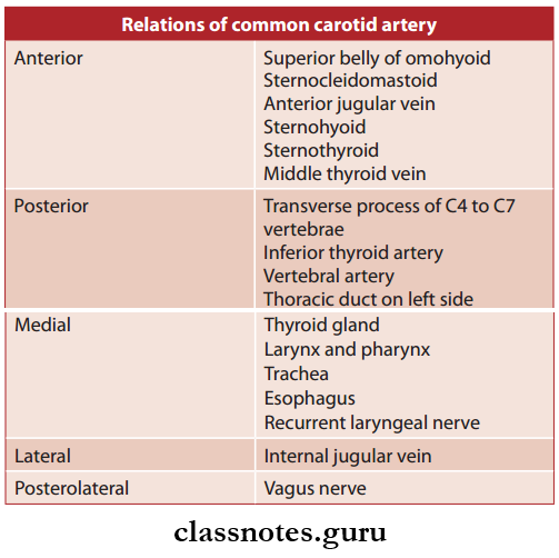
Relations of common carotid artery
Carotid Sinus
- It is a fusiform dilation at the termination of common carotid artery or the beginning of the internal carotid artery
- It receives rich innervation from the glossopharyngeal and sympathetic nerves
- It is a baroreceptor and regulates blood pressure.
Carotid Body
- Small oval reddish-brown structures situated behind the bifurcation of common carotid artery
- It receives rich innervation from glossopharyngeal, vagus, and sympathetic nerves similar to carotid sinus
- But carotid body act as a chemoreceptor and respond to the changes in O2 and CO2, the pH content of blood.
- Common Carotid Artery Applied
- The th carotid pulse is the most constant pulse of the body and is felt at 4cm above the sternoclavicular joint the level of cricoid cartilage.
Question 2. Write a short note on external carotid artery.
Answer:
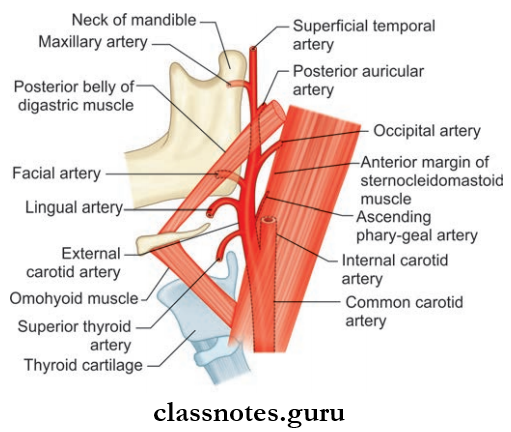
External carotid artery
- One of the terminal branches of common carotid artery
- Supplies the structures in front of the neck and external to head.
External Carotid Artery Course
- It extends upwards from the upper border of lamina of thyroid cartilage to a point behind the neck of mandible
- It is a slightly curved course where it is anteromedial to internal carotid artery in the lower part and anterolateral in the upper part
- At the point behind the neck of mandible, it terminates in the substance of parotid gland and divides into superficial temporal and maxillary arteries
- In the carotid triangle, its comparatively superficial and lies in under cover of the sternocleidomastoid, and above the carotid artery lie deep in substance of the parotid gland.
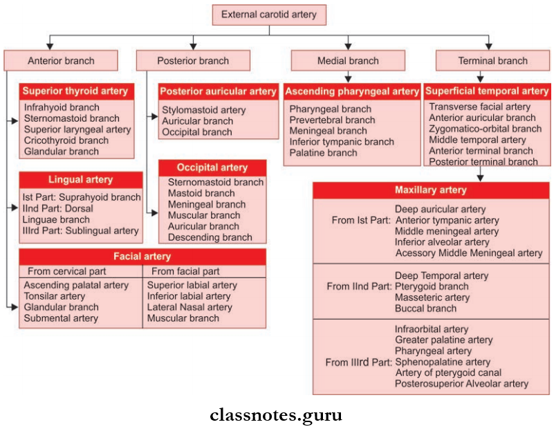
External Carotid Artery Branches
- Superior Thyroid Artery: Arises from the front of external carotid artery just below the level of greater cornu of hyoid bone and reaches upper pole of thyroid gland to supply it.
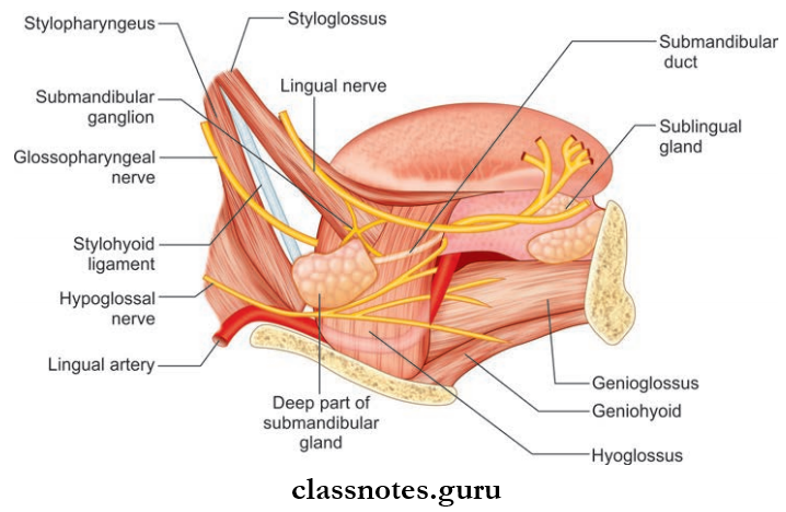
- Lingual Artery
- Arises from the front of the external carotid artery opposite to tip of greater cornu of hyoid bone and is divided into 3 parts by hyoglossus muscle
- It is the main artery to the tongue.
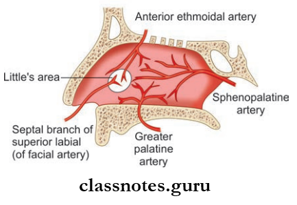
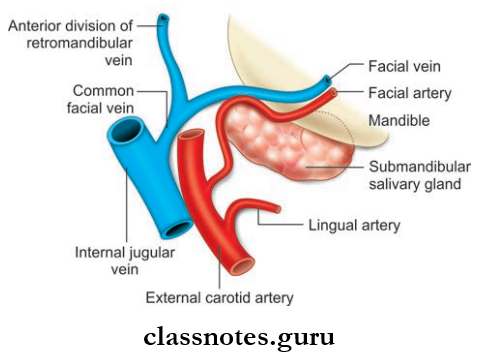
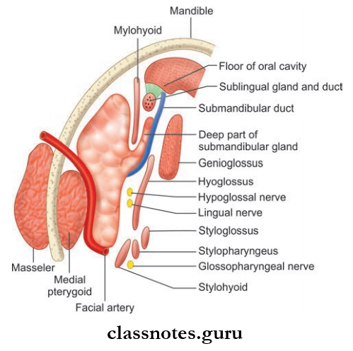
- Facial Artery
- Arises from the front of external carotid artery just above the tip of greater cornu of hyoid bone
- It has 2 parts: The cervical part and facial part.
- Occipital Artery
- Arises from the posterior aspect of external carotid artery at the same level of facial artery
- It supplies most of the back of scalp.
- Posterior Auricular Artery
- Also, arise from posterior aspect a little above the occipital artery
- It mainly supplies the ear.
- Ascending Pharyngeal Artery
- Slender artery arising from the medial aspect of the external carotid artery near its lower end.
- Superficial Temporal Artery
- Smaller terminal branches of the external carotid artery
- Divides into anterior and posterior branches to supply temple and scalp.
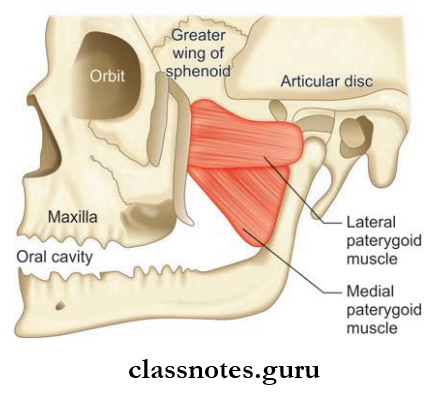
- Maxillary Artery
- Larger terminal branches of the external carotid artery
- Has three parts mandibular, pterygoid part and pterygopalatine part
- Supplies both jaws, temporal and infratemporal fossa, nose and paranasal sinuses, palate, external and middle ear, and dura mater.
External Carotid Artery Applied: The descending branch of the occipital artery provides the chief collateral circulation after ligation of external carotid artery.
Question 3. Write a note on the facial artery.
Answer:
Facial artery
It is a branch of external carotid artery and is the chief artery of the face.
Facial Artery Origin; It arises from front of the external carotid artery above the tip of greater cornu of hyoid bone.
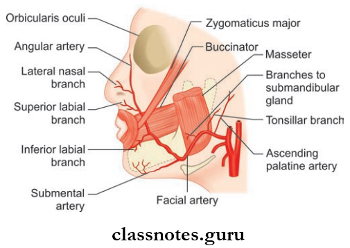
Facial Artery Course: It is divided into 2 parts
- Cervical and
- Facial part.
1. Cervical Part
- After a looped course in the submandibular region, it enters into the face by winding around lower border of body mandible by piercing deep cervical fascia
- Looped course—ascends upwards on superior constrictor deep to digastric and stylohyoid muscle and passes deep to ramus of mandible and grooves the posterior border of the submandibular gland and makes ‘s-shaped bend (fist bending over the submandibular gland, then up over the base of mandible).
2. Facial Part
- Begins at the lower border of body of mandible at anteroinferior angle of the masseter by piercing fascia colli and pass fist upwards and forward to a point 1.25 cm lateral to angle of mouth
- It ascends along the side of nose to medial canthus/medial angle of eye
- Near the medial angle it ends by anastomosing with nasal branch of ophthalmic artery (anastomosis between internal carotid artery and external carotid artery)
- The terminal part is known as angular artery.
Facial Artery Branches
- Branches of Cervical Part
- Ascending palatine artery which mainly supply the palate
- Tonsillar artery which supplies tonsil
- The glandular branch which supply the submandibular gland
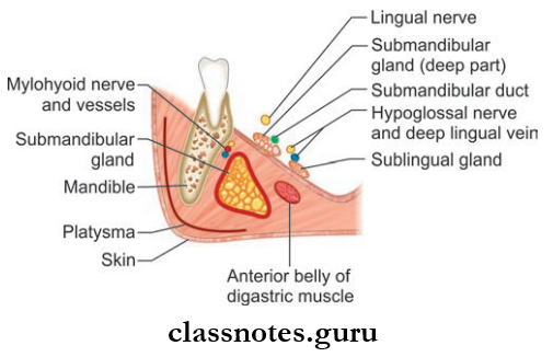
- Submental artery which supplies mylohyoid, submandibular and sublingual glands.
- Branches of Facial Part
- Inferior labial artery which supply the lower lip
- Superior labial artery which supply lower lip
- Lateral nasal artery which supply alae and dorsum of nose
- Muscular branches.
Facial Artery Applied: Tortuosity of facial artery prevents its wall from being stretched during movements of face.
Question 4. Write a short note on occipital artery.
Answer:
Occipital Artery Origin: Arises from external carotid artery opposite to the origin of facial artery.
Occipital Artery Course
- Runs backwards and upwards deep to the posterior belly of digastric crossing carotid sheath; 11th and 12th cranial nerves
- Then it runs deep to the mastoid process and the muscles attached to it
- It then crosses muscles at the apex of the posterior triangle and it pierces trapezius 2.5 cm from the midline
- It has a tortuous course in the superficial fascia of scalp.
Occipital Artery Branches
- Mastoid branches
- Meningeal branches
- Muscular branches
- Descending branch
- The superficial branch which anastomoses with superficial branch of transverse cervical artery
- The deep branch which anastomoses with vertebral and deep cervical arteries.
- Sternocleidomastoid branch.
Question 5. Write a short note on the maxillary artery.
Answer:
Large terminal branch of external carotid artery.
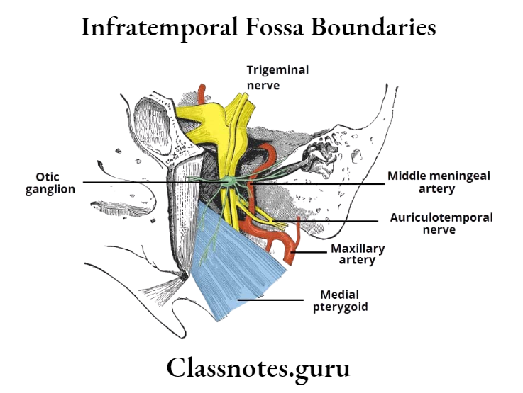
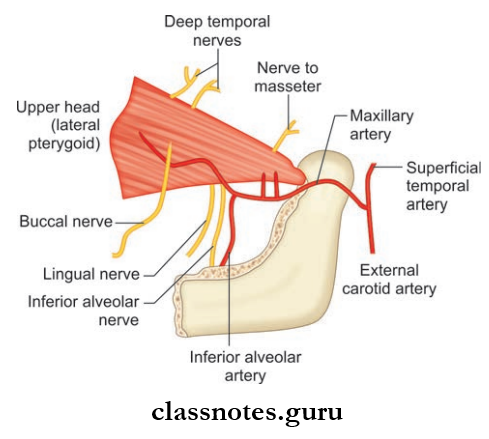
Maxillary Artery Course
- Arises behind the neck of mandible
- It then runs horizontally and forwards to the lower border of lower head of lateral pterygoid
- Then it runs upwards and forwards and cross the lower head of lateral pterygoid
- It then emerges between 2 heads of lateral pterygoid and enter the pterygopalatine fossa and terminates by dividing into branches.
Maxillary Artery Areas Supplied
- Upper and lower jaw
- Muscles of temporal and infratemporal fossa
- Nose and paranasal sinuses
- Palate and root of the pharynx
- External and middle ear
- Eustachian tube and dura mater.
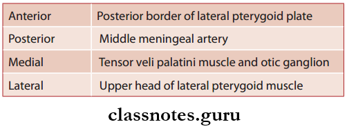
Maxillary Artery Parts: It is divided into 3 parts by lower head of the lateral pterygoid.
- The first part (mandibular part)
- Extends from origin to lower border of lateral pterygoid
- Lies between the neck of mandible and the sphenomandibular ligament.
- Second part (pterygoid part): From lower border to upper border of lower head of lateral pterygoid muscle.
- Third part (pterygopalatine part): It extends from upper border of lower head of lateral pterygoid to the pterygopalatine fossa.
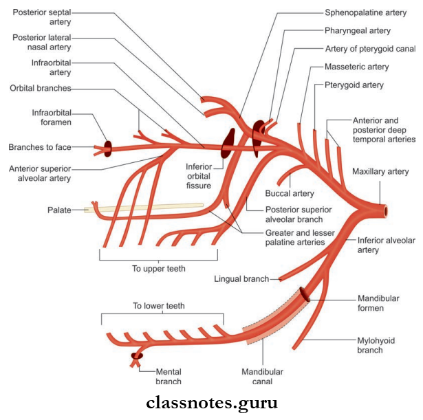
Maxillary Artery Branches
- Branches from 1st Part: Consist of five branches
- Deep auricular artery which supplies skin of external acoustic meatus and tympanic membrane
- Anterior tympanic artery which supplies inner surface of tympanic membrane
- The middle meningeal artery which supplies the meninges
- The accessory middle meningeal artery which supplies meninges and structures in the infratemporal fossa
- The inferior alveolar artery supplies the teeth, gums, and skin of chin.
- Branches from 2nd Part: Consist of four branches
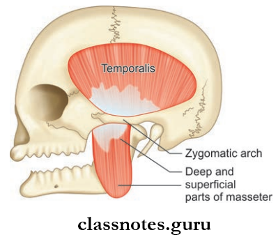
- Deep temporal artery which supplies temporalis muscle
- Pterygoid branches which supplies the medial and lateral pterygoid
- Masseteric artery which supplies masseter muscle
- Buccal branches which supply buccinators’ muscle.
- Branches from 3rd Part: Consist of six branches
- Posterior superior alveolar artery which supplies molar and premolar teeth and mucous membrane of maxillary air sinus
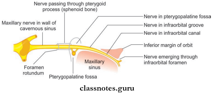
- Infraorbital artery which supplies lower orbital muscles, maxillay sinus, canine and incisor teeth of upper jaw, lacrimal sac, medial angle of eye, upper lip.
- Greater palatine artery which supplies roof of mouth and adjoining gum, soft palate, and tonsil
- The pharyngeal artery which supplies submucosal membrane of nasopharynx, auditory tube, and sphenoid sinus
- Artery of pterygoid canal which supplies the pharynx, auditory tube and tympanic cavity
- Sphenopalatine artery supplies lateral wall of nose, sphenoid and ethmoid sinus.
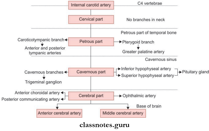
Question 6. Write a short note on internal carotid artery.
Answer:
Internal carotid artery
- One of the terminal branches of common carotid artery
- Considered as an upward continuation of common carotid artery and supplies structures inside the skull and orbit and forms the principal artery to brain and eye.
Internal Carotid Artery Origin: Begins at the upper border of lamina of thyroid cartilage at the level of C4 vertebrae as a continuation of common carotid artery.
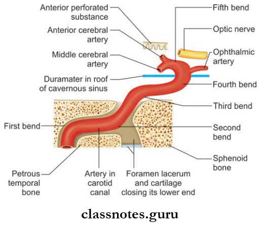
Internal Carotid Artery Course: It is divided into four parts for convenience.
- Cervical Part
- It ascends upward from origin to the base of skull and reach the lower end of the carotid canal and lies in front of transverse process of upper cervical vertebrae
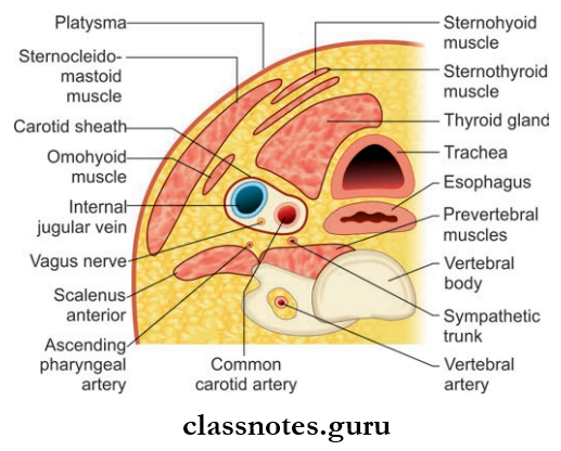
- In the neck, it is enclosed within carotid sheath along with internal jugular vein and vagus nerve
- The lower part of the artery lies superficial in the carotid triangle
- Upper part lies deep to posterior belly of the digastric, styloid process and parotid gland
- In the upper end, the jugular vein lies posterior and last four cranial nerves lie in between internal jugular vein and internal carotid artery.
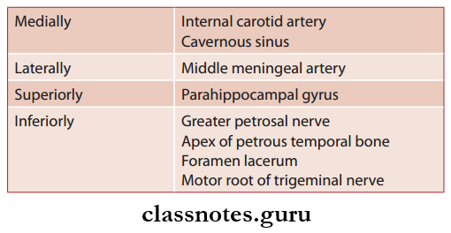
- Petrous Part
- Enters the petrous part of temporal bone through the carotid canal and runs upwards, forwards and medially
- It emerges at the apex of petrous temporal bone in the posterior wall of foramen lacerum and passes upwardsm to cranial cavity.
- Cavernous Part
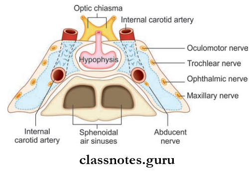
- From foramen lacerum it ascends and enters cavernous sinus. Here, it passes forwards alongside of sella turcica and medial wall of the sinus and abducens nerve lies inferolaterally
- In the anterior part of sinus artery pierce the dura in between anterior and middle clinoid process to reach beneath the cerebrum.
- Cerebral Part
- After emerging through the dura, it turns backward in subarachnoid space along the roof of sinus and lies inferior to optic nerve
- Later, it passes upwards by the side of optic chiasma and reaches anterior perforated substance of brain and divides into anterior and middle cerebral arteries.
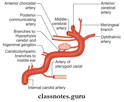
Internal Carotid Artery Branches
- No branches are given in the neck
- Branches from petrous part
- Caroticotympanic branches anastomose with posterior and anterior tympanic arteries
- The pterygoid branch which anastomoses with greater palatine artery.
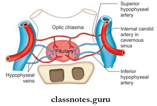
- Branches from cavernous part
- Cavernous branches to trigeminal ganglion
- Superior and inferior hypophyseal arteries to pituitary.
- Branches from cerebral part
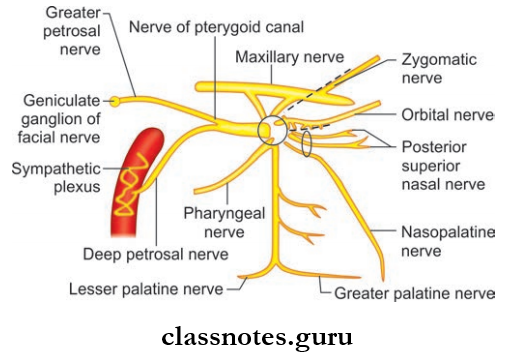
- Ophthalmic artery
- Anterior choroid artery
- Posterior communicating artery.
- Terminal branches
- Anterior cerebral artery
- Middle cerebral artery.
Internal Carotid Artery Applied
- The complete or partial obstruction of internal carotid arteries due to arteriosclerosis is the common cause of stroke or cerebral insufficiency.
- The curvatures of the petrous, cavernous, and cerebral parts of the internal carotid artery together form a ‘s-shaped figure which can be seen in the angiograms as a ‘carotid siphon’.
Question 7. Write a short note on ophthalmic artery.
Answer:
Ophthalmic artery
Branch of internal carotid artery.
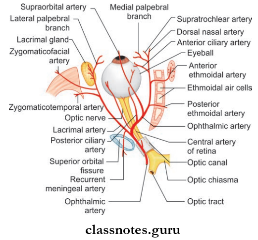
Ophthalmic Artery Course
- Arises from internal carotid artery and emerges from roof of cavernous sinus close to the optic canal
- It then enters the orbit through the optic canal inferolateral to optic nerve
- It then pierces duramater, ascends over the lateral side of optic nerve, and cross it from lateral to medial
- It then runs forwards along medial wall of the orbit and terminates by dividing into supratrochlear and dorsal nasal arteries at the medial angle of eye.
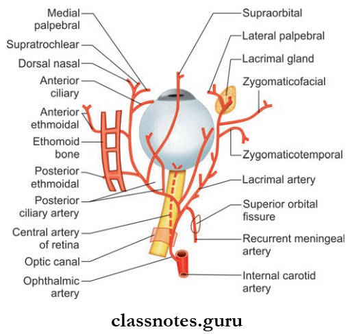
Ophthalmic Artery Branches
- Central artery of retina: Arises below optic nerve and run forward in dural sheath and pierces optic nerve and reaches optic disc and supply optic nerve and retina
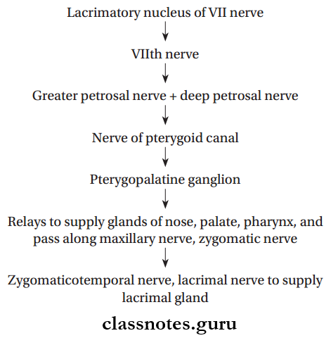
- Lacrimal artery: Arises just before the crossing of optic nerve. It passes along the upper part of lateral rectus and supply lacrimal gland, eyelids and conjunctiva
- Posterior ciliary arteries: Long and short types which supply sclera and choroid
- Supraorbital arteries: Accompany supraorbital nerve
- Posterior ethmoidal artery which supply the ethmoid sinus, nasal cavity, and dura
- Anterior ethmoidal artery which supply ethmoid sinus, medial and lateral wall of nose, and dura
- Dorsal nasal artery: Supplying the dorsum of nose
- Supratrochlear artery: Accompanying supratrochlear nerve and supply the forehead
- Medial palpebral branches: Supplying eyelids.
Question 8. Write a short note on subclavian artery.
Answer:
Subclavian artery
Main source of arterial supply to upper limb.
Subclavian Artery Origin
- Right subclavian artery arises from the brachiocephalic trunk behind the right sternoclavicular joint
- Left subclavian artery arises from arch of aorta and runs upward and makes groove on left lung and enters neck by passing behind left sternoclavicular joint.
Subclavian Artery Course
- On each side, it arches laterally across the cervical pleura into the fist rib posterior to scalenus anterior muscle
- For convenience, the subclavian artery is divided into 3 parts by scalenus anterior muscle:
- 1st part: From origin to medial border of scalenus anterior muscle
- 2nd part: Lies behind scalenus anterior muscle
- 3rd part: Lateral border of scalenus muscle to outer border of 1st rib.
- It continues as axillary artery at the outer border of 1st rib.
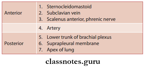
Subclavian Artery Relations
First Part (Anterior To Posterior)
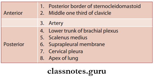
Second Part (Anterior To Posterior)
Third Part (Anterior to Posterior)
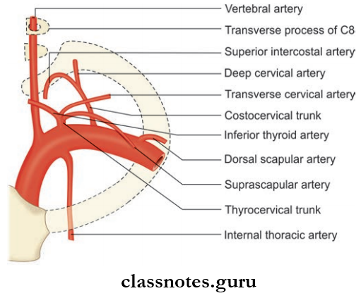
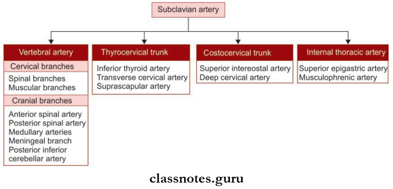
Subclavian Artery Branches: Gives four branches
- Vertebral artery
- Internal thoracic arteries
- Throcervical trunk
- Costocervical trunk
- Among these branches fist 3 arises from 1st part of the artery whereas costocervical trunk arises from 2nd part of artery
- Occasionally, a large branch arises from third part of artery called the dorsal scapular artery.
Subclavian Artery Applied
- In case of obstruction of subclavian artery proximal to the origin of vertebral artery, some amount of blood from the opposite vertebral artery will pass in a retrograde manner to the subclavian artery of affcted side through vertebral artery of that side to provide collaterals to upper limb on side of lesion.
- Hence, some amount of blood from brain is stolen by subclavian artery which is called as subclavian steal syndrome.
Question 9. Write a short note on the thyrocervical trunk.
Answer:
Thyrocervical Trunk
Branch from 1st part of subclavian artery.
Thyrocervical Trunk Origin: Arises from 1st part of the subclavian artery near medial border of the scalenus anterior muscle between phrenic and vagus nerves.
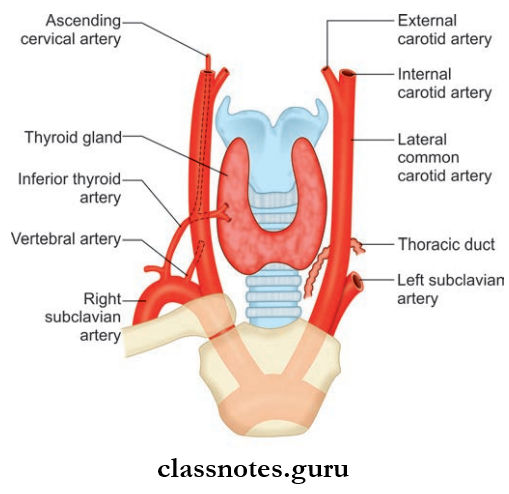
Thyrocervical Trunk Branches: Inferior thyroid artery which supply the thyroid gland and gives the following branches
- Ascending cervical artery
- Inferior laryngeal artery
- Branches to the pharynx, trachea, and esophagus.
- The suprascapular artery which takes part in anastomosis around scapula and also supplies muscle around the scapula, clavicle, and the acromioclavicular joint
- Transverse cervical artery which divides into superficial and deep branch of which the deep branch takes part in anastomosis around scapula and superfiial branch accompanies the spinal root of the accessory nerve.
Question 10. Write a short note on vertebral artery.
Answer:
Vertebral Artery
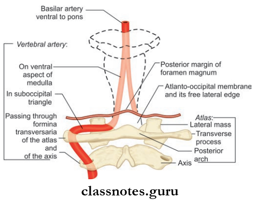
One of the two principal arteries that supply brain and also supplies the spinal cord, meninges, the surrounding muscles, and bones.
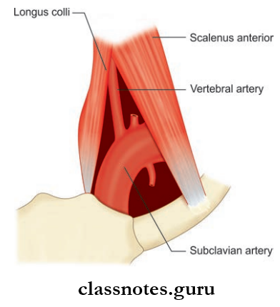
Vertebral Artery Origin: It arises from the posterior superior aspect of fist part of subclavian artery.
Vertebral Artery Course: It has a very long course and it is divided into four parts.
- Vertebral Artery Course First Part: Extends from the origin to foramen transversarium of sixth cervical vertebra.
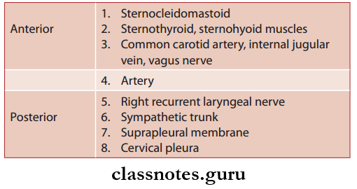
- Vertebral Artery Course First Part Relations: It lies in the triangular space between scalenus anterior and longus colli muscle called scalenovertebral triangle.
- Anteriorly Vertebral Vein
- Carotid sheath
- Inferior thyroid artery
- Thracic duct on left side.
- Posteriorly–Transverse Process Of C7 Vertebra
- Ventral rami of C7, C8 nerves
- Stellate ganglion.
- Vertebral Artery Course Second Part: Located within foramina transversaria of upper 6 cervical vertebrae.
- Vertebral Artery Course Second Part Relations
- Ventral rami of C2–C6 nerves posteriorly
- Accompanies vertebral venous plexus and large branch from the stellate ganglion.
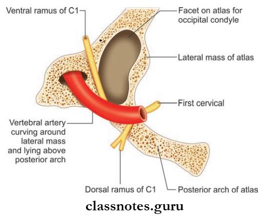
- Vertebral Artery Course Third Part:
- Extends from foramen transversarium of atlas to foramen magnum
- Lies in the suboccipital triangle
- The artery winds medially around the lateral process of atlas.
- Vertebral Artery Course Third Part Relations
- Anterior: Lateral mass of atlas
- Posterior: Semispinalis capitis
- Lateral: Rectus capitis lateralis
- Medial: Ventral ramus of C1 nerve
- Inferiorly: Dorsal ramus of C1 nerve and posterior arch of atlas.
- Vertebral Artery Course Fourth Part
- Intracranial part of artery
- Extends from posterior atlantooccipital membrane to lower border of Pontomedullary junction to form basilar artery
- In vertebral canal, it pierces dura and arachnoid and ascends in front of roots of hypoglossal nerve.
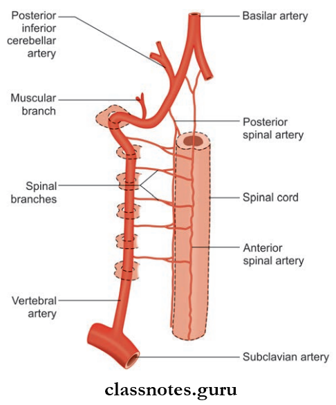
Vertebral Artery Branches
- Extracranial Branches
- Spinal branches arise from 2nd part and supply spinal cord, meninges, and vertebra
- Muscular branches arise from 3rd part and supply suboccipital muscles.
- Intracranial Branches
- Meningeal Branches: Supply bone and meninges of posterior cranial fossa
- Anterior Spinal Artery: Arises at termination and, supplies anterior 2/3rd of spinal cord and part of medulla
- Posterior Spinal Artery: Arises at the side of medulla and supplies posterior 1/3rd of the spinal cord
- Posterior Inferior Cerebellar Artery: Largest branch arises at the lower end of olive and supplies posterior lateral aspect of medulla, lower part of pons, and cerebellum
- Medullary artery: Supply medulla.
Question 11. Write a short note on subclavian vein.
Answer:
Subclavian vein
Subclavian vein is the continuation of the axillary vein.
Subclavian Vein Extent: Begin at the outer border of first rib and extends up to medial border of scalenus anterior.
Subclavian Vein Course
- Begin at the outer border of first rib and forms an arch across pleura below the subclavian artery
- It ends at the medial border of the scalenus anterior by joining the internal jugular vein to form the brachiocephalic vein.
Subclavian Vein Tributaries
- External jugular vein
- Dorsal scapular vein
- Thracic duct on left side and right lymphatic duct on right side.
Subclavian Vein Relations
- Anteriorly: Clavicle and subclavius muscle
- Posteriorly: Subclavian artery, scalenus anterior muscle, and phrenic nerve
- Inferiorly: First rib and pleura.
Subclavian Vein Applied: The subclavian vein is commonly used to measure intracardiac pressure.
Question 12. Write a short note on external jugular vein.
Answer:
External Jugular Vein Formation: Formed by the union of posterior division of retromandibular vein and posterior auricular vein just below the angle of mandible.
External Jugular Vein Course:
- External Jugular Vein Course then runs downwards under the cover of platysma,vertically across the sternocleidomastoid to pierce deep cervical fascia in posterior triangle near its anteroinferior angle to enter the supraclavicular space
- Then it terminates in subclavian vein.
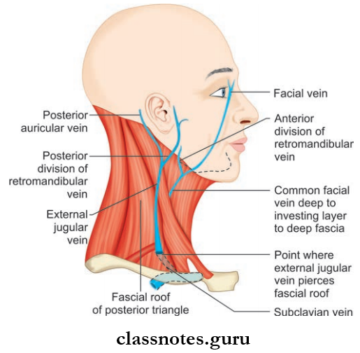
External Jugular Vein Tributaries
- Formative Tributaries:
- Posterior auricular vein
- Retromandibular vein
- TerminalTributaries:
- Transverse cervical vein
- Suprascapular vein
- Anterior jugular vein
- Other Tributaries:
- Posterior external jugular vein
- Oblique jugular vein.
Mnemonic
- External Jugular Vein: Tributaries
- PAST PR
- Posterior external jugular vein
- Anterior jugular vein
- Suprascapular vein
- Transverse cervical vein
- Posterior auricular vein
- Retromandibular vein.
Question 13. Write a short note on the internal jugular vein.
Answer:
Internal Jugular Vein
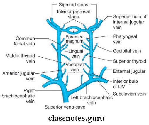
Internal Jugular Vein Formation: Begin as the direct continuation of sigmoid sinus at the base of skull below the jugular foramen.
Internal Jugular Vein Course: Then it descends vertically downwards and ends posterior to clavicle near its sternal end by joining the subclavian vein to form brachiocephalic vein.
Internal Jugular Vein Features
- The Origin And Termination Presents With 2 Dilatations:
- The dilatation present at the origin is called superior bulb which lies in jugular fossa of the temporal bone, beneath the flor of middle ear
- The dilatation present at the termination is called inferior bulb which lies in the lesser supraclavicular fossa.
Internal Jugular Vein Tributaries
- Inferior petrosal sinus
- Pharyngeal vein
- Common facial vein
- Lingual vein
- Superior thyroid vein
- Middle thyroid vein
- Thracic duct opens into the junction between the left internal jugular and left subclavian and right lymphaticm duct to right side simultaneously.
Internal Jugular Vein Relation
- Superfiial (anterolateral)
- Sternocleidomastoid
- Posterior belly of digastric
- Superior belly of omohyoid
- Parotid gland
- Styloid process
- Internal carotid artery, glossopharyngeal nerve, vagus, spinal accessory.
- Deep (posterior)
- Transverse process of atlas
- Cervical plexus
- Scalenus anterior
- The first part of subclavian artery.
- Medial
- Internal carotid artery
- Common carotid artery
- Vagus nerve.
Internal Jugular Vein Applied
- The internal jugular vein is commonly used for central venous access for measuring pulmonary artery pressure, prolonged intravenous feeding, and for the introduction of cardiac pacemaker
- The internal jugular vein is commonly injured during removal of tuberculous or neoplastic lymph nodes.
Mnemonics: Tributaries of internal jugular vein:
‘Medical Schools Let Confident People In’
From inferior to superior:
- Middle thyroid
- Superior thyroid
- Lingual
- Common facial
- Pharyngeal
- Inferior petrosal sinus.
Question 14. Write a short note on the brachiocephalic vein.
Answer:
Brachiocephalic vein
- Formed by the union of internal jugular vein and subclavian vein
- The right vein (2.5 cm) is shorter than left vein (6 cm).
Brachiocephalic Vein Course
- Right vein runs downwards vertically and the left vein runs downwards obliquely towards the right fist coastal cartilage behind upper half of manubrium sterni
- They unite at the lower border of 1st costal cartilage on right side to form superior vena cava.
Brachiocephalic Vein Tributaries
- Right Brachiocephalic
- Vertebral vein
- Internal thoracic vein
- Inferior thyroid vein
- First posterior intercostal vein.
- Left Brachiocephalic
- Vertebral vein
- Internal thoracic vein
- Inferior thyroid vein
- First posterior intercostal vein
- Left superior intercostal vein
- Thymic vein
- Pericardial vein.
Question 15. Write a short note on ophthalmic veins.
Answer:
Ophthalmic Veins
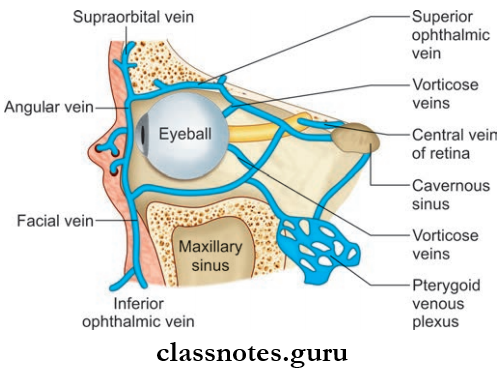
- They drain the orbit and receive tributaries which correspond to the branches of the ophthalmic artery
- The ophthalmic Veins Include:
- Superior Ophthalmic Vein: It accompanies ophthalmic artery and begins at medial palpebral ligament and run along with the ophthalmic artery and drains into the cavernous sinus after passing through superior orbital fissure.
- Inferior Ophthalmic Vein
- Runs below the optic nerve
- It drain to either the cavernous sinus directly or through superior ophthalmic vein.
Blood Vessels Of Head And Neck Multiple Choice Question And Answers
Question 1. The vertebral artery does not pass through the foramen transversarium of:
- First cervical vertebra
- Second cervical vertebra
- Sixth cervical vertebra
- Seventh cervical vertebra
Answer: 4. Seventh cervical vertebra
Question 2. All of the following arteries arise from the thyrocervical trunk except:
- Inferior thyroid artery
- Suprascapular artery
- Dorsal scapular artery
- Superficial cervical artery
Answer: 3. Dorsal scapular artery
Question 3. The subclavian artery is divided into 3 parts by
- Scalenus posterior muscle
- Scalenus medius muscle
- Scalenus anterior muscle
- Scalenus minimus muscle
Answer: 3. Scalenus anterior muscle
Question 4. The cervical part of the facial artery gives of all of the following branches except
- Tonsillar artery
- Inferior alveolar artery
- Branches to submandibular gland
- Submental artery
Answer: 1. Tonsillar artery
Question 5. Select the incorrect statement regarding the internal jugular vein:
- It begins as the direct continuation of the sigmoid sinus
- It presents 2 dilations
- It is crossed by two muscles on its superficial aspect
- Its lower part lies in the greater supraclavicular fossa
Answer: 4. Its lower part lies in the greater supraclavicular fossa