
Back Of Thigh Question And Answers
Question 1. Give a description of back of the thigh. What are its contents?
Answer:
Back Of The Thigh
The back of the thigh extends from the gluteal fold above to the back of the knee joint below. It is completely separated from the anterior compartment by the lateral intermuscular septum but incompletely separated by an ill-defined posterior intermuscular septum with the medial compartment.
Back Of Thigh Contents
- Muscles
- Hamstring muscles
- Short head of biceps femoris.
- Nerve
- Sciatic nerve.
- Arteries
- Arterial anastomosis in back of the thigh
- Perforating branches of the profundal femoris artery.
Read And Learn More: Anatomy Question And Answers
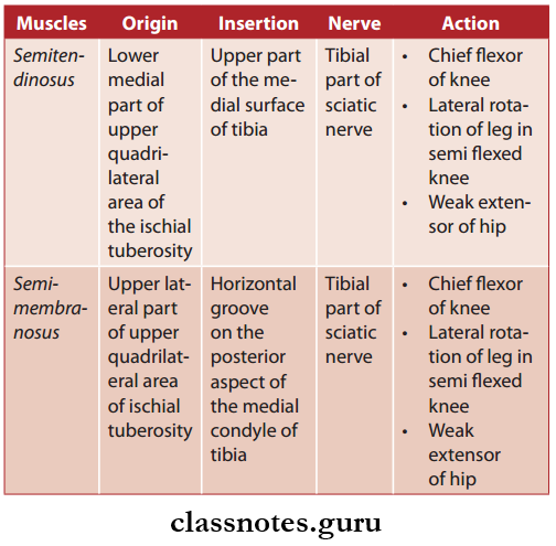
Question 2. Write a note on the hamstring muscles. Explain their origin, insertion, nerve supply, and actions.
Answer:
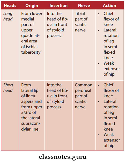
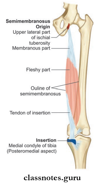
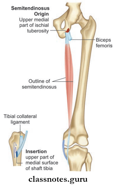
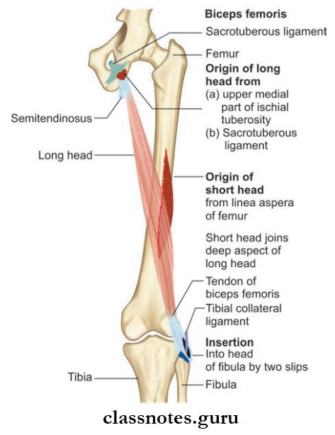
Hamstring Muscles
- Semitendinosus
- Semimembranosus
- Biceps femoris (only long head)
- Ischial head of adductor magnus.
- Characteristic features of hamstring muscles are All of them:
- Arises from the ischial tuberosity
- Inserted into either one bone of leg
- Supplied by the tibial part of the sciatic nerve
- Are floors of knee and extensors of the hip joint.
- The tibial collateral ligament of the knee joint morphologically represents the degenerated tendon of the adductor magnus muscle which is attached below to the tibia, so it is considered as hamstring muscle.
- The short head of biceps femoris is not considered as hamstring muscle.
Hamstring Muscles Biceps Femoris
Rest of Muscles of Posterior Compartment of Thigh
Hamstring Muscles Clinical Anatomy
- If hamstring muscles are paralyzed gluteus maximus muscle alone is not capable of maintaining erect poster, person cannot stand.
- In some persons hamstrings are short and they cannot touch the toe when extending the knee and they are not fit for gymnastics.
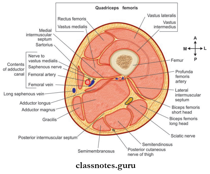
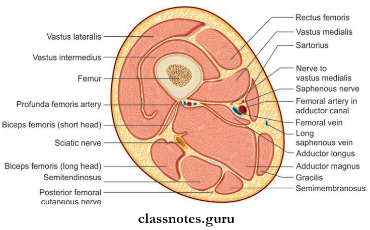
Question 3. Draw the cross-section at the level of mid-thigh.
Answer:
The Cross-Section At The Level Of Mid-Thigh
Question 4. Write a note on the popliteal fossa with its boundaries and contents. How popliteal artery, tibial nerve, and popliteal vein are arranged in the popliteal fossa?
Answer:
Popliteal Fossa With Its Boundaries
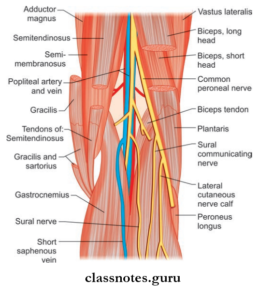
The popliteal fossa is a diamond-shaped depression, lying behind of knee joint, the lower part of the femur, and upper part of the tibia, best felt at the back of the semi-fixed knee joint.
Popliteal Fossa Boundaries
- Superomedially
- Semitendinosus and semimembranosus
- Inferomedially
- The medial head of the gastrocnemius
- Inferolaterally
- The lateral head of the gastrocnemius and plantaris muscle
- Floor/anterior wall
- From above downwards:
- The popliteal surface of the femur
- The capsule of knee joint and oblique popliteal ligament
- Popliteal fascia covering popliteus muscle
- The popliteal surface of the tibia
- From above downwards:
- Roof/posterior wall
- Deep fascial popliteal fascia
- Superficial fascia containing
- Small saphenous vein
- Cutaneous nerves
- The terminal part of posterior cutaneous nerve of thigh
- The posterior division of medial cutaneous nerve of the thigh
- Sural communicating nerve
Popliteal Fossa Contents
- Popliteal artery and branches
- Popliteal vein and branches
- Tibial nerve and branches
- Common peroneal nerve and branches
- Popliteal pad of fat
- The terminal part of posterior cutaneous nerve of thigh
- Descending genicular branch of obturator nerve
- Terminal part of the short saphenous vein.
Arrangements Of The Popliteal Artery, Tibial Nerve And Popliteal Vein In Popliteal Fossa
- In the upper part of the popliteal fossa from medial to lateral side: Artery, vein, nerve (AVN)
- In the middle part of the popliteal fossa from superficial to deep: Nerve, vein, artery (NVA)
- In the lower part of the fossa from medial to lateral
- side: Nerve, vein, artery (NVA).