
Urinary Bladder And Urethra Question And Answers
Question 1. Explain in detail about the external features and relations of the urinary bladder in males and female.
Answer:
Urinary Bladder
- Hollow muscular organ acting as a temporary reservoir of urine
- It receives urine through the ureter and is passed out via the urethra by micturition
Urinary Bladder In Male And Female Location:
In adults, an empty bladder lies in the front part of the lesser pelvis below the peritoneum and behind the pubic symphysis
- In male, it lies in front of the rectum
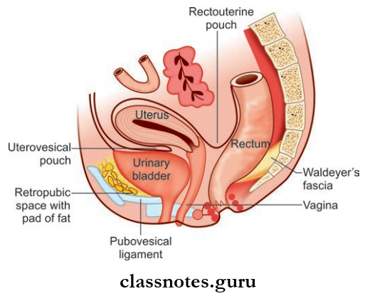
- In female, it lies in front of the uterus
Urinary Bladder Anatomy Important Questions
Urinary Bladder In Male And Female Shape: Empty bladder is tetrahedral, distended bladder is globular
Read And Learn More: Abdomen And Pelvis
Urinary Bladder In Male And Female Capacity: Normally 120–320 ml of urine is present in the urinary bladder, maximum capacity – 500 ml.
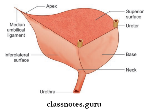
Urinary Bladder In Male And Female External Features
- Empty Bladder Has A Tetrahedral Shape, It Has:
- Apex
- Base
- Neck
- The surfaces—superior and two inferolateral surfaces
- Four borders—anterior, posterior, and two lateral.
Urinary Bladder Apex Or Anterior Angle
- Urinary Bladder is the meeting point of the superior and two inferolateral surfaces
- Lies posterior to the upper margin of the pubic symphysis
- It provides attachment to the median umbilical ligament.
Urinary Bladder Base Or Posterior Surface
- Urinary Bladder is almost like an inverted triangle
- Urinary Bladder has broad and narrow ends
- The broad end (posterior border) is directed superiorly
- The narrow end is directed inferiorly.
Urinary Bladder Apex Or Anterior Angle Urinary Bladder Base Or Posterior Surface Relations Of Base
- Male
- The Upper Part Of Base
- Rectovesical pouch
- Coils of intestine
- Lower Part Of Base
- Seminal vesicles
- Termination of vas deferens
- The Upper Part Of Base
- Female
- Uterine cervix
- Vagina
Urethra Anatomy MCQs With Answers
Urinary Bladder Neck Or Inferior Angle
- The lowest and most fixed part of the urinary bladder lies about 3–4 cm behind the lower part of the pubic symphysis
- It is the meeting point of inferolateral surfaces and the narrow end of the base
- It is pierced by the internal urethral meatus and continues as the urethra
- Circular fiers of the detrusor muscle get modified at the neck to form an internal urethral sphincter.
Urinary Bladder Neck Or Inferior Angle Relations
- Male
- Base of prostate
- Female
- Pelvic fascia
- Urogenital diaphragm
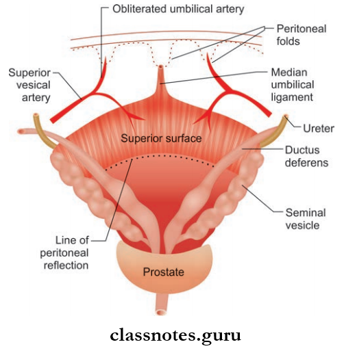
Urinary Bladder Superior Surface
- Triangular in shape
- Bounded on each side by inferolateral surface
- Base of this triangle is formed by the posterior border which lies between two ureteric orifices.
Urinary Bladder Superior Surface Relations
- Male
- Peritoneum
- Coils of intestine
- Sigmoid colon
- Female
- Peritoneum
- Uterine cervix (supravaginal part)
Urinary Bladder Inferolateral Surfaces
- Two in number
- Directed downward, forward, and medially
- They meet each other in the midline and are separated by the anterior border
- Lateral borders separate inferolateral surfaces from superior surfaces
- They are devoid of peritoneum.
Urinary Bladder And Urethra Viva Questions
Urinary Bladder Inferolateral Surfaces Relations
- Common For Both Males And Females.
- Anteriorly
- Retropubic space/Cave of Retzius
- Puboprostatic ligaments in males and pubovesical ligaments in female
- Pubis
- Posteriorly
- Obturator internus muscle
- Levator ani muscle
- Anteriorly
Question 2. Write a note on the ligaments supporting the urinary bladder.
Answer:
The urinary bladder is supported by true and false ligaments:
1. True Ligaments: These are the condensation of pelvic fascia around the base and neck of the bladder and developmental remnants:
- Median Umbilical Ligament
- Remnant of urachus
- Extends from apex to umbilicus
- Medial And Lateral Puboprostatic (m)/Vesical Ligaments (f)
- Formed from the condensation of pelvic fascia and smooth muscle fibers
- They fit the neck of the bladder and also forms the floor of the Cave of Retzius
- They Are Closely Related To Vesical Venous Plexus
- Medial Puboprostatic (m)/Vesical Ligaments (f)
- Extend from neck of the bladder to pubic symphysis
- Directed downwards and backward
- Lateral Puboprostatic (m)/Vesical Ligaments (f)
- Extend from the neck of the bladder to the tendinous arch of the obturator fascia
- Directed medially and backward
- Medial Puboprostatic (m)/Vesical Ligaments (f)
- Lateral Ligament
- Condensation of pelvic fascia
- Extend from inferolateral surface to tendinous arch of pelvic fascia
- Posterior Ligaments
- Condensation of pelvic fascia
- Extend from neck and base of the bladder to lateral pelvic wall
- They contain vesical venous plexus
- Directed backward and upwards along the vesical venous plexus.
2. False Ligaments: They are raised peritoneal folds that do not form any support to urinary bladder, they are:
- Median umbilical fold formed by median umbilical ligament
- Medial umbilical fold formed by obliterated umbilical arteries
- Lateral false ligament formed by the peritoneum of the paravesical fossa
- Posterior false ligament formed by the peritoneum of urogenital folds.
Anatomy Of Urinary Bladder And Urethra Exam Questions
Question 3. Write a note on the interior of the urinary bladder and what is trigone of the bladder is.
Answer:
Interior Of The Urinary BLadder
- In an empty bladder, the mucus membrane is pale in color
- Most of the mucosa appears as irregular folds or rugae since they are loosely attached to the muscular layer
- But over a triangular area, located in the lower part of the base of the urinary bladder, the mucosa is fully attached to the muscular coat
- This does not show rugae or irregular fold
- This area is known as the trigone of the bladder.
Trigone Of Bladder
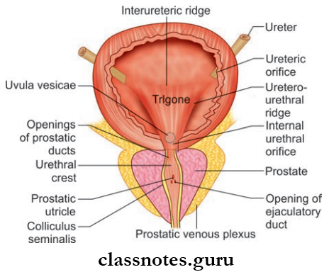
- Trigone Of Bladder has the shape of an equilateral triangle
- Trigone Of Bladder Has:
- Apex/Anterior Angle
- Directed downward and forward
- The internal urethral orifice is present at the apex
- Posterolateral Angles
- Two in number
- Ureters open at these angles
- These openings are 5 cm apart in the distended bladder and 2.5 cm apart in the empty bladder
- Apex/Anterior Angle
- The upper margin of the trigone is formed by fibers of the inner longitudinal muscle coat of the ureter
- Two sides of trigone are formed by ureters-ureteral ridges (modification of inner longitudinal muscle coat of ureter)
- In male, there is a slight elevation of trigone, above and behind the internal urethral orifice (which is produced by the projection of the median lobe of the prostate).
Trigone Of Bladder Applied Anatomy
- In BPH, the median lobe enlarges and blocks the internal urethral meatus, leading to urinary retention
- The Mucosa of trigone is more vascular and sensitive than other parts of urinary bladder.
Question 4. Write a short note on blood supply, lymphatic drainage, nerve supply, and development of urinary bladder.
Answer:
Urinary Bladder Arterial Supply
- Male: Branches of superior vesical and inferior vesical arteries
- Female: Branches of superior vesical and vaginal arteries, and minor contribution from uterine arteries.
Venous Drainage of Urinary Bladder
- Veins of the bladder from the vesical venous plexus
- The vesical venous plexus drains into internal iliac veins
- The vesical venous plexus communicates with the prostatic venous plexus.
Lymphatic Drainage of Urinary Bladder: Lymphatics drain into external iliac lymph nodes.
Nerve Supply Of Urinary Bladder
- Sympathetic Nerves: Derived from T11, T12, L1, L2 spinal segments through the vesical venous plexus
- Parasympathetic Nerves: Derived from S2, S3, and S4 spinal segments.
Development of Urinary Bladder
- Cloaca Is Subdivided By Urorectal Septum Into:
- Anterior primitive urogenital sinus
- Posterior anorectal canal
- The Cranial and largest part of the urogenital sinus is called the vesicourethral canal
- The vesicourethral canal forms most of the urinary bladder
- Trigone of the bladder is formed by absorption of the mesonephric duct
- The apex of the bladder is derived from the urachus
- Splanchnopleuric mesoderm gives rise to muscular and serous walls of the bladder
- Ectopia vesicae: Congenital anomaly in which the posterior wall of the bladder is exposed to the outside, and the anterior wall of the bladder is missing
- Hourglass bladder: The urinary bladder becomes divided into upper and lower parts by a middle constriction, thus giving the appearance of an hourglass.
Urinary Bladder And Urethra Short Questions And Answers
Question 5. Describe about male urethra and write briefly about the external urethral orifice.
Answer:
Male Urethra
- It is 18–20 cm long
- Extent: Internal urethral orifice to external urethral orifice
- Shape:
- Erected Penis: J-shaped
- In the Flaccid State Of The Penis: S-shaped
- Division
Based On The Location, The Urethra Is Divided Into 3 Parts, Namely Prostatic Part, Membranous Part, And Penile Part
Urethra Prostatic Part
- Urethra Prostatic is the widest and most dilatable part
- Urethra Prostatic is 3–4 cm long
- This portion passes through the prostate
- Internally The Posterior Wall Of The Prostatic Part Shows The Following Features:
- Urethral Crest
- Urethral Crest is a midline ridge, present throughout the posterior wall of the prostatic part of the urethra
- This ridge projects into the lumen, giving the lumen a crescentic appearance on the transverse section
- Colliculus Seminalis Or Verumontanum
- Colliculus Seminalis is an elevation in the middle of the urethral crest
- Colliculus Seminalis has a slit-like orifice, which is the opening for the prostatic utricle
- Ejaculatory ducts open on both sides of the opening for prostatic utricle
- Prostatic Sinuses
- They are shallow depressions present on both sides of urethral crest
- Each sinus has 15–20 openings for the prostatic glands.
- Urethral Crest
Urethra Membranous Part
- The Urethra Membranous is the shortest and least dilatable part of the urethra
- Urethra Membranous is about 1.5–2 cm long
- Urethra Membranous passes through the urogenital diaphragm
- Runs downwards and just 2.5 cm above pubic symphysis, it pierces the perineal membrane
- The wall of the membranous urethra has a muscular coat provided by the sphincter urethrae muscle
- Urethra Membranous act as a voluntary external sphincter of the bladder
- The membranous part also contains numerous mucus glands.
Urethra Penile Part
- Also called the spongy part
- It is 15–16 cm long (flaccid penis)
- This portion passes through the corpus spongiosum of the penis
- Extent: Membranous urethra to the external urethral orifice
Urinary Bladder And Urethra Clinical Questions
Urethra Penile Part Course:
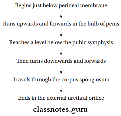
- During its course, the penile part shows two dilatations, they are:
- Intrabulbar Fossa: At the bulb of penis
- Navicular Fossa: At the glans penis
Urethra Other Features
- All portions of the penile urethra except that at the terminal navicular fossa contain urethral glands called Littre’s glands
- These glands open into the penile part through small pit-like mucus recesses called urethral lacunae
- But the roof of navicular fossa contains one lacunae called lacuna magna.
External Urethral Orifice
- The narrowest part of the urethra, about 6 mm long
- It is like a sagittal slit
- It is limited on both sides by a small labium.
Epithelial Lining Of Urethra
- Part Of The Urethra
- Prostatic Part
- Above seminal colliculus
- Below seminal colliculus
- Membranous part
- Spongy urethra above navicular fossa
- Stratified columnar epithelium
- Navicular fossa
- Epithelial Lining
- Transitional epithelium
- Stratified columnar epithelium
- Stratified columnar epithelium
- Stratified squamous epithelium
- Prostatic Part
Blood Supply Of Urethra
- Arterial Supply
- Urethral Artery: Branch of the internal pudendal artery
- Dorsal penile artery.
- Venous Drainage
- The penile part of the urethra drains into → Dorsal veins of the penis → Internal pudendal vein → Prostatic venous plexus
- Prostatic and membranous part of urethra → Prostatic and vesical venous plexus → Internal iliac veins.
- Lymphatic Drainage
- Prostatic and membranous urethra → Internal iliac nodes
- Penile part of urethra → Deep inguinal nodes.
- Nerve Supply
- Sympathetic Supply: From L1 and L2 spinal segments through superior hypogastric plexus
- Parasympathetic Supply: From S2, S3, S4 segments through pelvic splanchnic nerves
- The terminal part of the urethra is supplied by somatic nerves derived from the urethral branches of the pudendal nerve.
Urinary Bladder And Urethra MBBS Notes
Question 6. Write a brief about the female urethra.
Answer:
Female Urethra
- Shorter than the male urethra
- The female urethra corresponds to prostatic and membranous parts of the male urethra
- Extent: From the neck of the bladder to the external urethral orifice in the vestibule of the vagina
- Anterior Relations: Anterior wall of the vagina and pubic symphysis
- The urethral wall is made of an inner longitudinally arranged smooth muscle layer and outer circularly arranged sphincter urethrae.
Urinary Bladder And Urethra Multiple Choice Questions
Question 1. Lymphatics from penile part of urethra drain into which of the following lymph nodes?
- Internal iliac nodes
- Deep inguinal nodes
- Sacral nodes
- Para-aortic lymph nodes
Answer: 2. Deep inguinal nodes
Question 2. The __________ space is located between the body of the urinary bladder and the pubic symphysis:
- Retropubic space
- Vesicouterine space
- Uterosacral space
- Rectopubic space
Answer: 1. Retropubic space
Urinary Bladder And Urethra NEET PG Questions
Question 3. A student while passing a catheter into male urethra suddenly injured it. The rupture is in?
- Prostate gland
- Spongy part of urethra
- Membranous part of urethra
- Prostatic part of urethra
Answer: 3. Membranous part of urethra
Question 4. Pubovesical ligament holds the ___________________ of the urinary bladder in place:
- Fundus
- Body
- Base
- Neck
Answer: 4. Neck
Question 5. The urinary bladder is attached to the anterior abdominal wall via the________ligament:
- Median umbilical
- Medial umbilical
- Lateral umbilical
- Round
Answer: 1. Median umbilical