
Specialized Radiographic Techniques Important Notes
- Xero radiography uses photoconductive selenium plates instead of film.
- It produces images with high contrast, resolution, edge, enhancement, positive and negative display.
- Computer tomography (Axial tomography or computerized axial transverse scanning)
- CT scanner consists of a radiographic tube that emits a finely collimated fan-shaped X-ray beam that is directed to a series of scintillation detectors or ionization chambers.
- The CT image is recorded and displayed as a matrix of individual blocks called voxels.
- Each square of the image matrix is called a pixel.
- For image display each pixel is assigned with a CT number representing density.
- These numbers are also known as Hounsfield units, which may range from – 1000 to +1000
- Each constitutes a different level of optical density.
- To convert a two-dimensional CT image into a three-dimensional CT image, each rectangular solid voxel is dimensionally altered into multiple cuboidal voxels.
- This process is called INTERPOLATION.
- IT Creates sets of evenly shaped cuboidal voxels (Aubervilliers) that occupy the same volume as the original voxel.
- Computed tomography is useful in evaluating structures in and adjacent to salivary glands.
- It distinguishes both soft and hard tissues as well as minute differences in soft tissue densities.
- It is useful in assessing acute inflammatory processes and abscesses as well as cysts, mucoceles, and neoplasias.
- Salivary gland radiology:
- Arcelin: Introduced sialography in 1913. Jacobvisi introduced the sialography technique.
- Contrast agents used in sialography are
- Water soluble – Eg: Pyridone, Singoaffin
- Fat-soluble – Eg: Lipidiol, Ethiodol
- Water-based contrast agents are used for chronic inflammatory lesions
- Oil-based contrast agents are used in neoplasms.
- Different projection after injection of contrast agent.
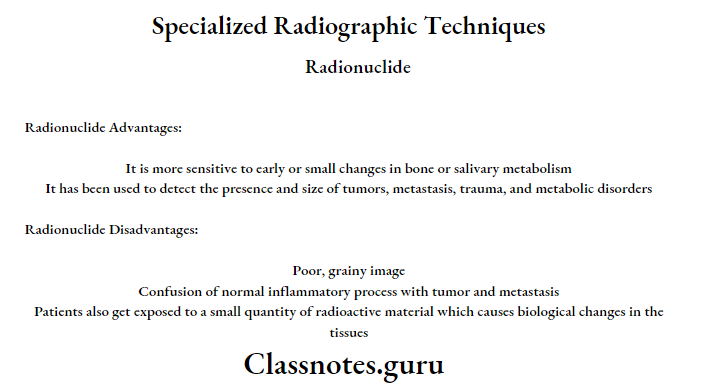
- Nuclear medicine/ scintigraphy provides a functional study of salivary glands.
- The isotope used in the technique is “99 Tc – per technetate”.
- All major salivary glands can be studied at once by scintigraphy.
- It is especially advantageous for conditions in which sialography is contraindicated as well as for patients whose ducts can not be cannulated.
- Ultrasonography is a relatively inexpensive, widely available painless, easy-to-perform, and non-invasive technique.
- The primary application of ultrasonography is for the differentiation of solids from cystic ones.
- Radiographic techniques
Specialized radiographic techniques in dentistry

Read And Learn More: Oral Radiology Question and Answers
Specialized Radiographic Techniques Short Answers
Question 1. Indications and contraindications of sialography.
Answer.
Indications:
- Detection of calculus or foreign bodies
- Determination of the extent of destruction of the gland secondary to obstructing calculi or foreign bodies
- Detection of fistula, diverticula, or strictures
- Determination and diagnosis of recurrent swellings and inflammatory processes
- Demonstration of a tumor and the determination of its location, size, and origin
- Selection of a site for biopsy
- Outline of the plane of the facial nerve
- Detection of residual stones
- Sialography can be used for therapeutic procedures
Contradictions:
- Patients with known sensitivity to iodine
- During the presence of acute inflammation
- It may interfere with subsequent thyroid function tests
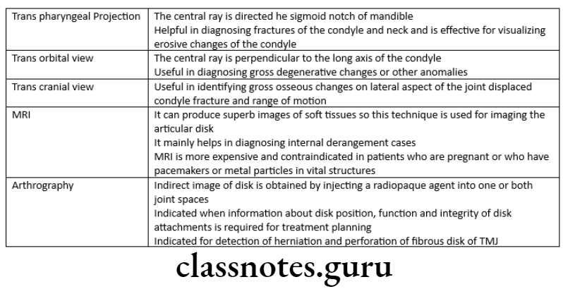
Question 2. Technique for transcranial view of TMJ
Answer.
Transcranial projection:
Structure Seen:
- Useful in detecting arthritis of the articular surfaces
- To evaluate the joint’s bony relationship
Film Position:
- The cassette is placed against the patient’s ear and centered over the TMJ of interest
- It is placed parallel to the sagittal plane
Patient’s Position:
- The sagittal plane must be vertical
- The ala tragus line should be parallel to the floor
- The view is taken with
- Open mouth
- Rest position
- Closed mouth
Central Ray:
- It differs according to the technique
- Postauricular
- Point of entry is 1/2 “behind and 2” above the auditory meatus
- Grewcock approach
- The path of entry is through point 2 above the auditory meatus
- Gill’s approach
- Point of entry is 1/2 “anterior and 2” above the auditory meatus
- Angulation: +20º To +25º
- Point Of Exit: TMJ of interest
- Postauricular
Advanced imaging techniques in oral radiology
Question 3. Indication of sub mento vertex view.
Answer.
Indication of sub mento vertex view
- To demonstrate the base of the skull
- To examine the position and orientation of the condyle and sphenoid sinus
- To reveal the fracture in the zygomatic arch of the maxilla
- To assess the medial and lateral pterygoid plates
Question 4. Radionuclide imaging.
Answer.
Method:
- Radioactive substances should be injected intravenously into the patient
- Rectilinear scanner or gamma scintillation camera records the gamma emission from the patient
- The camera uses a scintillation crystal that can fluorescence on interaction with gamma rays emitting from the radioactive substances
- The emitting light fluorescence is detected by a photomultiplier tube that magnifies and amplifies the signals many times to produce an image
Atom Used:
- Iodine
- Gallium
- Selenium
- Technetium
Question 5. Digital imaging.
Answer.
Digital imaging
- The use of digital technology results in a 50 to 90% reduction in patient radiation exposure because of the greater sensitivity of the digital receptor
Digital Imaging Types:
- Direct digital radiography
- Indirect digital radiography
Digital imaging Uses:
- It can be used to view the images where multiple images are required for analyzing
- In endodontic practice, the root canal length, working length, and distance between obturating material and the root apex
- In periodontics, to assess and measure the height of the alveolar bone
- It can be used in a patient who is un cooperative for regular radiographic techniques
- To evaluate the bony changes in the pathology of jaws
- To detect early dental caries
Extraoral radiographic techniques
Question 6. Sialography – indications
Answer.
Sialography – indications
- Detection of calculus or foreign bodies
- Determination of the extent of destruction of the gland secondary to obstructing calculi or foreign bodies
- Detection of fistula, diverticula, or strictures
- Determination and diagnosis of recurrent swellings and inflammatory processes
- Demonstration of a tumor and the determination of its location, size, and origin
- Selection of a site for biopsy
- Outline of the plane of the facial nerve
- Detection of residual stones
- Sialography can be used for therapeutic procedures
Viva Voce
- Transpharyngeal projection is used for viewing the lateral surface of the condylar head and neck
- Reverse Towne’s projection is used for viewing the posterior aspect of both the condylar head and neck
- Selenium plate is used in xeroradiography
- Water’s view is used to view maxillary sinus