
Regressive Alteration Of The Teeth Important Notes
- Types Of Secondary Dentin
- Physiological
- Secondary Dentin is a regular, uniform layer of dentin around the pulp chamber that is laid down throughout the life of the tooth as a result of age and tooth eruption
- Reparative secondary dentin
- Reparative secondary dentin is the dentin that forms around the pulp chamber in response to irritation or attrition
- Physiological
- Forms Of Pulp Calcifications
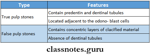
- Discrete pulp stones (denticles)
- True denticles
- They are made up of localized masses of calcified tissue that resemble dentin because of their tubular structure
- They are further divided into
- Free denticles and
- Attached denticles
- False denticles
- Composed of localized masses of calcified material
- They do not exhibit dentinal tubules
- True denticles
- Diffuse calcification
- Mostly seen in the root canals
- Discrete pulp stones (denticles)
Regressive Alteration Of The Teeth Short Question And Answers
Question 1. Describe in detail regressive alterations of teeth
Answer:
Regressive Alterations Of Teeth:
- Regressive Alterations Of Teeth are a group of retrogressive changes in the teeth occurring due to nonbacterial causes and resulting in
- wear and tear of tooth structure along with Impairment of function
- Regressive Alterations Of Teeth are
- Attrition
- Abrasion
- Erosion
Question 2. Classify resorption of teeth. Describe external resorption.
Answer:
Resorption Of Teeth: Resorption of teeth can be defined as chronic progressive damage or loss of tooth structure due to the action of odontoclasts
Resorption Of Teeth Classification:
- Physiological
- Resorption of roots of deciduous teeth
- Pathological
- External resorption
- Internal resorption
Resorption Of Teeth External Resorption:
- Pathological resorption that begins peripherally on the surface of the tooth root and moves toward the pulp is called external resorption
Resorption Of TeethCauses:
- Periapical inflammation
- Reimplantation of teeth
- Tumors and cysts
- Excessive mechanical or occlusal forces
- Impaction of teeth
Resorption Of Teeth Radiographic Features:
- Loss of continuity in peripheral and external outlines of teeth
- Root canals are intact
- Presence of blunting of root apex
- Larger lesions produce a moth-eaten appearance due to irregular or uneven destruction of tooth
- External resorption in apical regions appears as if the root canal-filling materials are projecting beyond the apex
- Obliteration of PDL space
Read And Learn More: Oral Pathology Questions and Answers
Resorption Of Teeth Complications:
- Loss of vitality of the tooth
- Risk of fracture of the tooth
- Ankylosis of teeth
Question 3. Hypercementosis
Answer:
Hypercementosis Definition: It is a prominent thickening of cementum on the root surfaces of the tooth due to excessive cementogenesis
Hypercementosis Etiology:
- Accelerated elongation of a tooth
- Periapical inflammation
- Tooth repair
- Paget’s disease of bone
- Mechanical stimulation- orthodontic forces
- Functionless or unerupted teeth
Hypercementosis Types:
- Localized
- Generalized
- Clinical Features:
- Asymptomatic
- Identified on routine radiograph
Hypercementosis Radiographic Appearance:
- Thickening and apparent blunting of the roots
- Rounding of the root apex
- Well-defined periodontal space
- Histological Features:
- Excessive deposition of cellular and acellular cementum
- This occurs over the entire root or only at the root apex
- This cementum is arranged in concentric layers
Hypercementosis Complications:
- Obliteration of periodontal space
- Ankylosis of tooth
- Difficulty in extraction
- Concrescence of teeth
Question 4. Age changes in pulp
Answer:
Age Changes In Pulp
- Age Changes In Pulp
- Decreased cellularity
- Increased vascularity
- Increased fibrosis
- Impaired pulpal response to tissue injury
- Decrease in pulpal volume
- Pulp calcification- pulp stones
- Pulp Stones/ Denticles:
- Deposition of calcified mass within the dental pulp for no apparent reason is called pulp calcification
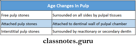
Age Changes In Pulp Types:
- Depending on the microscopic structure
- Depending upon location
Question 5. Internal resorption or Pink tooth of mummery
Answer:
Internal Resorption Or Pink Tooth Of Mummery
Internal resorption refers to the resorption process that starts internally within the tooth itself and the dentin is gradually resorbed from the pulpal side toward the periphery
Internal Resorption Or Pink Tooth Of Mummery Etiopathogenesis:
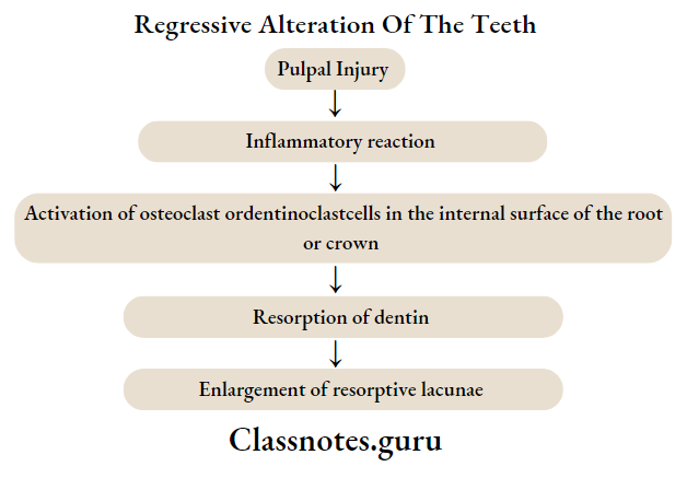
- Hyperplastic pulpal tissues occupy the spaces created due to dentinal re4sorption
Internal Resorption Or Pink Tooth Of Mummery Clinical Feature:
- Can involve the crown portion or root portion of teeth
- Any tooth can ho Involved
- Initially asymptomatic
- Later there Is the appearance of the pink-hued area on the crown of the tooth
- Also known as the pink tooth of mummery
- The affected tooth is vital
Internal Resorption Or Pink Tooth Of Mummery Radiographic Features:
- The involved tooth shows a round or ovoid radiolucent area In the central portion of the tooth
- Well well-defined, spherical-shaped, radiolucent area occurs in dentin
- The external outline of the tooth is intact
Internal Resorption Or Pink Tooth Of Mummery Types:
- Internal inflammatory resorption
- Internal replacement resorption
Internal Resorption Or Pink Tooth Of Mummery Treatment:
- Extirpation of pulp
- Endodontic treatment
- Extraction
Question 6. Age changes in dentin
Answer:
Age Changes In Dentin
- Formation of secondary dentin
- It causes
- Reduction in size of pulp chamber
- Obliteration of pulp chamber
- It causes
- Dentinal sclerosis
- This occurs due to the continued production of peritubular dentin
- It causes
- More brittle roots
- Increased translucency of roots
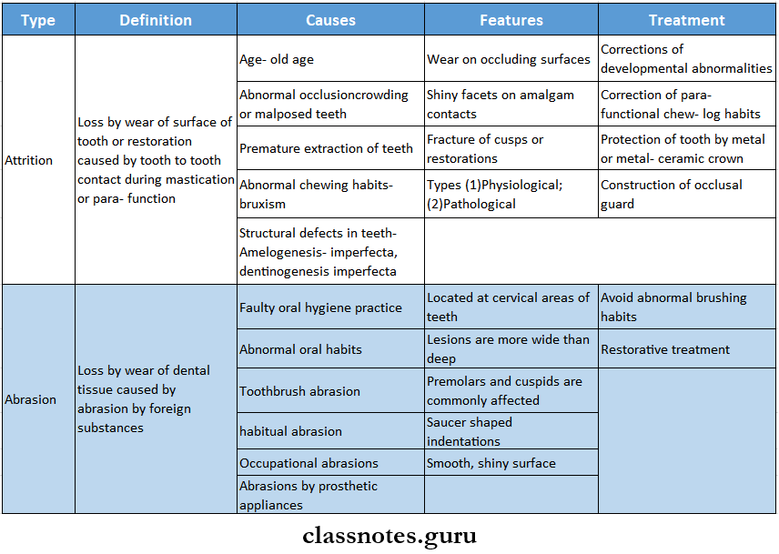
Question 7. Attrition
Answer:
Attrition Definition Loss by wear of surface of tooth or restoration caused by tooth tooth-to-tooth contact during mastication or parafunction
Attrition Clinical Features:
- Wear on occluding surfaces
- Shiny facets on amalgam contacts
- Fracture of cusps or restorations
Attrition Types:
- Physiological-Causes
- Old age- increasing age causes attrition
- Pathological-causes
- Abnormal occlusion
- Premature extraction of teeth
- Abnormal chewing habits
- Structural defects in teeth
Question 8. Abrasion
Answer:
Abrasion Definition: Loss by wear of dental tissue caused by abrasion by foreign substances
Abrasion Clinical Features:
- Located at cervical areas of teeth
- Lesions are more wide than deep
- Premolars and cuspids are commonly affected
- Saucer shaped indentations
- Smooth, shiny surface
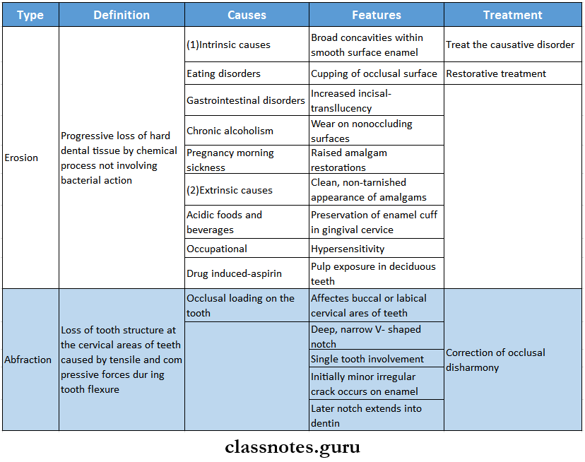
Question 9. Erosion
Answer:
Erosion Definition Progressive loss of hard dental tissue by a chemical process not involving bacterial action
Erosion Causes:
- Intrinsic causes
- Eating disorders
- Gastrointestinal disorders
- Chronic alcoholism
- Pregnancy morning sickness
- Extrinsic causes
Erosion Clinical Features:
- Broad concavities within smooth surface enamel
- Cupping of the occlusal surface
- Increased incisal translucency
- Wear on non-occluding surfaces
- Raised amalgam restorations
- The clean, non-tarnished appearance of amalgams
- Preservation of enamel cuff in gingival service
- Hypersensitivity
- Pulp exposure in deciduous teeth
Question 10. Pink Tooth
Answer:
Pink Tooth
Pink Tooth occurs due to internal resorption
Pink Tooth Clinical Features:
- Can involve the crown portion or root portion of teeth
- Any tooth can be involved
- Initially asymptomatic
- Later there is the appearance of the pink-hued area on the crown of the tooth
- So it is known as the pink tooth of mummery
- The affected tooth is vital
Question 11. Cementicles
Answer:
Cementicles
- The calcified bodies found in the periodontal ligament are called cementless
- These bodies are seen in older individuals
Cementicles Appearance:
- May remain free in the connective tissue
- May join with the cementum
- May fuse into large calcified masses
Cementicles Origin: Degenerated epithelial cells from the nidus for the calcification of cementless
Question 12. Hypercementosis
Answer:
Hypercementosis Definition: It is a prominent thickening of cementum on the root surfaces of the tooth due to excessive cementogenesis
Hypercementosis Etiology:
- Accelerated elongation of a tooth
- Periapical inflammation
- Tooth repair
- Paget’s disease of bone
- Mechanical stimulation- orthodontic forces
- Functionless or unerupted teeth
Hypercementosis Types:
- Localized
- Generalized
Hypercementosis Clinical Features:
- Asymptomatic
- Identified on routine radiograph
Hypercementosis Radiographic Appearance:
- Thickening and apparent blunting of the roots
- Rounding of the root apex
- Well-defined periodontal space
Question 13. Secondary dentin
Answer:
Secondary Dentin
- Secondary Dentin is a narrow band of dentin bordering the pulp
- Secondary Dentin represents continued, slower deposition of dentin by odontoblasts
- There is greater deposition on the roof and floor of the chamber than on other parts
- Secondary Dentin contains fewer tubules which sclerose more readily
- This reduces the overall permeability of the dentin
- Due to the regular arrangement of dentinal tubules, it is also called regular secondary dentin
Question 14. Factors causing external resorption of teeth
Answer:
Factors Causing External Resorption Of Teeth
- Periapical inflammation
- Reimplantation of teeth
- Tumors and cysts
- Excessive mechanical or occlusal forces
- Impaction of teeth
- Idiopathic
Question 15. Dead tracts
Answer:
Dead Tracts
- Dentinal tubules are emptied by complete retraction of the odontoblast process from the tubule or through death of the odontoblast
- The dentinal tubules then become sealed off so that in-ground section air-filled tubules appear by transmitted light as black dead tracts
- They are most often in coronal dentin
- Frequently bound by bands of sclerotic dentin
- These areas demonstrate decreased sensitivity
- They are the initial step in the formation of sclerotic dentin
Regressive Alteration Of The Teeth Viva Voce
- Attrition is defined as the physiological wearing away of tooth surface as a result of tooth to tooth contact
- Abrasion is the pathological wearing away of tooth substance through some abnormal mechanical process.
- Erosion is defined as the irreversible loss of dental hard tissue by a chemical process that does not involve bacteria
- Abfraction is loss of tooth surface at cervical areas of teeth caused by tensile and compressive forces during tooth flexure
- Dentinal sclerosis is a regressive alteration in tooth substance that is characterized by calcification of the dentinal tubules
- Sclerotic dentin under carious lesion is harder than adjacent normal dentin
- Secondary dentin is the dentin that is formed and deposited in response to normal/ slightly abnormal stimulus after root completion
- Hypercementosis is the excessive deposition of cementum on the root surface
- Cementicles are small foci of calcified tissue not necessarily true cementum which lie free in the periodontal ligament of the lateral and apical root areas.