
Rectum And Anal Canal Question And Answers
Question 1. Describe in detail the anatomical features and relations of the rectum.
Answer:
Rectum:
- Part of large intestine between sigmoid colon and anal canal
- Lies in the true pelvis
- Location: Posterior part of lesser pelvis, in front of the lower three pieces of sacrum and coccyx
- Length: 12 cm
- Rectum Diameter:
- Upper Part: 4 cm (same as sigmoid colon)
- Lower Part: Dilated—rectal ampulla
- Rectum Extent:
- Upper end—continuous with a sigmoid colon at the level of S3 vertebrae
- Lower end—lies a little below and in front of the tip of the coccyx
Rectum And Anal Canal Important Questions
Read And Learn More: Abdomen And Pelvis
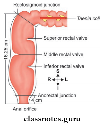
Rectum Course:
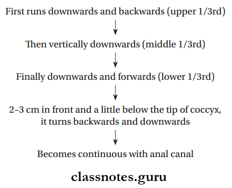
Rectum Curvatures: There are two anterior-posterior curvatures and three lateral curvatures.
Anteroposterior Curvature
- Sacral Curvature: It follows the concavity of the sacrum and coccyx
- Perineal Curvature: It is the backward bend of the anorectal junction
Lateral Curvature
- Upper Lateral Curvature: It is convex to the right at the S3–S4 junction
- Middle lateral curvature: It is convex to the left at the sacrococcygeal junction and is most prominent
- Lower Lateral Curvature: It is convex to the right at the level of tip of the coccyx
Rectum Mucosal Folds: The mucous membrane of rectum shows a number of longitudinal and transverse folds.
- Transverse Folds/Houston’s Valves:
- Permanent folds
- Located against the concavities of lateral curvatures of the rectum
They Are Four In Number:
- First, Fold: lies near the upper end close to the rectosigmoid junction, projects from the right or left wall of rectum
- Second Fold
- Lies 2.5 cm above 3rd fold
- Projects from the left wall of the rectum
- Third Fold: largest fold
- Projects from the anterior and right walls of the rectum
- Lies at the level of the upper end of the ampulla.
- Fourth fFold
- Lies 2.5 cm below the third valve
- Projects from the left wall of the rectum.
Rectum And Anal Canal Viva Questions
Rectum Longitudinal Folds
- Transitory/temporary folds
- Seen in the lower part of the rectum
- Disappears when rectum distended
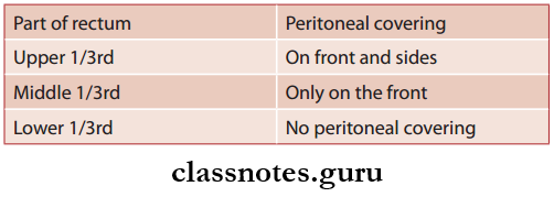
Rectum Peritoneal Relations
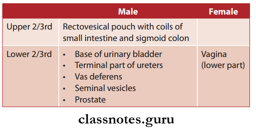
Rectum Visceral Relations
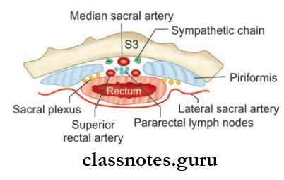
- Rectum Anterior Relations
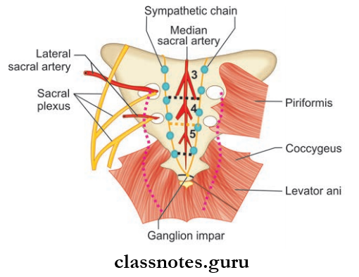
- Rectum Posterior Relations
- Lower part of the sacrum
- Coccyx
- Muscles
- Piriformis muscles (right and left)
- Coccygeus muscles
- Levator ani muscles
- Blood Vessels
- Median sacral vessels
- Lateral sacral vessels
- Superior rectal vessels
- Ganglion impar
- Pelvic splanchnic nerves and sympathetic chains
- Fascia of Waldeyer.
Rectum Arterial Supply
- Superior rectal artery
- Middle rectal arteries
- Inferior rectal arteries
- Median sacral artery
Rectum Venous Drainage: Internal and external venous plexus of rectum and anal canal:
- Superior rectal vein
- Middle rectal vein
- Inferior rectal vein
Rectum Lymphatic Drainage
Rectum And Anal Canal Short Questions And Answers
- The upper half of the rectum—pararectal and sigmoid nodes, inferior mesenteric nodes.
- The lower half of the two rectums—internal iliac nodes.
Rectum Nerve Supply
- Sympathetic supply—derived from L1, L2 spinal segments
- Parasympathetic supply—S2, S3, S4 spinal segments.
Question 2. What are the supports of rectum?
Answer:
The Supports Of Rectum
- Lateral Ligaments Of The Rectum:
- Present on each side of the rectum
- Formed by condensation of pelvic fascia
- Puborectalis sling of the pelvic diaphragm
- Fascia of Waldeyer
- Pelvic floor-formed by levator and muscles
- The rectovesical fascia of Denonvilliers
- Pelvirectal and ischiorectal fat—surrounds the rectum
- Pelvic peritoneum and related vascular pedicles
- Perineal body.
Rectum Applied Anatomy
- Prolapse Of Rectum: The pelvic diaphragm is an important support for the rectum
- When the pelvic diaphragm is weakened (for example, damage during parturition), the rectum can prolapse out of anus.
Question 3. Describe in detail the anatomical features and relations of the anal canal.
Answer:
The Anatomical Features
- Terminal part of the large intestine
- Length: 3.8 cm
- Location: In the anal triangle between the ischiorectal fossa
- Extent: Anorectal junction to the anal orifice
- Direction: Downwards and backward
- It is surrounded by inner involuntary and outer voluntary sphincters
- They keep the lumen closed.
Interior Of Anal Canal
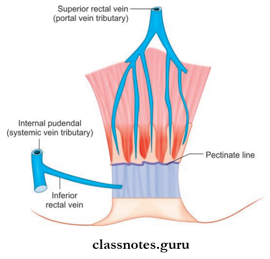
- The pectinate line divides the anal canal into upper and lower parts
- The pectinate line, also called the watershed line, is a transverse line, running along the lower limit of anal valves
- It is a dividing line between endodermal and ectodermal parts of anal canal
- The lower part of the anal canal can again be subdivided into upper and lower regions by the Hilton’s line.
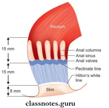
Interior Of Anal Canal Upper Part
- Length: 15 mm
- Extent anorectal junction to pectinate line
- Lined by a mucous membrane:
- Reddish in color
Anatomy Of Rectum And Anal Canal Exam Questions
Interior Of Anal Canal Shows The Following Features:
- Anal Columns Of Morgagni
- There are 6–10 longitudinal folds seen in the mucous membrane
- They contain terminal radicles of the superior rectal artery and vein.
Anal Columns of Morgagni Applied Anatomy: Radicles are well developed in the right anterior, right posterior, and left lateral positions. As a result, piles are more common at these sites.
Anal Valve of Morgagni: They are transverse folds of the mucous membrane, which connect the lower ends of adjacent anal columns.
Anal Valve of Morgagni Applied Anatomy: Passage of hard stools could injure the anal valve leading to anal fissure formation.
- Anal Sinuses
- They are vertical recesses above each anal valve and between anal columns
- Anal glands often into the floor of the anal sinuses.
Interior Of Anal Canal Lower Part
- Upper Region/Pecten
- Known as transition zone
- It is 15 mm long
- Extent: Pectinate line to Hilton’s line
- Lined by nonkeratinized stratified squamous epithelium
- Mucosa is less mobile than the upper part
- Mucosa appears bluish in color due to the presence of rich venous plexus underneath
- White Line Of Hilton
- It is whitish in color compared to bluish color of pecten, so known as white line of Hilton
- It corresponds to inter sphincteric groove in the wall of anal canal.
- Lower Region
- Shortest part of anal canal
- It is 8 mm long
- Extent: White line of Hilton to anal verge
- It is lined by true skin with sweat and sebaceous glands
Rectum Relations
- Anteriorly
- Male
- Membranous urethra
- Bulb of penis
- Female: Lower end of vagina
- Both: Perianal body
- Male
- Posteriorly
- Anococcygeal ligament
- Tip of coccyx.
- Laterally: ischiorectal fossa
Rectum Anal Musculature: Anal musculature is divided into four groups, namely
- Internal sphincter
- External sphincter
- Anorectal ring
- The conjoint longitudinal muscle layer
1. Internal Sphincter
- Involuntary
- Made up of thickened circular smooth muscle coat, surrounding upper 2/3rd of the anal canal
- Extent: From anorectal junction to Hilton’s line
- Above it is continuous with a circular muscle coat of rectum
Rectum And Anal Canal Clinical Anatomy Questions
2. External Sphincter
- Voluntary
- Made up of striated muscle
- Surrounding the entire length of the anal canal
External Sphincter Is Divisible Into Three Parts:
- Deep
- Superficial
- Subcutaneous
- Deep Part
- Located outer to the internal sphincter
- Deep Part has no bony attachment
- Few fires from deep part are attached to the anorectal raphe
- Superficial Part
- The superficial Part lies below the deep part
- Extends up to inter-sphincteric groove
- Superficial Part is the only part of the external sphincter with bony attachments
- Origin from the last piece of coccyx
- Insertion on either side of perineal body
- It does not completely encircle the anal canal (does not support the anal canal in the midline posterior)
- Subcutaneous Part
- Lies below the internal sphincter in the perianal space
- Subcutaneous Part encircles the lowest part of anal canal below inter sphincteric groove
- Subcutaneous Part also has no bony attachment.
3. Anorectal Ring
- Muscular Ring Present At The Anorectal Junction Made Up Of:
- Puborectalis part of levator ani
- Fibers of the deep part of the external sphincter
- Fibers of internal sphincter
- Anorectal ring forms a sling from the pubic bones
- Contraction of the puborectalis pulls the anorectal junction forward, which increases the angulation between the rectum and anal canal (an important factor in the continence mechanism).
4. Conjoint Longitudinal Muscle Layers
- It is the continuation of the longitudinal muscle layer of the rectum which fuses with few fiers of puborectalis (conjoint)
- It separates the internal and external sphincters
- As it goes downwards, this layer becomes ferroelastic and breaks into a number of fibrous septa at the level of the white line of Hilton
- The fibrous septa spread out fanwise and get attached to the skin around the anus
- Most medial septum forms—anal intermuscular septum
- Most lateral septum forms—perianal fascia.
Rectum And Anal Canal Notes For MBBS
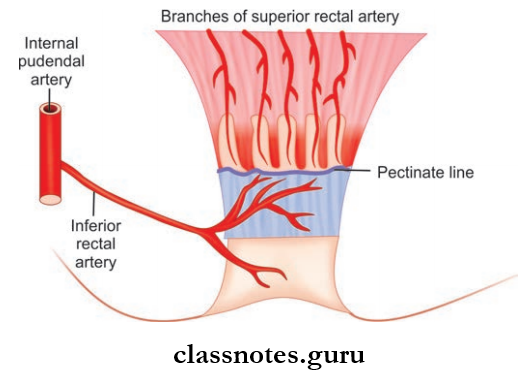
Blood Supply Of Anal Canal
- Arterial Supply
- Superior Rectal Artery: above the pectinate line
- Below The Level Of Pectinate Line: inferior rectal artery.
- Venous Drainage
- Above The Level Of Pectinate Line: superior rectal vein, from there to portal vein
- Below The Level Pf Pectinate Line: inferior rectal vein.
- Lymphatic Drainage
- Upper Part: Internal iliac nodes
- Lower Part: Horizontal group of superficial inguinal nodes.
- Nerve Supply
- Above The Pectinate Line:
- Sympathetic (L1 and L2) through inferior hypogastric plexus
- Parasympathetic (S2, S3, S4) through pelvic splanchnic nerve
- Below The Pectinate Line: somatic supply through inferior rectal nerve
- Internal Sphincter:
- Sympathetic Nerves: contract
- Parasympathetic Nerves: relax
- External Sphincter: inferior rectal nerve and perineal branch of S4 nerve.
- Above The Pectinate Line:
Question 4. Which are the surgical spaces related to the anal canal?
Answer:
The Surgical Spaces Related To The Anal Canal
- Ischioanal space or ischiorectal space.
- Perianal Space: Lies below the level of Hilton’s line between the perianal fascia and skin. A perianal abscess occurs here.
- Submucous Space: Lies above Hilton’s line between the internal anal sphincter and mucous membrane lodges the internal rectal venous plexus.
Question 5. Write a note on the development of the rectum and anal canal.
Answer:
The Development Of The Rectum And Anal Canal
- The upper part of the rectum develops from the endoderm of hindgut
- The lower part of the rectum and upper part of the anal canal is developed from the anorectal canal
- The lower part of the anal canal developed from the proctodeum.
Rectum And Anal Canal NEET PG Questions
Rectum And Anal Canal Multiple Choice Questions And Answers
Question 1. Lymph from the superior rectum drains into ______ nodes:
- Superficial inguinal
- External iliac
- Lumbar
- Sacral
Answer: 3. Lumbar
Question 2. Which lymph nodes drain the lower anal canal?
- External iliac
- Deep inguinal
- Paraaortic
- Superfiial inguinal
Answer: 4. Superfiial inguinal
Question 3. Which of the following is true about the internal anal sphincter?
- Is skeletal muscle
- Has longitudinal fibers
- Has no bony attachment
- None of these
Answer: 3. Is skeletal muscle
Rectum And Anal Canal Labeled Diagram With Questions
Question 4. Which of the following pelvic organs is not a content of the spermatic cord?
- Rectum
- Pelvic appendix
- Ovary
- Urinary bladder
Answer: 1. Rectum
Question 5. Conjoint longitudinal coat of the anal canal is formed by the fusion of?
- Pubococcygeus with longitudinal muscle coat of rectum
- Iliococcygeus with longitudinal muscle coat of rectum
- Internal with external anal sphincter
- Deep with the superficial part of the external anal
Answer: 1. Pubococcygeus with longitudinal muscle coat of rectum
sphincter