
Posterior Abdominal Wall Question And Answers
Question 1. What is thoracolumbar fascia? What are its attachments?
Answer:
Thoracolumbar Fascia
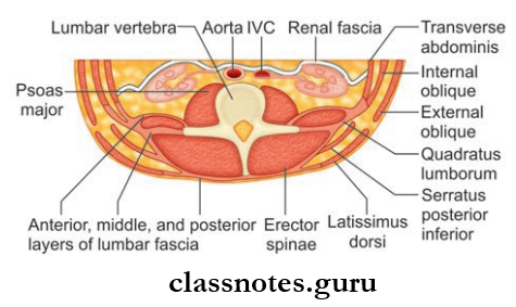
- It is the deep fascia covering the deep muscles on the posterior aspect of the trunk
- It attaches erector spinae to the posterolateral surface of vertebral bodies
- Thoracolumbar Fascia Can Be Divided Into Two Parts:
- Lumbar Part
- Thoracic Part.
Read And Learn More: Abdomen And Pelvis
1. Thoracolumbar Fascia Lumbar Part
- Made Up Of 3 Strong Layers Of Deep Fascia, Namely:
- Anterior Layer: The layer
- Middle And Posterior Layers: Thick and strong layers
- Between the anterior and middle layers lies the quadratus lumborum muscle
- Between the middle and posterior layers lies the erector spinal muscle and transversal spinal muscle
- Laterally the 3 layers fuse to form an aponeurotic sheet
- Internal oblique and transversus abdominis muscles gets there origin from this aponeurotic sheet.
Posterior Abdominal Wall Important Questions
Thoracolumbar Fascia Attachments
- Anterior Layer
- Superiorly: Forms the lateral arcuate ligament
- Inferiorly: Iliac crest
- Medially: Transverse process of lumbar vertebrae
- Middle Layer
- Superiorly: Lower border of 12th rib
- Inferiorly: Iliac crest
- Medially: Tips of the transverse process of lumbar vertebrae, intertransverse ligaments
- Posterior Layer
- Superiorly: Thracic part of the thoracolumbar fascia
- Inferiorly: Iliac crest
- Medially: Spinous process of lumbar vertebrae.
2. Thoracolumbar Fascia Thoracic Part
Thoracolumbar Fascia Attachments
- Superiorly: Continuous with the superficial lamina of investing layer of cervical fascia
- Laterally: Angles of ribs
- Medially: Spinous process of thoracic vertebrae.
Question 2. Write a note on psoas major.
Answer:
Psoas Major
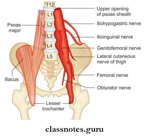
- The Psoas major is one of the important muscles in the posterior abdominal wall
- Other Muscles Are Psoas minor, iliacus, and quadratus lumborum.
Posterior Abdominal Wall Viva Questions
Psoas Major Origin
- Psoas arises from 14 fleshy Slips:
- Five Slips: Each slip arises from bodies and intervertebral discs between two adjacent vertebrae, from T12 – L5
- Five Slips: Each slip arises from the anterior surface and lower borders of the transverse process of five lumbar vertebrae (L1 – L5)
- Four Slips: Each slip arises from tendinous arches connecting the constricted parts of the lumbar vertebrae.
Psoas Major Insertion
- Psoas major descends along the pelvic brim
- Passes anterior to the inguinal ligament and anterior to the hip joint
- Enter the thigh
- And ends on the medial side of a tendon (the lateral side of this tendon receives fiers of iliacus)
- This tendon is inserted into the anterior surface of tip of the lesser trochanter
- Since psoas major and iliacus have a common insertion and action they are together called Iliopsoas.
Psoas Major Nerve Supply: Ventral rami of L2, L3, and L4 spinal nerves.
Psoas Major Actions
- Chief flexor of thigh
- Maintain stability at hip
- Lateral flexion of the trunk on same side.
Psoas Major Relations:
- At Abdomen
- Anterolaterally
- Kidney
- Ureter
- Renal vessels
- Gonadal vessels
- Psoas fascia
- Medial arcuate ligament
- Psoas minor
- Medially
- Lumbar vertebral bodies and vessels
- Posteriorly
- Lumbar plexus
- Transverse process of lumbar vertebrae
- Anterolaterally
Anatomy Of Posterior Abdominal Wall Exam Questions
- At Thigh
- Anteriorly
- Femoral artery
- Fascia lata
- Posteriorly
- Synovial bursa separating capsule of the hip joint from the muscle
- Medially
- Femoral vein
- Pectineus muscle
- Laterally
- Iliacus muscle
- Femoral nerve
- Anteriorly
Question 3. What is psoas sheath?
Answer:
Psoas Sheath
- Psoas Sheath is a fascial sheath enclosing the psoas major muscle
- Psoas Sheath is derived from psoas fascia.
Question 4. Write a note on cisterna chyli.
Answer:
Cisterna Chyli
- Elongated lymphatic sac
- Length: 5–7 cm
- Breadth: 4 mm
- Vertebral level: L1 – L2
- Location: Between aorta and azygos vein, in front of L1 and L2
- It is overlapped by right crus of the diaphragm
- Formed by union of right and left lumbar lymph trunks
- It continues superiorly as the thoracic duct
Psoas Sheath Tributaries:
- Two lymph vessels from lower intercostal lymph nodes (open superiorly).
- Right and left intestinal lymph trunks (opens in the middle)—arising from preaortic lymph nodes.
- Right and left lumbar lymph trunks (opens inferiorly)—arising from lateral aortic lymph nodes.
Posterior Abdominal Wall Short Questions And Answers
Posterior Abdominal Wall Multiple Choice Questions
Question 1. Thoracolumbar fascia:
- Is also known as lumbar ventral fascia
- Encloses all the intrinsic muscles of the back
- Encloses the trapezius, rhomboids, and serratus anterior muscles
- Terminates at the first rib
Answer: 2. Encloses all the intrinsic muscles of the back
Posterior Abdominal Wall Anatomy MCQs
Question 2. Which is not true about the psoas major?
- It arises from the lower border of T12–L5 vertebrae and intervertebral discs between them
- It crosses the pelvic brim and passing deep to the inguinal ligament, gets attached to the lesser trochanter of the femur
- It is supplied by L1, L2, L3
- It causes flexion and lateral rotation movements at hip joint
Answer: 4. It causes flexion and lateral rotation movements at hip joint
Question 3. The cisterna chyli lies adjacent to the:
- T12 vertebral body on the right side, posterior to the aorta
- T12 vertebral body on the left side, anterior to the aorta
- L1 vertebral body on the left side, anterior to the aorta
- L1 vertebral body on the right side, posterior to the aorta
Answer: 4. L1 vertebral body on the right side, posterior to the aorta