
Pericardium And Heart Question And Answers
Question 1. Write a short note on the pericardium.
Answer:
Pericardium
- The Pericardium is a firoserous sac, which surrounds the heart and the root of great vessels
- Located in the middle mediastinum
- Pericardium Has Two Parts:
- Fibrous Pericardium: The outer layer of the pericardium formed by tough connective tissue
- Serous Pericardium: The inner layer, it is subdivided into:
- Parietal layer
- Visceral layer.
Read And Learn More: Thorax Anatomy
Pericardium Contents
- Heart
- Great vessels: Ascending aorta, pulmonary trunk, superior vena cava, inferior vena cava, and pulmonary veins.
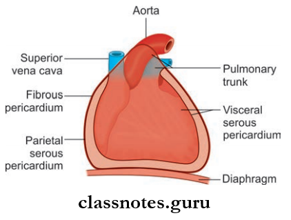
Fibrous Pericardium
- Fibrous Pericardium is a cone-shaped sac
- Fibrous Pericardium Has An:
- Apex
- Blunt
- Continuous with the adventitia of great vessels
- Lies at the level of the sternal angle
- Base
- Broad
- Attached is the central tendon of the diaphragm
Pericardium Relations:
- Anteriorly: Attached to the sternum through sternopericardial ligaments (This attachment helps to retain the position of the heart in the thoracic cavity)
- Posteriorly: Principal bronchi, esophagus, descending thoracic aorta
- Blood supply: Pericardiophrenic vessels
- Nerve supply: Phrenic nerves.
Serous Pericardium: The inner layer. It has two parts:
- Parietal Layer
- It is fused with the inner surface of the fibrous pericardium
- It is continuous with a visceral layer around the roots of great vessels
- Visceral Layer (Epicardium)
- It is fused with the heart
- Deficient only at the cardiac grooves, where it is separated from the heart by the cardiac blood vessels.
Pericardial Cavity
- The Pericardial Cavity is a narrow space formed between the fibrous and serous pericardium
- Capacity—300 ml
- The Pericardial Cavity contains a small amount of serous fluid
- This serous fluid acts as a lubricant, which lubricates the opposed surfaces of fibrous and serous pericardium.
- The arterial supply of fibrous pericardium and the parietal layer of the serous pericardium is by branches of:
- Internal thoracic artery
- Musculophrenic artery
- Descending thoracic aorta
- The visceral layer of the serous pericardium is supplied by coronary arteries
- Venous Drainage
- Veins Drain Into:
- Azygos system of veins
- Internal thoracic vein
- Nerve Supply:
- Fibrous pericardium and parietal pericardium:
- Phrenic nerve (sensitive to pain)
- Visceral pericardium: Autonomic nerves of the heart (insensitive to pain).
- Veins Drain Into:
Pericardium Applied Anatomy
- Pain sensation from the parietal pericardium is carried by somatic afferent fiers of phrenic nerves, so pain during pericarditis or any other irritation to the pericardium is referred to:
- Supraclavicular region of the shoulder
- Lateral part of the neck
- Pericardial Effusion: Accumulation of flid in the pericardial sac.
Pericardium anatomy and function
Question 2. What are the sinuses of the pericardium and what is its use?
Answer:
Sinuses Of Pericardium: The visceral layer of serous pericardium/epicardium is arranged at the roots of great vessels in the form of two tubes:
- Arterial Tube: Arterial tube encloses the pulmonary trunk and ascending aorta
- Venous Tube: Venous tube encloses the superior and inferior vena cava and pulmonary veins.

Transverse Sinus
- There is a horizontal passage present between the arterial and venous ends of the heart tubes, known as transverse sinus
- Located on the upper part of the posterior surface of the heart
- It is a tubular recess between the right and left sides of the pericardial cavity posteriorly
Transverse Sinus Boundaries:
- Anteriorly: Ascending aorta, pulmonary trunk
- Posteriorly: Superior vena cava, pulmonary veins
- Inferiorly: Upper surface of left atrium
- Superiorly: Bifurcation of the pulmonary trunk
- On Each Side: Opens into the pericardial cavity.
Oblique Sinus
- Oblique Sinus is a space located on the posterior surface of the heart between the venous tubes of four pulmonary veins
- The oblique Sinus is called a cul de sac (a passage with access only at one end) since it is closed on all sides except inferiorly
- Oblique Sinus lies behind the left atrium
Oblique Sinus Boundaries:
- Anteriorly: Left atrium
- Posteriorly: Parietal pericardium
- Right side: Right pulmonary veins and inferior vena cava
- Left side: Left pulmonary vein
- Superiorly: Upper margin of the left atrium
- The presence of an oblique sinus allows free pulsation of the left atrium.
Sinuses Applied Anatomy
During cardiac surgery, the transverse sinus provides the space to clamp the ascending aorta and pulmonary trunk, in order to insert tubes of heartlung machines.
Question 3. Describe the external features of the heart.
Answer:
External Features Of Heart
- The heart is a conical hollow muscular pump
- Structurally and functionally it consists of two halves right and left
- Each half has an atrium and ventricle, thus it has 4 chambers.
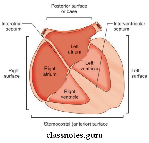
External Features of Heart Margins
- Right Margin: Formed by the rounded surface of the right atrium
- Left Margin: Formed by the rounded surface of the left ventricle.
External Features of Heart Surfaces
- Anterior/Sternocostal Surface
- Posterior Surface/base
- Right And Left Surface
- Diaphragmatic Surface/Inferior Surface.
1. Anterior/Sternocostal Surface
- Anterior/Sternocostal Surface Formed By:
- Right atrium
- Right ventricle
- Left ventricle (minor contribution)
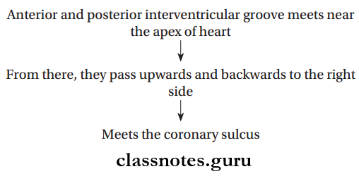
- The right atrium and ventricle are separated from each other by the atrioventricular groove or the coronary sulcus (lodges the coronary artery)
- Ventricles are separated anteriorly by the anterior interventricular groove
- The sternocostal surface is separated from the diaphragmatic surface by a sharp border (Inferior border)
- The point where the inferior border meets the left border is known as the apex
- The upper border is formed by the left atrium.
2. Posterior Surface/Base
- Posterior Surface/Base Formed by:
- Left atrium
- Posterior part of the right atrium (small contribution).
3. Diaphragmatic Surface
- Diaphragmatic Surface Formed by:
- Left ventricle (2/3rd contribution)
- Right ventricle (1/3rd contribution)
- Two ventricles are separated by the posterior interventricular groove.
Anterior Part of Coronary Sulcus
- Right Hhalf: It is easily visible on the sternocostal surface
- Course: Runs downwards and to the right between right atrium and right ventricle
- Left Half: Its view is hidden by the aorta and pulmonary trunk.
Posterior Part of Coronary Sulcus
Lies in the diaphragmatic surface at the junction of the right ventricle and right atrium and left ventricle and left atrium.
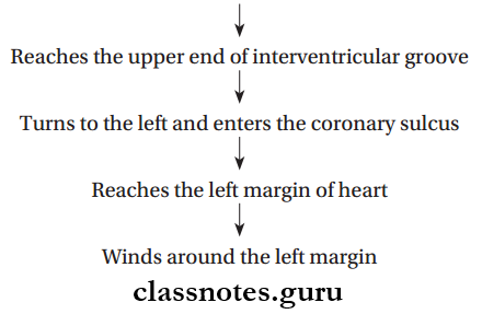
The Course Of Interventricular Grooves
Fibrous and serous pericardium
Question 4. Write a note on the development of the heart tube.
Answer:
Development Of Heart Tube
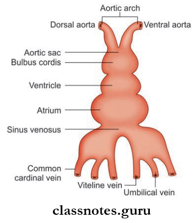
- Mesodermal in origin
- During 3rd week angioblastic cords are formed from intraembryonic mesoderm
- Angioblastic cords are paired endothelial strands formed in the cardiogenic area
- Cords undergo canalization and form heart tubes
- Firstly heart has right and left endothelial tubes which fuse together to form a single tube
- Single tube undergoes dilatation separated by constrictions from top to bottom
- These dilatations from above to below are later identified as:
- Bulbus Cordis Has Three Parts:
- Truncus Arteriosus: Distal 1/3rd part, forms ascending aorta and pulmonary trunk. It is continuous with the aortic sac distally.
- Conus: Middle 1/3rd, forms outflow tracts of ventricles
- The proximal 1/3rd part forms the primitive right ventricle
- The primitive ventricle forms the trabeculated part of the left ventricle
- Primitive atrium
- Sinus Venous:
- Sinus venous has prolongations at the caudal end called right and left horns
- Each horn is joined by a vitelline vein, umbilical vein, and common cardinal vein.
Question 5. Explain in detail the external and internal features of the right atrium.
Answer:
Features Of Right Atrium
- The right upper chamber of the heart
- It Receives Venous Blood Mainly From:
- Superior vena cava
- Inferior vena cava
- Coronary sinus
- It pumps blood into the right ventricle through the tricuspid valve
- The tricuspid opening is guarded by the tricuspid valve or atrioventricular valve
- Right Atrium Forms:
- The sternocostal surface of the heart
- Base of heart
- Right border
- A portion of the upper border.
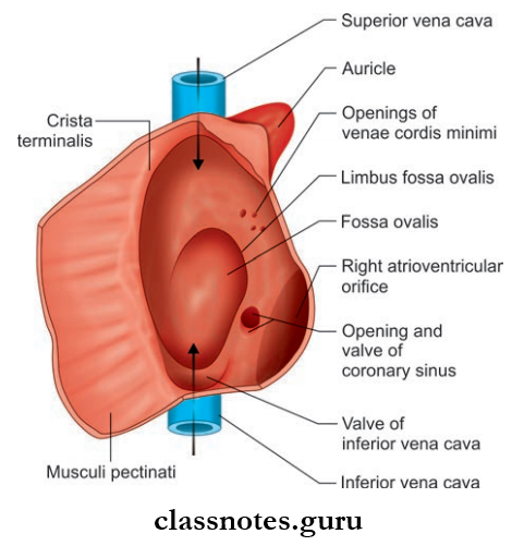
External Features Of Right Atrium
- The right atrium is a vertically elongated chamber
- An ear-like projection arises from an anterosuperior part of the right atrium, known as the right auricle
- The right auricle has a notched margin and it covers the ascending aorta and infundibulum of the right ventricle
- The Interior of the right auricle is sponge-like
- Right atrium receives a superior vena cava at its upper end and an inferior vena cava at its lower end
- On the right border of the atrium, a sulcus passes from the superior vena cava to the inferior vena cava, known as sulcus terminalis
- The upper part of this sulcus lodges the SA node
- Sulcus terminalis is formed by an internal muscular ridge known as crista terminalis.
Internal Features Of Right Atrium: The Interior of the heart can be divided into two parts:
- Sinus venarum/Smooth posterior part: Derived from right horn of sinus venosus
- Atrium proper/Rough anterior part: Derived from the primitive atrial chamber.
- Sinus Venarum/Smooth Posterior Part
- The superior vena cava opens at its upper end and the inferior vena cava at its lower end
- Opening of the inferior vena cava is bounded by a semilunar valve called as eustachian valve/valve of the inferior vena cava
- It is actually rudimentary
- In embryonic life, it transmits blood directly from the inferior vena cava to the left atrium through the foramen ovale
- Opening for coronary sinus is present between openings of inferior vena cava and right atrioventricular orifice, which is also guarded by a valve known as the valve of the coronary sinus
- Venae cordis minimi opens into the right atrium through numerous small openings in the wall of the atrium.
- Atrium Proper/Rough Anterior Part
- Also called the pectinate part
- It contains the right auricle
- Its wall shows the presence of transversely running muscular ridges called the musculi pectinate
- Ridges arise from the crista terminalis, run forward and downward towards the av orifice (teeth of a comb-like appearance)
- Some of them enter the right auricle and form a network.
Tributaries Of Right Atrium
- Superior vena cava
- Inferior vena cava
- Coronary sinus
- Anterior cardiac veins
- Venae cordis minimi
- Right marginal vein
Question 6. From where does the right atrium develop?
Answer:
Development Of Right Atrium
- Mainly formed from the right half of the primitive atrium
- The rough anterior part develops from the right horn of the sinus venosus
- The sinus venous and right half of the atrioventricular canal get absorbed into the right atrium
- The smooth posterior part along with the right auricle develops from the primitive atrium.
Question 7. What are the features of the interatrial septum and how is it formed?
Answer:
Interatrial Septum
- Interatrial Septum is derived from septum primum and secundum
- When looked at from the right atrium, this septum shows some gross features:
- Fossa Ovalis
- Oval depression on the lower part
- Represents the site of embryonic septum primum
- Annulus Ovalis/Limbus Fossa Ovalis
- Prominent margin of fossa ovalis
- Represents the lower curved edge of the septum secundum.
- Fossa Ovalis
Development of Interatrial Septum
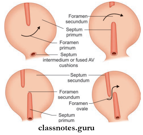
- Interatrial Septum develops from two septa (septum primum and septum secundum) arising from the roof of the atrial chamber
- Septum primum arises from the roof of the atrial chamber and grows downward towards the septum intermedium
- Initially, there will be a gap between septum primum and septum intermedium known as foramen primum
- Finally, septum primum fuses with septum intermedium
- Before the fusion, the upper part of the septum primum breaks down leaving a free upper edge
- A new foramen is created known as foramen secundum
- Another septum called septum secundum starts to grow from the right of the septum primum, towards the septum intermedium
- Septum secundum overlaps the upper margin of septum primum, creating an oblique passage between septum primum and secundum called foramen ovale
- In fetal life, foramen ovale allows the blood to flow from right to left atrium
- Finally, by the fusion of septum secundum and septum primum, the foramen ovale is obliterated
- The upper and lower half of the interatrial septum are formed by the septum secundum and primum respectively.
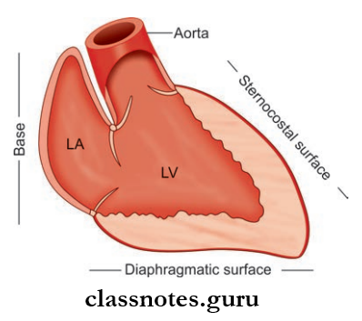
Question 8. Write briefly about the gross features of the left atrium.
Answer:
left atrium Gross Features
- Quadrangular shaped thin-walled cavity
- Situated posteriorly
- Also contains the left auricle
- The Left Atrium Forms The Part Of:
- The upper border of the heart
- The sternocostal surface of the heart
- Left surface and left border
- Left 2/3rd of the base of the heart.
Internal Features Of Left Atrium
- Most of the wall is smooth, this part derived from absorbed pulmonary veins
- Musculi pectinate only present in the left auricle, derived from the primitive atrial chamber of the heart tube
- It receives 4 pulmonary veins in its upper lateral part
- Anteroinferiorly, the left atrium opens into the left ventricle through the left atrioventricular orifice
- This orifice is guarded by the mitral valve.
Pericardial cavity anatomy
Question 9. What are the similarities and differences between right and left ventricles?
Answer:
Question 10. Write briefly on the valves of the heart.
Answer:
Heart Valves
- Valves Of Aortic Orifices: Tricuspid and mitral valve (Bicuspid valve)
- Valves Of Pulmonary And Aortic Valves/Semilunar Valves: Pulmonary valve and aortic valve
Tricuspid Valve Of Heart: Made up of three cusps attached to a fibrous ring
- Anterior Cusp: Attached to the superolateral part of the margin of the right AV orifice
- Posterior Cusp: Attached to the inferolateral part of the margin
- Septal Cusp: Attached to the medial margin
Bicuspid Value Of Heart: Made up of two cusps attached to a fibrous ring
- Anterior cusps: Attached to the upper right part of the margin of the left AV orifice
- Posterior cusp: Attached to the lower left part of the margin of the left AV orifice
Free margins and ventricular surfaces of cusps are connected to papillary muscles through chordae tendineae.
Pulmonary Orifice Of Heart
- Longer 3 cm in diameter
- Location: Superior and left to the tricuspid orifice (aortic orifice intervenes between pulmonary orifice and tricuspid orifice)
- Guarded by pulmonary valve
- Has three semilunar cusps:
- Cusps are triangular in shape
- Made up of a double fold of endocardium with fibrous tissue enclosed in it
- Has two free margins
- Free margins meet at the apex of the cusp
Aortic Orifice Of Heart
- Smaller, 2.5 cm in diameter
- Location: Anterior and right to mitral orifice
- Guarded by aortic valve
- Same
Question 11. Write a note on the interventricular septum.
Answer:
Interventricular Septum
- Separates right and left ventricle
- The position corresponds to anterior and inferior interventricular grooves
- The right side is convex, it bulges into the right ventricle
- The right side of the septum is directed anteriorly and to the right, and the left side is directed backward and to the left
- Right and left AV orifices are separated by the posterosuperior border of the septum
- The septum is thick and muscular, except at a small area near the posterior margin, where it is membranous
- The septal cusp of the tricuspid valve is attached to the membranous part and separates the septum into:
- Anterior part: Separating right and left ventricle
- Posterior part: Separating left ventricle from right atrium.
Interventricular Septum Applied Anatomy
- Fallot’s Tetralogy Congenital condition is characterized by:
- Stenosis of the pulmonary trunk
- Large ventricular septal defect
- Overriding of aortic orifice above VSD
- Right ventricular hypertrophy due to high BP in RV
- Leading to the right to the left shunt of blood through VSD, resulting in severe cyanosis.
Question 12. Describe in detail the blood supply of the heart. What is cardiac dominance?
Answer:
Blood Supply Of Heart
- Coronary arteries supply blood to the heart
- There are two of them:
- Right coronary artery
- Left coronary artery
- Originate from the ascending aorta
- Run in the coronary sulcus
- They can be called the largest vasa vasorum if we consider the heart as a modified blood vessel
- The pattern of branching of coronary arteries shows variation in different individuals.
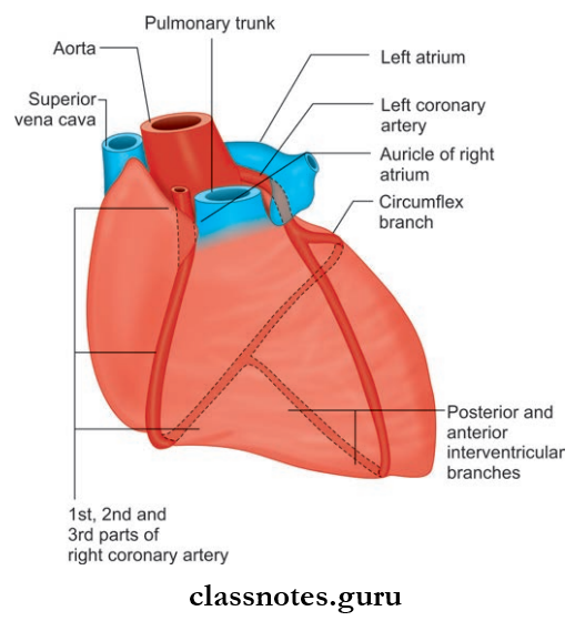
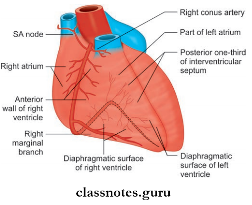
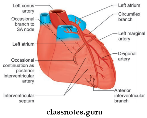
The Most Common Pattern Is Described Below:
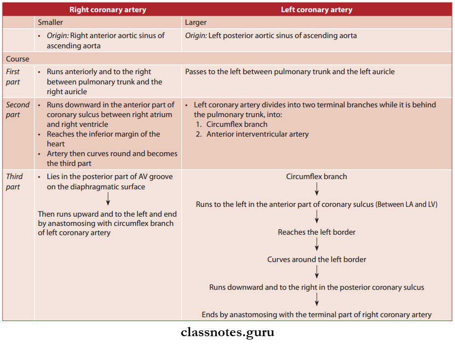
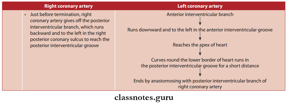
Branches Of Coronary Arteries
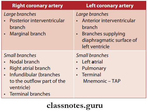
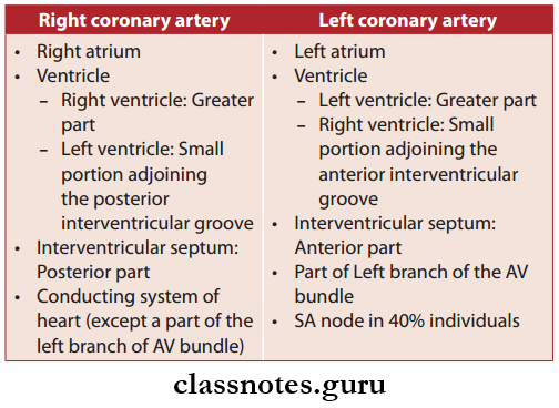
Areas of Distribution
Cardiac Dominance
- Normally, in 90% of the cases, the right coronary artery gives off the posterior interventricular branch, such hearts are called right dominant heart
- But in 10% of the population, the left coronary artery gives off the posterior interventricular branch, such hearts are called the left dominant heart.
Cardiac Dominance Applied Anatomy
- The left coronary artery is more prone to blocks, so in left-dominant individuals, blocks may be fatal
- Coronary Angiography: Coronary arteries and their branches are visualized by this technique, to determine the sites of narrowing.
Question 13. Write a short note on the veins of the heart.
Answer:
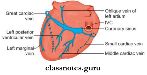
Veins Of The Heart They Are Namely:
Coronary sinus, Great cardiac vein, middle cardiac vein, small cardiac vein, right marginal vein, an oblique vein of the left atrium, a posterior vein of the left ventricle, anterior cardiac vein, and vena cordis minimi.
1. Veins Of The Heart Coronary Sinus
- The largest vein of the heart
- 3 cm long
- Most of the veins draining the heart open into the coronary sinus
- Located in the left posterior coronary sulcus
- Its right end opens into the posterior wall of the right atrium
- Tributaries:
- Great Cardiac Vein
- Small Cardiac Vein
- Middle Cardiac Vein
- Posterior Vein Of The Left Ventricle
- Oblique Vein Of Left Atrium
- Right Marginal Vein (Sometimes).
- Tributaries:
Great Cardiac Vein
- Seen mainly on the sternocostal surface
- It ascends in the anterior interventricular groove, parallel to the anterior interventricular branch of the left coronary artery
Small Cardiac Vein:
- It runs in the right posterior coronary sulcus, accompanying the right coronary artery
- Opens into the right end of the coronary sinus.
Middle Cardiac Vein:
- Begins near the apex of the heart
- Runs backward on the diaphragmatic surface
- It lies in the posterior interventricular groove accompanying the posterior interventricular branch of the right coronary artery
- Opens into the middle part of the coronary sinus.
Posterior Vein Of The Left Ventricle:
- Runs backward on the diaphragmatic surface of the ventricle
- Opens into the middle of the coronary sinus.
Oblique Vein Of Left Atrium:
- Derived from left common cardinal vein
- Runs in the posterior surface of the left atrium
- Opens into the left end of the coronary sinus.
Right Marginal Vein: Opens into the coronary sinus or into a small cardiac vein.
2. Anterior Cardiac Vein
- 3 or 4 in number
- Runs parallel to each other on the anterior wall of the right ventricle
- Opens through the anterior wall of the right atrium.
3. Venae Cordis Minimi
- Smallest cardiac vein or the Thbesian vein
- Present in all chambers of the heart
- More on the right side
- Drain directly into the chambers of the heart.
Pericardium histology
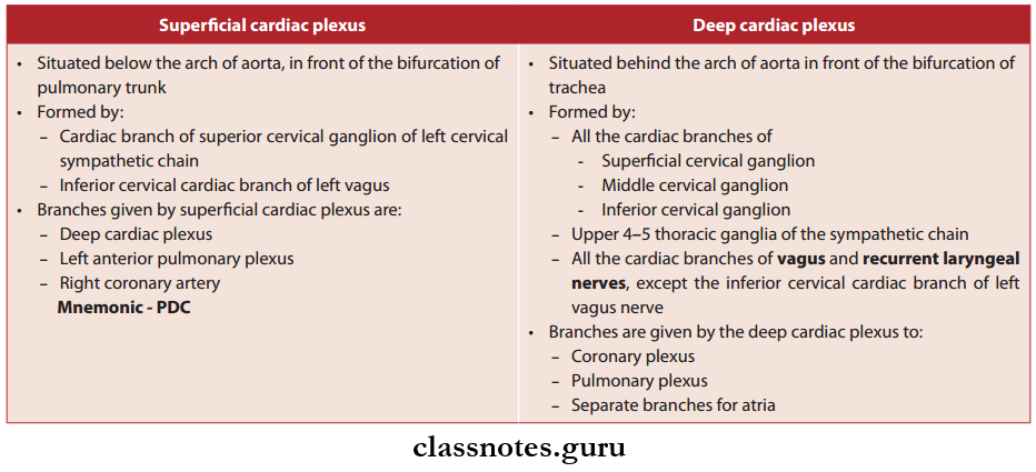
Quetsion 14. What are cardiac plexuses?
Answer:
Cardiac Plexuses
- The heart has both sympathetic and parasympathetic supply
- Sympathetic Fibers:
- Derived from upper 4–5 thoracic spinal segments
- Cardioacceleratory (increase HR)
- Also dilates coronary arteries.
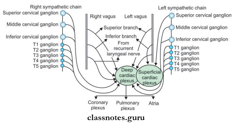
- Parasympathetic Fibers:
- Derived from the vagus nerve
- Cardioinhibitory
- Constricts coronary arteries.
- Both sympathetic and parasympathetic fibers supply the heart through the superficial and deep cardiac plexus
- Branches from the cardiac plexus run along the coronary arteries and supply the myocardium.
Question 15. What is the lymphatic drainage of the heart?
Answer:
Heart Drainage
- Lymphatics from the heart accompany coronary arteries
- Forms two trunks:
- The right trunk, drains into brachiocephalic nodes
- Left trunk, drains into inferior tracheobronchial nodes.
Question 16. Write briefly on the conduction system of the heart.
Answer:
Conduction System

- Right bundle branch → Pass through the right side of the interventricular septum → Reaches the right ventricle → Divides into Purkinje fibers
- Left bundle branch → Pass through the left side of the interventricular septum → Reaches the left ventricle→ Divides into Purkinje fibers.
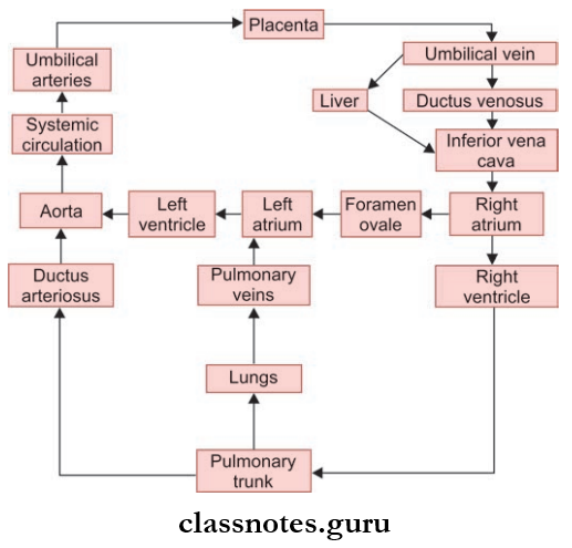
Question 17. Draw a flowchart depicting fetal circulation.
Answer:

Pericardium And Heart Multiple Choice Questions
Question 1. The only part of the heart not covered by serous pericardium is:
- The posterior wall of the left atrium
- The right wall of the oblique sinus
- Infundibulum of the pulmonary trunk
- The floor of the transverse sinus
Answer: 4. Floor of the transverse sinus
Question 2. Which of the following does not open into the right atrium?
- Anterior cardiac vein
- Small cardiac vein
- Coronary sinus
- Venae cordis minimi
Answer: 2. Small cardiac vein
Pericardium histology
Question 3. The apex of the heart is:
- Entirely formed by the left ventricle
- Formed by the right ventricle
- The greater part by the right ventricle
- The greater part formed by the left ventricle
Answer: 1. Entirely formed by the left ventricle
Question 4. All the following openings in the right atrium are guarded by valves except:
- Coronary sinus
- Superior vena cava
- Inferior vena cava
- Atrioventricular opening
Answer: 2. Superior vena cava
Question 5. The atrioventricular node lies at:
- Membranous interventricular septum
- Crista terminalis
- Interatrial septum
- Muscular interventricular septum
Answer: 3. Interatrial septum