
Perineum And True Pelvis Question And Answers
Question 1. What is perineum and what are its boundaries?
Answer:
Perineum
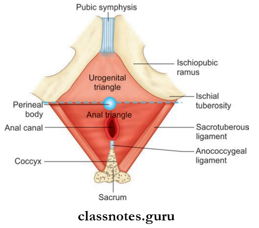
Lowest region of the trunk in the erect position, lying below the pelvic diaphragm.
Superficial Boundary Of Perineum
- Anteriorly
- Male: Scrotum
- Female: Mons pubis
- Posteriorly: Buttocks
- Each side: Upper medial aspect of thigh
Deep Boundary Of Perineum
- Anteriorly: Lower margin of the pubic symphysis
- Posteriorly: Tip of the coccyx
- Each side: Sacrotuberous ligament, ischial tuberosity, conjoint ischiopubic rami
Read And Learn More: Abdomen And Pelvis
Question 2. What is the urogenital and anal triangle?
Answer:
Perineum Is Divided Into Two Triangles, Namely:
- Anteriorly—urogenital triangle
- Posteriorly—anal triangle
- The apex of both triangles are directed opposite to each other
- The base of both triangles are formed by a horizontal plane passing through the anterior end of ischial tuberosities
- The perineal body is located at the midline of the base of both triangles.
Pelvis And Perineum Anatomy Questions And Answers
Question 3. Write a note on the boundaries and contents of the superficial perineal pouch.
Answer:
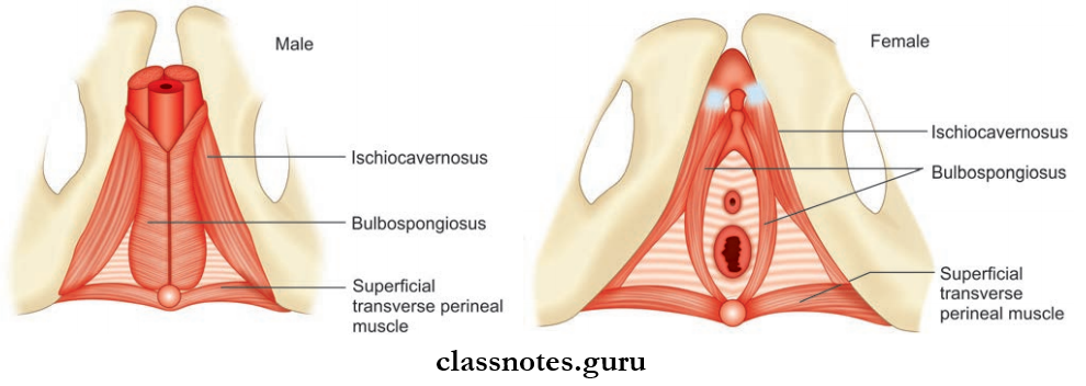
Superficial Perineal Pouch Boundaries: Same for both males and females
- Superiorly: Perineal membrane
- Inferiorly: Membranous layer of the superficial fascia of the perineum (Colles fascia)
- Laterally: Conjoint ischiopubic rami
- Posteriorly: The pouch is closed by the fusion of superior and inferior walls
- Anteriorly: The pouch is open, and is continuous with the anterior abdominal wall through scrotum and penis (in males).
Superficial Perineal Pouch Contents
- Male
- Root of penis
- Duct of bulbourethral glands
- Superficial transverse perineal muscles
- Urethra
- Branches of internal pudendal artery and nerve
- Female
- Root of clitoris
- Urethra
- Greater vestibular glands
- Superficial transverse perineal muscles
- Branches of internal pudendal artery and nerve
Question 4. Write a note on the boundaries and contents of the deep perineal pouch.
Answer:
Deep Perineal Pouch
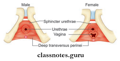
Deep Perineal Pouch Boundaries: Same for both males and females
- Superiorly: Superior fascia of urogenital diaphragm
- Inferiorly: Perineal membrane (Inferior fascia of urogenital diaphragm)
- Laterally: Conjoint ischiopubic rami
- Posteriorly: It is limited by the fusion of two fascial layers
- Anteriorly: It is closed due to the fusion of superior and inferior fascial layers.
Deep Perineal Pouch Contents
- Male
- Membranous urethra
- Bulbourethral glands on both sides of the urethra
- Two striated muscles: Sphincter urethrae and deep transverse perineal muscles
- Dorsal nerve of the penis
- Branches of internal pudendal artery and nerve
- Female
- Urethra
- Vagina
- Two Striated Muscles:
- Sphincter urethra and deep transverse perineal muscles
- The dorsal nerve of the clitoris
- Internal pudendal artery and its terminal branches
Pelvis And Perineum Important Questions
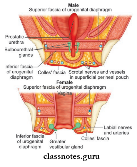
Question 5. What is the urogenital diaphragm?
Answer:
Urogenital Diaphragm
- Urogenital is a muscular sheet consisting of sphincter urethrae muscle and deep transverse muscle
- Urogenital is enclosed between the superior and inferior fascia of the urogenital diaphragm
- Structures Piercing Urogenital Diaphragm:
- Male: Membranous urethra
- Female: Urethra and vagina
- It Acts As A Support For:
- Male: Prostate and neck of the urinary bladder
- Female: Vagina and neck of the urinary bladder
- It also reinforces the pelvic diaphragm at the urogenital hiatus.
Pelvis And Perineum MCQs With Answers
Question 6. Write a note on the perineal membrane.
Answer:
Perineal Membrane
- Inferior fascia of the urogenital diaphragm
- It lies between deep and superficial perineal pouches
- Triangular in shape
- Apex is directed anteriorly
- Base is directed posteriorly
Perineal Membrane Relations
- Superiorly: Deep perineal pouch
- Inferiorly: Superficial perineal pouch
Perineal Membrane Attachments
- Apex: Attached to the arcuate ligament of the pubis as transverse perineal ligament
- Laterally: Ischiopubic rami
- Base: Perineal body
- The posterior border is continuous with the fascia over deep transverse perineal muscles
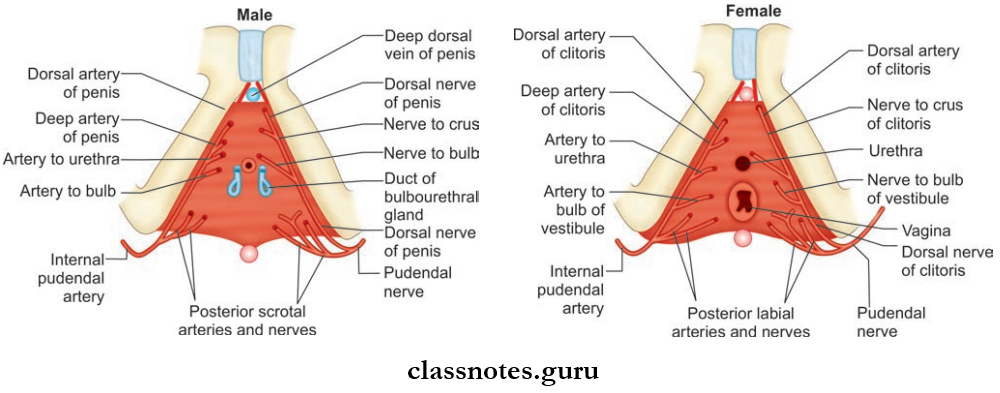
Perineal Membrane Is Pierced By:
- Male
- Urethra in the midline
- Ducts of bulbourethral glands
- Artery to the bulb of penis
- Deep artery and dorsal artery of penis
- Urethral artery
- Dorsal nerve of penis
- Two posterior scrotal nerves and vessels on each side of the base
- Female
- Urethra
- Vagina
- Arteries to the bulb of the vestibule on each side of the vagina
- Deep artery of clitoris
- Dorsal nerves and vessels of the clitoris
- Posterior labial nerves and vessels
- The perineal membrane of the female is very thin because it is pierced by the vagina in addition to other structures.
Pelvis And Perineum Viva Questions
Question 7. Write briefly on the perineal body.
Answer:
Perineal Body
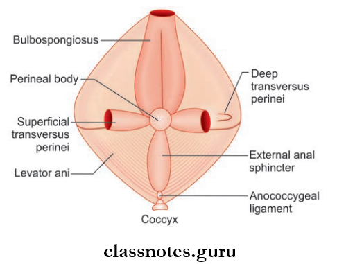
- Perineal Body is a mass of fibromuscular tissue situated in the midline at the junction of the urogenital triangle and anal triangle
- In males, it lies between the bulb of the penis and the anal canal
- In females, it lies between the anal canal and the lower part of the posterior wall of the vagina
- It Provides Attachment To 10 Muscles Of The Perineum Which Are:
- Right and left superficial transverse perineal muscles
- Right and left deep transverse perineal muscles
- Right and left bulbospongiosus muscles
- Two levator ani muscles
- One sphincter ani muscle
- One longitudinal muscle coat of anal canal.
Perineal Body Applied Anatomy
- In males, the perineal body supports the prostate and anal canal
- In females, the perineal body is an important contributor for the maintenance of pelvic diaphragm
- Sometimes during episiotomy, the perineal body can get a cut which results in prolapse of the uterus, urinary bladder, and rectum.
Anatomy Of Pelvis And Perineum Exam Questions
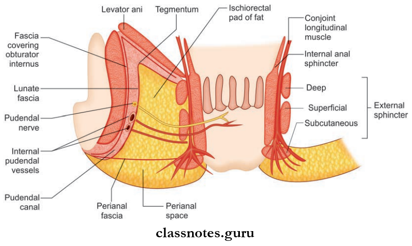
Question 8. What is the pudendal canal?
Answer:
Pudendal Canal
- Fascial canal within the obturator fascia lining the lateral wall of the ischiorectal fossa
- Also known as Alcock’s canal
- Situated about 1 inch above the ischial tuberosity
- Extent: From lesser sciatic notch to the posterior boundary of perineal pouches
- The canal is bounded by obturator fascia and lunate fascia
Pudendal Canal Contents:
- Pudendal nerve
- Internal pudendal vessels.
Question 9. Write a note on the pelvic diaphragm.
Answer:
Pelvic Diaphragm
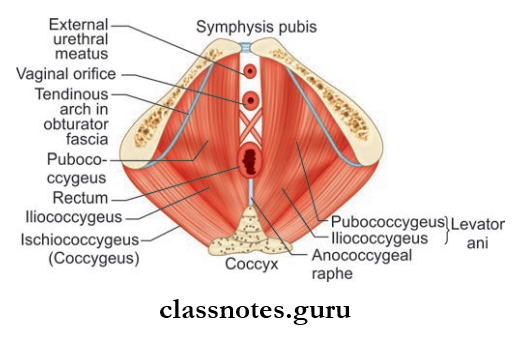
- V-shaped flor of true pelvis
- Also called pelvic floor
- It separates the pelvis from the perineum
- Formation: By the right and left levator ani and coccygeus muscles enclosed in the superior and inferior layers of the fascia of the pelvic diaphragm
- Structures Passing Through It:
- Males: Urethra and anorectal junction
- Females: Vagina, urethra, and anorectal junction
These Structures Pass Through Two Openings:
Hiatus Urogenital:
- Hiatus Urogenital is a triangle-shaped opening formed between the anterior fibers of levator ani muscles
- The urethra in males and females and the vagina in females pass through it
Hiatus Rentals:
- Hiatus Rentals is a circular opening formed between the anococcygeal raphe and perineal body
- Anorectal Junction Passes Through It
- The pelvic diaphragm is covered in its superior and inferior aspects by the superior and inferior fascia of the pelvic diaphragm.
1. The Superior Fascia Of The Pelvic Diaphragm
- Attached anteriorly to the superior ramus of the pubis and to the back of the body of pubis
- Superior Fascia Of The Pelvic Is continuous:
- Anteriorly: With sacrococcygeal ligament
- Posteriorly : With fascia on the piriformis
- Laterally: With obturator fascia
Pelvis And Perineum Short Questions And Answers
2. Inferior Fascia Of The Pelvic Diaphragm
- Also covers the medial wall of the ischiorectal fossa
- Laterally, it is continuous with the obturator fascia.
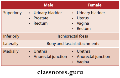
Relations Of Pelvic Diaphragm
Question 10. Describe in detail about the functions, boundaries, and contents of the ischiorectal fossa. Name the three recesses in relation to the ischiorectal fossa.
Answer:
The Ischiorectal Fossa
- Now called the ischioanal fossa, since it is located between ischial tuberosity and anal canal, not in the rectum
- It is a wedge-shaped fat-filed space on either side of the anal canal
Ischiorectal Fossa Functions:
- Allows distension of anal canal during defecation
- Allows dilatation of vagina during parturition.
Ischiorectal Fossa Boundaries
- Apex
- Formed by the junction of fascia covering obturator internus and inferior fascia of pelvic diaphragm
- Apex directed anteromedially towards the pubic symphysis
- Laterally: Fascia covering obturator internus muscle and ischial tuberosity
- Medially:
- On The Upper Part: Fascia covering external anal sphincter
- On The Lower Part: Fascia covering levator ani muscle
- Posteriorly:
- Lower margin of gluteus maximus muscle
- Sacrotuberous ligament
- Anteriorly: Posterior margin of perineal pouches
- Base/Floor: Deep transverse perineal fascia.
Ischiorectal Fossa Contents
- Ischiorectal pad of fat
- Pudendal canal and its contents (pudendal nerve, internal pudendal vessels)
- Posterior scrotal vessels and nerves
- Perineal branch of 4th scrotal nerve
- Inferior rectal branches of the pudendal nerve
- Perforating cutaneous nerve
- Lymphatic trunks.
TThere Are 3 Recesses Or Narrow Extensions Of The Ischiorectal Fossa, Namely:
- Anterior Recess: Extends from the urogenital diaphragm to body of the pubis
- Posterior Recess: Lies deep to the sacrotuberous ligament
- Horseshoe Recess:
- Lies posterior to the anal canal
- Connects the two ischioanal/rectal fossa.
Question 11. Write a note on celiac ganglion.
Answer:
Celiac Ganglion
- Largest ganglion in the body
- Two in number
- Location: On each side of the celiac trunk
- Shape: Irregular
- Division: Each ganglion is divided into:
- Large upper part—receives greater splanchnic nerve
- The smaller lower part or aorticorenal ganglion receives lesser splanchnic nerve.
Question 12. Write a note on the formation and branches of the celiac plexus.
Answer:
The Formation And Branches Of Celiac Plexus
- A dense network of nerve fibers connecting the two celiac ganglia
- It is the largest major autonomic plexus
- Location: In front of the abdominal aorta around the celiac trunk and around the root of the superior mesenteric artery
- Vertebral level: T12 – L1
- Celiac Plexus Is Formed By The Following Incoming Fibers:
- Preganglionic sympathetic fibers through greater and lesser splanchnic nerves
- Postganglionic sympathetic fibers from celiac ganglion
- Preganglionic vagal fibers from the posterior vagal trunk containing fiers from both right and left vagus, with predominant fiers from the right vagus
- Sensory fibers from the diaphragm reach the plexus along the inferior phrenic artery
Celiac Plexus Branches: The Celiac plexus gives rise to a number of secondary plexus, which surround the branches of the aorta. These are:
- Hepatic plexus
- Phrenic plexus
- Suprarenal plexus
- Left gastric plexus
- Splenic plexus
- Renal plexus
- Testicular plexus
- Ovarian plexus
- Superior mesenteric plexus
- Intermesenteric plexus
- Inferior mesenteric plexus
Pelvis And Perineum Clinical Anatomy Questions
Question 13. Write a brief on the sacral plexus.
Answer:
Sacral Plexus
- Sacral Plexus is a network of nerve fibers that supplies the skin and muscles of the pelvis and lower limb
- Location: On the surface of the posterior pelvic wall, anterior to the piriformis muscle
- Sacral Plexus is derived from the anterior rami of spinal nerves— L4, L5, S1, S2, S3, and S4
- Sacral Plexus also receives contributions from the lumbar spinal nerves L4 and L5 to form the lumbosacral trunk
- Each anterior rami divides into anterior and posterior branches.
Sacral Plexus Branches:
- Superior gluteal nerve
- Inferior gluteal nerve
- Sciatic nerve
- Posterior femoral cutaneous nerve
- Pudendal nerve
- Other branches: Nerve to piriformis, nerve to obturator internus, nerve to quadratus femoris.
- Anterior branches supply the flexor muscles of the lower limb
- Posterior branches supply the extensor and abductor muscles
Perineum And True Pelvis Multiple Choice Question And Answers
Question 1. The contents of the ischiorectal fossa are all except:
- Pudendal nerve
- Pudendal artery
- Perineal branch of the obturator nerve and nerve to obturator internus
- None
Answer: 3. Perineal branch of the obturator nerve and nerve to obturator internus
Question 2. The contents of the ischiorectal fossa lie in the:
- Medial wall
- Lateral wall
- The floor of the fossa
- Near the apex
Answer: 2. Lateral wall
Pelvis And Perineum MBBS Notes
Question 3. What structure represents the posterolateral boundary of the perineum?
- Ischial tuberosity
- Sacrospinous ligament
- Gluteus maximus
- Sacrotuberous ligament
Answer: 4. Sacrotuberous ligament
Question 4. Where is celiac plexus situated?
- Anteromedially to the sympathetic chain
- Posteromedially to left sympathetic chain
- Posteriorly to the abdominal aorta
- Anteriorly to abdominal aorta
- Posteriorly to arch of aorta
Answer: 4. Posteriorly to arch of aorta
Question 5. Th membranous layer of superfiial fascia of perineum is called:
- Fascia lunata
- Colles’ fascia
- Scarpa’s fascia
- Camper’s fascia
Answer: 2. Colles’ fascia
Question 6. The pelvic diaphragm is comprised of which of the following muscles?
- Piriformis, obturator internus, levator ani, and ischiococcygeus
- Obturator internus, levator ani, and ischiococcygeus
- Levator ani and ischiococcygeus
- Levator ani
Answer: 3. Levator ani and ischiococcygeus