Palatine Bones
Palatine Bones Terminology
‘Palatine’ bone is so named because of its contribution to the ‘hard palate’. The word ‘palate’ is derived from ‘plate’ because it forms a ‘plate-like’ partition between nasal and oral cavities.
Palatine Bones Location
Each palatine bone is located between the maxilla and pterygoid process of the sphenoid in the posterior part of the nasal cavity.
Palatine Bones Features And Attachments
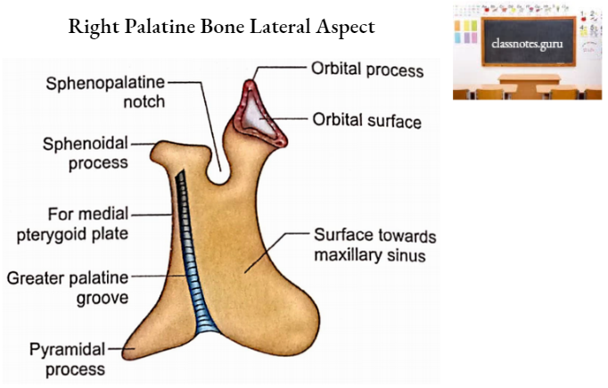
Each palatine bone is ‘L’ shaped in appearance and consists of two plates (horizontal and perpendicular) and three processes (pyramidal, orbital, and sphenoidal).
Palatine Bones Plates
1. Horizontal Plate
It projects medially from the lower end of the perpendicular plate. It has two surfaces (nasal and palatine) and four borders (anterior, posterior, lateral, and medial).
- Surfaces
- Nasal Surface
- It faces superiorly.
- It is concave from side to side.
- It forms the posterior part of the floor of the nasal cavity.
- Palatine Surface
- It faces inferiorly.
- With the corresponding surface of the opposite side, it forms posterior 1/4th of the hard palate.
- Near its posterior border, this surface presents a curved ridge called a palatine crest. This crest and the area behind it give attachment to palatine aponeurosis.
- Nasal Surface
- Borders
- Anterior Border
- It articulates with the posterior border of the palatine process of the maxilla to form a palatomaxillary suture.
- Posterior Border
- It is concave.
- It is free.
- This gives attachment to palatine aponeurosis (the aponeurosis is also attached to the palatine crest and the area behind it).
- Its medial end projects backward and with that of the opposite side forms the posterior nasal spine. To this spine is attached the musculus uvulae.
- Lateral Border
- It is attached to the lower border of the perpendicular plate.
- Its lower end is marked by a greater palatine groove.
- Medial Border
- It articulates with that of the opposite bone to form an interpalatine suture.
- Articulating medial borders of horizontal plates of two palatine bones project upwards to form a nasal crest.
- The nasal crest articulates with the posterior part of the lower border of the vomer and is continuous anteriorly with the nasal crests of maxillae.
- Anterior Border
2. Perpendicular Plate
It has two surfaces (maxillary and nasal) and four borders (anterior, posterior, superior, and inferior).
- Surfaces
- Maxillary Surface
- It faces laterally.
- Its major part is rough to articulate with the nasal surface of the maxilla.
- Its upper and posterior part is smooth and forms the medial wall of the pterygopalatine fossa.
- Its anterior part is also smooth and forms the posterior part of the medial wall of the maxillary sinus.
- Its posterior part shows a vertical groove (greater palatine groove) which is converted into a greater palatine canal by the maxilla in the articulated skull. Greater palatine vessels and nerves pass through the greater palatine canal.
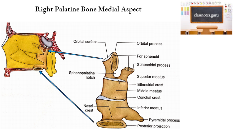
- Nasal Surface
- It faces medially.
- It has two horizontal crests. The lower crest is called the conchal crest because it articulates with the inferior concha. The upper one is named as the ethmoidal crest because of its articulation with the middle concha of the ethmoid.
- The area below the conchal crest forms the inferior meatus of the nose.
- The area between the two crests contributes to the middle meatus of the nose.
- The area above the ethmoidal crest takes part in the formation of the superior meatus of the nose.
- Maxillary Surface
- Borders
- Anterior Border
- Its lower part articulates with the maxillary process of the inferior concha and assists in the formation of the medial wall of the maxillary sinus.
- Its upper part forms the posterior boundary of the maxillary hiatus.
- Posterior Border
- It articulates with the anterior border of a medial pterygoid plate of the sphenoid.
- Superior Border
- It supports the orbital process in front and the sphenoidal process behind.
- Between the orbital and sphenoidal processes is the sphenopalatine notch which is converted into a sphenopalatine foramen by the inferior surface of the body of the sphenoid.
- Sphenopalatine foramen is the communication between the pterygopalatine fossa and the posterior part of the superior meatus of the nose.
- Sphenopalatine foramen transmits sphenopalatine vessels and posterior superior nasal nerves.
- Inferior Border
- It is continuous with the lateral border of the horizontal plate.
- In front of the pyramidal process, it is marked by the lower end of the greater palatine groove.
- Anterior Border
Palatine Bones Processes
1. Pyramidal Process
- It projects downwards, backward, and laterally from the junction of two plates of palatine bone.
- It fits into the pterygoid fissure of the pterygoid process of sphenoid.
- Its posterior surface completes the lower part of the pterygoid fossa.
- Its lateral surface is rough anteriorly and smooth posteriorly. The rough part articulates with maxillary tuberosity. The smooth part forms the lower part of the infratemporal fossa.
- Its inferior surface presents lesser palatine foramina for lesser palatine nerves and vessels.
2. Orbital Process
It projects upwards and laterally from the anterior part of the upper border of the perpendicular plate. A constricted neck connects it with the perpendicular plate.
It has three articular surfaces (anterior, posterior, and medial) and two non-articular surfaces (superior and lateral).
- Articular Surfaces
- Anterior Surface
- It articulates with maxilla.
- Posterior Surface
- It articulates with a sphenoidal body.
- Medial Surface
- It articulates with ethmoidal bulla.
- Anterior Surface
- Non-Articular Surfaces
- Superior Surface
- It forms the posterior part of the floor of the orbit.
- Lateral Surface
- It forms part of the medial wall of the pterygopalatine fossa. The border between lateral and posterior surfaces is prolonged downwards as the anterior boundary of the sphenopalatine notch.
- Superior Surface
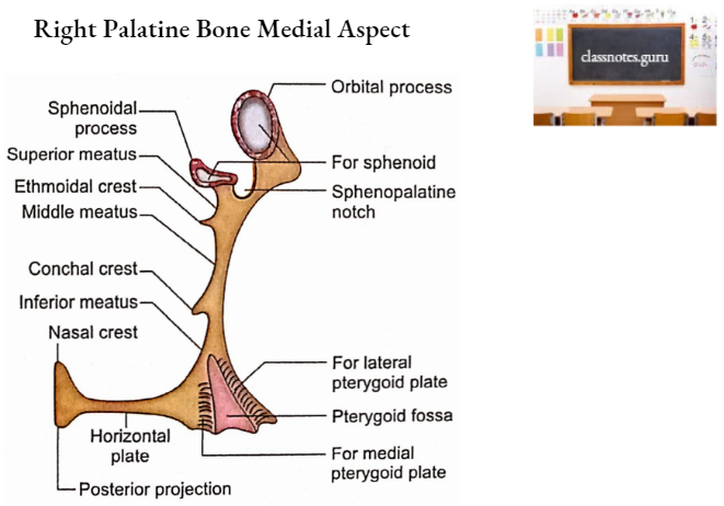
3. Sphenoidal Process
It is directed upwards and medially from the posterior part of the upper border of the perpendicular plate. It has three surfaces (superior, inferomedial, and lateral) and three borders (posterior, anterior, and medial).
- Surfaces
- Superior Surface
- It articulates with the undersurface of the sphenoidal concha and root of the medial pterygoid plate.
- It is grooved to complete the palatovaginal canal.
- Inferomedial Surface
- It contributes to the roof and lateral wall of the nasal cavity.
- Lateral Surface
- Its posterior part articulates with the medial pterygoid plate.
- Its anterior part contributes to the medial wall of the pterygopalatine fossa.
- Superior Surface
- Borders
- Posterior Border
- It articulates with the vaginal process of the medial pterygoid plate.
- Anterior Border
- It forms the posterior boundary of the sphenopalatine notch.
- Medial Border
- It articulates with a lot of vomer.
- Posterior Border
Palatine Bones Ossification
- Palatine bone ossifies in the membrane.
- A single center appears in the perpendicular plate during the 8th week of intrauterine life.
- The ossification spreads into the processes and horizontal plate.
- At birth, the height of the perpendicular plate is equal to the transverse width of the horizontal plate.
- The length of the perpendicular plate becomes double the transverse width of the horizontal plate at puberty.
Palatine Bones Applied Anatomy
- The palatine bone may be involved in the fracture of the mid-facial skeleton.
- Palatine bone receives adequate blood supply from periosteal arteries and, therefore, all the fragments of fractured bone retain a periosteal blood supply.
- Palatine bone is clothed in mucosa over large areas of its surfaces. Its fracture may open into the nasal or oral cavities or maxillary sinus with the potential risk of infection.
- Le Fort fractures of the midfacial skeleton always involve the perpendicular plates of palatine bones.
- Guerin’s fracture (Le Fort 1 fracture) involves the lower 1/3rd of the perpendicular plates of palatine bones. In cases of Le Fort 2 and 3 fractures, the upper parts of the perpendicular plates are affected.
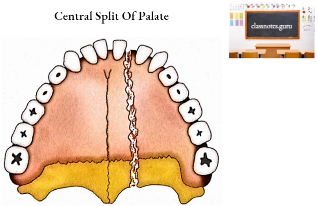
- A horizontal plate of palatine bone may be fractured in an uncommon central split of the palate. It is actually paramedian in nature because median sutures (intermaxillary and interpalatine) are relatively strong.