
Object Localisation Techniques Long Essays
Question 1. Enumerate intra-oral radiographic techniques. Describe the procedure of localizing impacted left maxillary canine.
Answer:
Intra-Oral Radiographic Techniques:
- Intra-oral radiographic Techniques are:
- Paralleling technique
- Bisecting angle technique
Right Angle Technique:
- This is used to localize the impacted left maxillary canine
- It is also called Miller’s right-angle technique
- It uses two radiographic projections taken at right angles to each other
Intra-Oral Radiographic Technique:
- The periapical film is exposed using a proper technique to show the position of the object in super inferior and anteroposterior relationship
- Next, occlusal film is exposed, directing the central X-ray beam perpendicular to the film
- These two radiographs are compared
Read And Learn More: Oral Radiology Question and Answers
Intra-Oral Radiographic Uses
- Locates the maxillary impacted canine
- Diagnosed fracture of the mandible
- Locates any displacement
Object Localisation Techniques Short Essays
Question 1. Object localization techniques.
Answer.
Object localization techniques
- Intraoral localization techniques are used to locate the position of a tooth object in the jaws
- Indications:
- Foreign bodies
- Impacted teeth
- Unerupted teeth
- Retained roots
- Salivary stones
- Jaw fractures
- Broken needles
- Root position
- Filling materials
Object localization techniques in dental radiology
Radiographic Techniques:
- Maxillary area
- Incisor zone
- Stereoscopic
- Lateral profile
- Occlusal
- Cuspid zone
- Stereoscopic
- Lateral profile
- Occlusal
- Bicuspid and molar zone
- Periapical
- Occlusal
- Incisor zone
- Mandibular area
- Incisor zone
- Periapical
- Lateral profile
- Occlusal
- Posterior zone
- periapical
- Occlusal
- Third molar zone
- Periapical
- Lateral oblique
- Oblique occlusal
- Incisor zone
- Methods
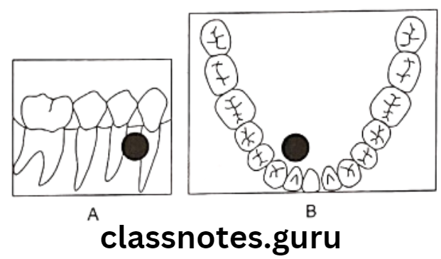
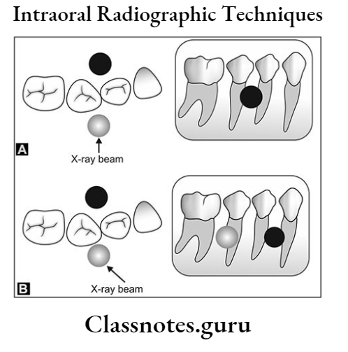
Buccal and lingual objects shift positions when the direction of the X-ray beam is changed. A Buccal and lingual are superimposed in the original radiograph B If the tube head is shifted in the distal direction, the buccal object moves mesially and the lingual object moves distally (same direction = Lingual; Opposite Direction = Buccal)
Buccal object rule in radiography
The right angle technique A – The object appears to be located in the bone on the periapical radiograph. B – The occlusal radiograph reveals that the object is located in the soft tissue lingual to the mandible
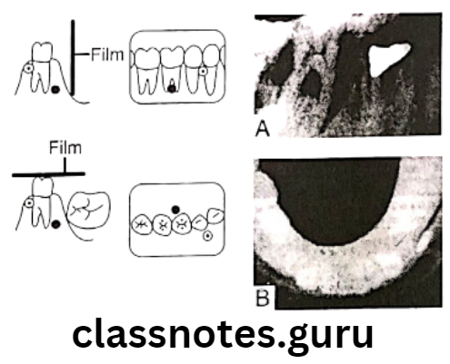
Right-angled localization technique. Two films are exposed at right angles to each other to identify the location of an object. The periapical radiograph A – will demonstrate the superior-inferior and anterior-posterior position of the objects. A cross-sectional occlusal radiograph B – will demonstrate the anteroposterior and buccal lingual positions. Thus these two radiographs will demonstrate all three dimensions of an area, and the location of objects can be identified
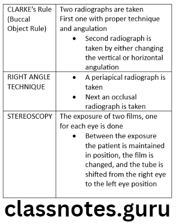
Question 2. Indications and interpretation of Clarke’s technique.
Answer.
Clarke’s Technique Indications:
- Foreign bodies
- Impacted teeth
- Unerupted teeth
- Retained roots
- Salivary stones
- Broken needles
- Jaw fractures
- Filling materials
- Root position
Clarke’s Technique Method:
- Two radiographs are taken
- The first one with proper technique and angulation
- The second radiograph is taken by either changing the vertical or horizontal angulation
Clarke’s Technique Interpretation:
- When the dental structure or object seen in the second radiograph appears to have moved in the same direction as the shift of the position-indicating device, the structure is said to be positioned lingually
- If the object appears to have moved in the opposite direction, then the object is said to be positioned buccally
Clarke’s Technique Slob Rule:
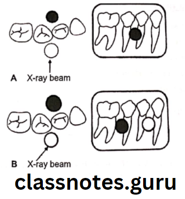
Buccal and lingual objects shift positions when the direction of the X-ray beam is changed. A – Buccal (cross-hatched circle and lingual (black circle) are superimposed in the original radiograph B – if the tube head is shifted in the distal direction, the buccal object moves mesially and the lingual object moves distally (same direction = Lingual; Opposite Direction = Buccal)
Radiographic localization techniques in dentistry
Object Localisation Techniques Short Answers
Question 1. SLOB technique.
Answer.
SLOB Technique Method:
- Two radiographs are taken
- The first one with proper technique and angulation
- The second radiograph is taken by either changing the vertical or horizontal angulation
SLOB Technique Interpretation:
- When the dental structure or object seen in the second radiograph appears to have moved in the same direction as the shift of the position-indicating device, the structure is said to be positioned lingually
- If the object appears to have moved in the opposite direction, then the object is said to be positioned buccally
Methods of locating impacted teeth in radiographs
SLOB Technique Slob Rule:
- Same side Lingual Opposite side Buccal:
Buccal and lingual objects shift positions when the direction of the X-ray beam is changed. A – Buccal (cross-hatched circle and lingual (black circle) are superimposed in the original radiograph B – If the tube head is shifted in the distal direction, the buccal object moves mesially and the lingual object moves distally (same direction = Lingual; Opposite Direction = Buccal)