Normal Anatomic Structures Short Essays
Question 1. Anatomical landmarks in the upper posterior periapical radiograph
Answer.
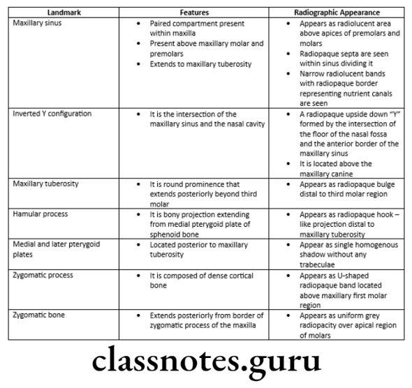
Anatomic Landmarks In the Maxillary Posterior Region Are:
- Maxillary sinus
- Inverted Y – configuration
- Maxillary tuberosity
- Hamular notch
- Medial and lateral pterygoid plates
- Zygomatic process
- Zygomatic bone
Normal anatomical landmarks in radiographs
Question 2. Normal radiographic anatomy of teeth and supporting structures.
Answer:
Anatomic Landmarks In Maxillary Anterior Region Are:
- Incisive foramen
- Superior foramina of incisive canal
- Median palatine suture
- Lateral fossa
- Nasal fossa
- Nasal septum
- Floor of nasal cavity
- Anterior nasal spine
- Inferior nasal concha
- Nasolacrimal canal
- Nose
- Inverted Y
Read And Learn More: Oral Radiology Question and Answers
Anatomic Landmarks In the Maxillary Posterior Region Are:
- Maxillary sinus
- Inverted Y – configuration
- Maxillary tuberosity
- Hamular notch
- Medial and lateral pterygoid plates
- Zygomatic process
- Zygomatic bone
Anatomic Landmarks In the Mandibular Region
- Genial tubercle
- Nutrient canal
- Submandibular gland fossa
- Sublingual gland fossa
- Retromolar triangle.

Anatomical structures in dental radiology
Normal Anatomic Structures Short Answers
Question 1. Normal anatomical landmarks in the maxillary anterior region.
Answer.
Normal anatomical landmarks in the maxillary anterior region

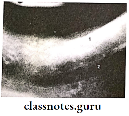
Question 2. Radiographic appearance of the mandibular canal.
Answer.
The radiographic appearance of the mandibular canal
- Radiographically, it appears as a radiolucent band
- It is outlined by two thin radiopaque lines that represent the cortical walls of the canal
- It may appear below or superimposed on the Mandibular molar teeth
- It extends from the Mandibular foramen to the mental foramen
- It houses the inferior alveolar nerve and the blood vessels
- It is a tube-like passage through the bone that travels the length of the mandible
Normal anatomy of oral cavity
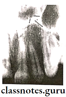
Question 3. Line of tennis.
Answer.
Line of tennis
- It is the intersection of the maxillary sinus and the nasal cavity
- On radiograph, it appears as
- A radiopaque upside-down “Y” formed by the intersection of the floor of the nasal fossa and the anterior border of the maxillary sinus
- It is located above the maxillary canine
Question 4. Lamina dura.
Answer.
Lamina dura
- This is the wall of the tooth socket that surrounds the tooth
- It is made up of dense cortical bone
- On radiograph, it appears as
- A thin radiopaque line that surrounds the root of the tooth
- It is continuous with the shadow of the cortical bone at the alveolar crest
- It is slightly thicker than the trabeculae of the cancellous bone in the area
- When the X-ray beam is directed through the relatively long expanse of the structure, the lamina dura appears radiopaque and well-defined
- When the beam is directed more obliquely, the lamina dura appears more diffuse
- The thickness and density of the lamina dura will vary with the amount of occlusal stresses
- It is wider and denser around the roots of teeth in heavy occlusion
- It is thinner and less dense around teeth not subjected to the occlusion function
- A double lamina dura image appears if the mesial or distal surfaces of the root present two elevations in the path of the X-ray beam
- The presence of an intact lamina dura around the tooth indicates a vital pulp.
- However, in some cases, its absence may be normal
Anatomic landmarks in intraoral radiographs
Question 5. Anatomical landmarks of the maxillary
Answer:
Anatomic Landmarks In the Maxillary Anterior Region Are:
- Incisive foramen
- Superior foramina of the incisive canal
- Median palatine suture
- Lateral fossa
- Nasal fossa
- Nasal septum
- The floor of the nasal cavity
- Anterior nasal spine
- Inferior nasal concha
- Nasolacrimal canal
- Nose
- Inverted Y
Anatomic Landmarks In the Maxillary Posterior Region Are:
- Maxillary sinus
- Inverted Y-configuration
- Maxillary tuberosity
- Hamular notch
- Medial and lateral pterygoid plates
- Zygomatic process