
Morphology And Physiology Of Bacteria Important Notes
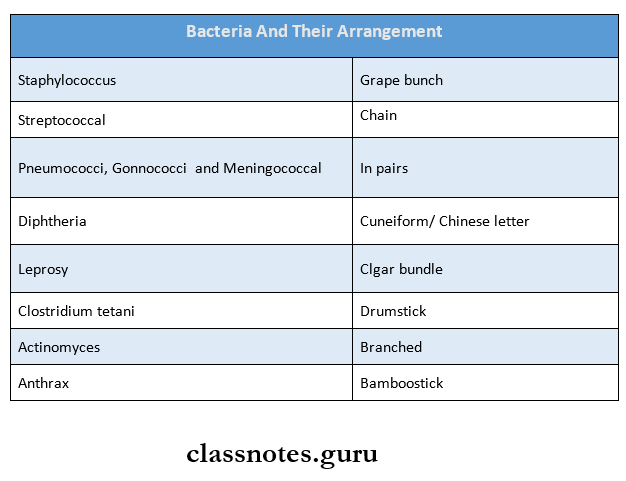
1. Bacteria And Their Arrangement
Read And Learn More: Microbiology Question and Answers
2. Contents Of Bacterial Cell
- Cell
- Cytoplasmic membrane
- Cytoplasm
- Nucleus
- Capsule
- Flagella
- Fimbriae
- Spore
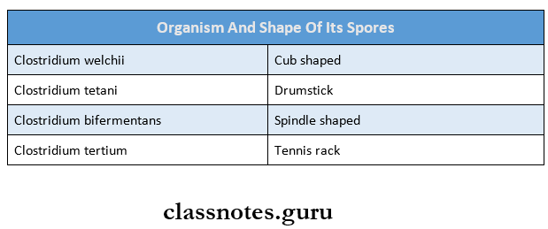
3. Organism And Shape Of Its Spores
Morphology And Physiology Of Bacteria Long Essays
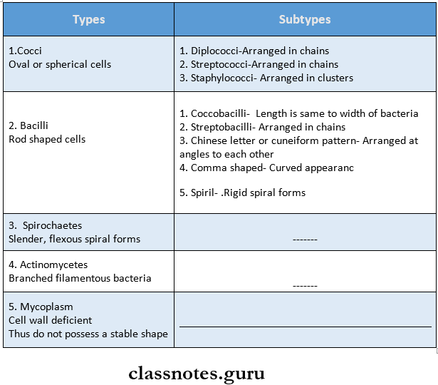
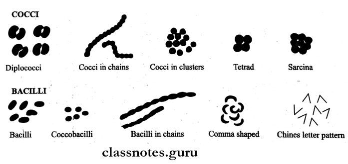
Question 1. Classify bacteria depending on their shape.
Answer:
Morphology And Physiology Of Bacteria
Depending Upon The Shape, Bacteria Are Classified Into:
Morphology And Physiology Of Bacteria Short Essays
Question 1. Bacterial Spore
Answer:
Bacterial Spore
- Spores are highly resistant resting stages of the bacteria formed in unfavorable environmental conditions.
- Sporulation is not a method of reproduction.
Morphology Of Spore
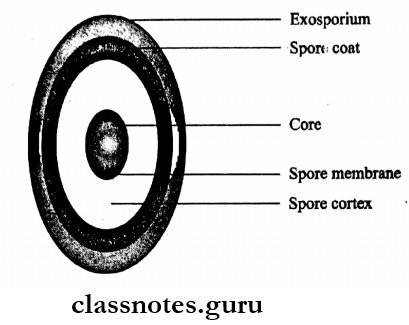
Spores consist of the following structures Cell
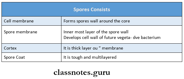
- Some of the spores contain exosporium
- It is the loose outer covering
Morphology And Physiology Of Bacteria
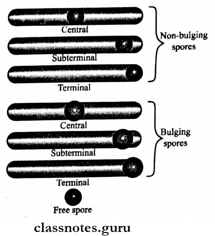
Bacterial Spore Shape And Position:
- Bacterial Spore Position
- Spores may be
- Central
- Subterminal (or)
- Terminal
- Spores may be
- Bacterial Spore Shape:
- They may be
- Spherical
- Oval
- Depending on the diameter, they may be
- Bulging
- Non-bulging.
- They may be
Bacterial Spore Properties:
- Resistance
- Spores are extremely resistant to ordinary boiling, disinfectants, and heating.
- Used for Sterilization
- Spores of some species of bacteria act as indicators for proper sterilization
- Gemination
- Gemination is the process of spore conversion into vegetative cells under favorable conditions.
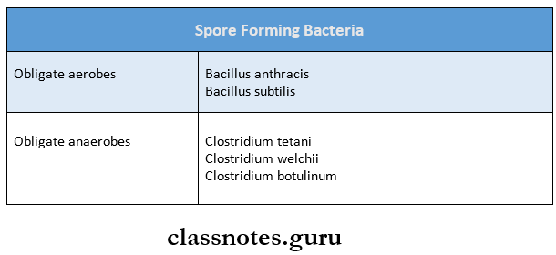
Bacterial Spore Demonstration:
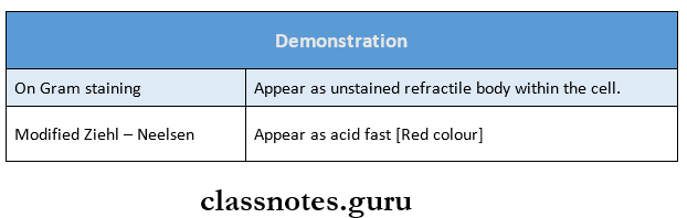
Spore Forming Bacteria are:
Question 2. Gram staining/Gram stain?
Answer:
Gram Staining/Gram Stain
- Gram stain is the most widely used stain in bacteriology.
- Gram staining is the essential procedure used in the identification of bacteria and is frequently the only method required to study their morphology.
Gram Staining Method:
- Gram staining involves four basic steps.
- The primary staining with a pararosaniline dye such as crystal violet, methyl violet (or) gentian violet for one minute
- Application of gram’s iodine [dilute solution of io¬dine) over the slide for one minute.
- Decolorization with an organic solvent such as ethanol, acetone (or) aniline for 10-30 seconds
- Counterstaining with a dye of contrasting color such as carbon fuchsin, safranin, or neutral red for 30 seconds
Differentiation on gram staining: It is called a differential stain because it differentiates between gram-positive and gram-negative bacteria
Gram Staining Mechanism:
Morphology And Physiology Of Bacteria
1. The exact mechanism is not understood.
2. It may be attributed to the following:
- Permeability of bacterial cell wall and cytoplasmic membrane:
- The protoplasm of the Gram-positive cells is more acidic than that of Gram-negative cell
- Due to this, it retains the basic dyes strongly
- Now when iodine is added to it, the acidic nature of the protoplasm increases
- As a result, iodine combines with the dye and forms a dye-iodine complex and fixes the dye in the bacterial cell
- The Cytoplasmic membrane of Gram-positive cells is less permeable to this complex
- Thus the dye-iodine complex gets trapped within the cell
- In contrast, the Gram-negative cell wall has increased permeability
- This leads to the outflow of the complex during decolorization
- The integrity of the cell wall
- If the cell wall is damaged, the Gram-positive bacteria becomes Gram-negative
Question 3. Acid Fast stain/Ziehl Neelsan Stain?
Answer:
Acid Fast stain/Ziehl Neelsan Stain
- The acid-fast stain was discovered by Ehrlich and subsequently modified by Ziehl and Neelsen.
- Some organism retains carbol fuchsin even when colorized with acid. Such organisms are called acid-fast organisms.
- Example: Mycobacteria
Acid-fast Stain Method:
- Pour carbol fuchsin satin on a slide containing a fixed smear
- Gently heat the underside of the slide till it produces steam
- Leave carbol fuchsin over the slide for 5-10 minutes along with intermittent heating
- Don’t allow the slide to dry out for it adds stain to it and reheat
- Wash the slide in tap water
- Decolorize the smear with 20% sulphuric acid and wash it With water
- Repeat the procedure till the pink/ red color stops coming out
- Counter-stain the smear with 2% methylene blue for 1¬2 minute
- Wash it with water and air dry it
- Observe under microscope
Acid-fast Stain Microscopic Examination: Acid-fast bacilli appear red in the blue background of pushing cells and epithelial cells
Acid-fast Stain Principle:
- Acid fastness depends on
- The high content of lipids, fatty acids, and higher alcohols found in the cell wall of Mycobacterium
- The integrity of the cell wall
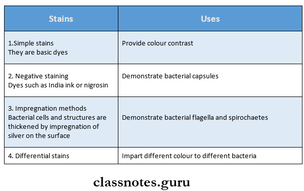
Question 4. Name four staining techniques in microbiology.
Answer:
Commonly Used Staining Techniques In Microbiology Are As Follows.
Question 5. Bacterial cell wall
Answer:
Bacterial Cell Wall
- The cell wall is a tough and rigid structure
- It surrounds the bacteria like a shell
Bacterial Cell Wall Functions:
- Accounts for the shape of the cell
- It takes part in cell division
- Protects the cell against osmotic damage
- Provide rigidity
- It possesses a target site for antibiotics, lysosomes, and bacteriophages
Morphology And Physiology Of Bacteria
Bacterial Cell Wall Structure:
- The rigid part of the cell wall is peptidoglycan
- It has the following components
1. Lipoprotein Layer It connects the peptidoglycan to the outer membrane
2. Outer Membrane
- It contains certain proteins named outer merm brane proteins
- These are target sites for bacteriocins
3. Lipo-polysacharride .
- This layer consists of lipid A to which polysaccha¬ride is attached
- The toxicity is associated with lipid A
- The polysaccharide determines a major surface an¬tigen to 0 antigen
- It contains endotoxin in gram-negative bacteria
- It is composed of 3 regions as follows
- Region 1 – polysaccharide portion (0 antigen specificity)
- Region 2 – core polysaccharide
- Region 3 – Lipid A portion (responsible for tox¬icity)
4. Periplasmic Space
- It is the space between the inner and outer membrane
- It contains various binding proteins
5. Peptidoglycan

Morphology And Physiology Of Bacteria Short Question And Answers
Question 1. Name capsulated bacteria
Answer:
Name Capsulated Bacteria
- Streptococcus pneumonia
- Klebsiella sp.
- Bacillus antacids
- Cryptococcus neoformans. (a fungus)
Question 2. Fimbriae
Answer:
Fimbriae
- Fimbriae is also called pili
- These are hair-like appendages projecting from the cell surface as straight filaments.
Fimbriae Types:
- Common pili
- Sex of F (fertility) pili
- Col 1 (colicin) pili
Fimbriae Functions:
- Adhesion
- Transfer of genetic material
Morphology And Physiology Of Bacteria
Fimbriae Demonstration:
- Fimbriae are demonstrated by
- Electron microscopy
- Haemagglutination test
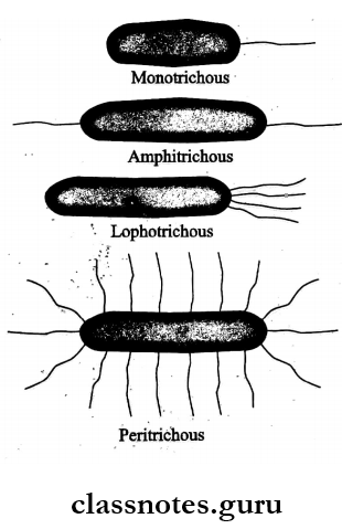
Question 3. Flagella
Answer:
Flagella: Flagella are cytoplasmic appendages protruding through a cell wall.
Flagella Structure:
- They are thread-like structures
- Size:
- Length – 5-20 micrometers
- Diameter- 0.01-0.02 micrometer
Flagella Parts:
- It is composed of three parts
- Filament
- It lies external to the cell
- It is connected to the hook at the cell surface ‘250
- Hook
- The hook-basal body is embedded in the cell envelope
- Basal body.
- It contains outer and inner rings
- Filament
Flagella Composition:
- The flagella is made up of flagellin, a protein
- Specific flagellar antibodies are produced in high titers
Flagella Functions:
- These are organs of locomotion.
- Flagellar antibodies are used for serodiagnosis
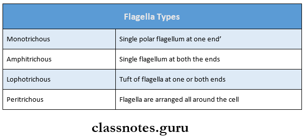
Flagella Types:
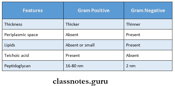
Question 4. Differences between Gram-positive and Gram-negative cell walls:
Answer:
Differences Between Gram-Positive And Gram-Negative Cell Walls
Morphology And Physiology Of Bacteria Viva Voce
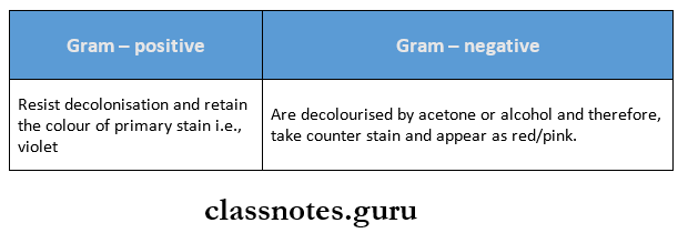
- Gram-positive bacteria appear violet and Gram-negative bacteria appear red on staining
- In acid-fast staining, a positive reaction gives a red color while a negative reaction gives a blue color
- All cocci except Neisseria are Gram-positive
- Flagella is a locomotory organ
- The nucleus of bacteria consists of plasmids or episomes
- Bacteria without cell walls are called Mycoplasma