Maxillae Terminology
Maxilla is a Latin word meaning ‘cheek’ or ‘jaw’. The word is commonly used in reference to the upper jaw.
Maxillae Location
- There are two maxillae which form a major part of the upper facial skeleton.
- The whole of the upper jaw is formed by two maxillae.
- The junction of two maxillae is marked by intermaxillary suture visible in the hard
palate and face in the midline.
Maxillae Features And Attachments
Each maxilla consists of a body and four processes (zygomatic, frontal, alveolar and palatine).
Maxillae Body
It has four surfaces (anterior, infratemporal, orbital and nasal).
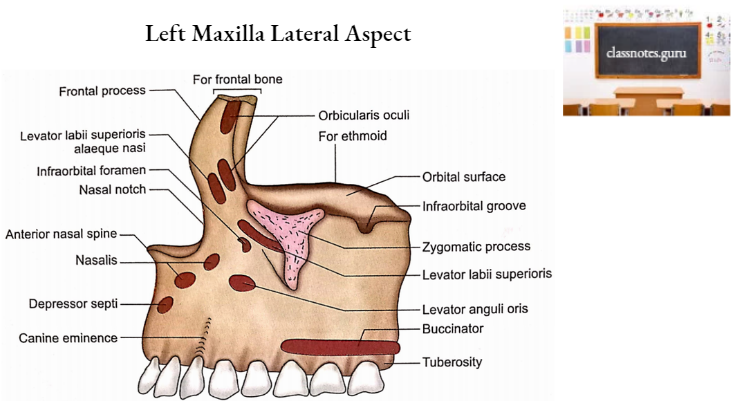
1. Anterior Surface
- It is directed forward and laterally.
- There is a vertical elevation at the site of the socket for the canine root. This is called canine eminence.
- Medial to canine eminence is a depression called incisive fossa which gives origin to depressor septi.
- The anterior surface below the incisive fossa gives attachments to the incisivus superior and orbicularis oris.
- Just above the incisive fossa, there is the attachment of the nasalis muscle.
- Lateral to canine eminence is another fossa called canine fossa. Levator anguli oris originates from the canine fossa.
- Above the canine fossa is a foramen called infraorbital foramen. It transmits the infraorbital nerve and vessels.
- Above the infra-orbital foramen is a sharp infra-orbital margin which gives origin to levator labii superioris.
- Its upper part is limited medially by a deep notch called a nasal notch.
2. Infratemporal Surface
- It faces backwards and laterally.
- It forms anterior wall of infratemporal fossa.
- It shows 2-3 openings of alveolar canals which transmit posterior superior alveolar nerves and vessels.
- Its anteroposterior part is marked by maxillary tuberosity which articulates with the pyramidal process of palatine bone.
3. Orbital Surface
- It forms the floor of the orbit.
- Running forward is an infraorbital groove in the middle of its posterior part. The groove continues with the infraorbital canal which opens on the anterior surface as the infraorbital foramen. It is meant for infraorbital nerves and vessels.
- Anteromedially it gives origin to the inferior oblique muscle.
- It has three borders (medial, posterior and anterior).
Medial Border
It is marked anteriorly by a lacrimal notch. Behind this notch, this border provides attachments to lacrimal bone, orbital plate of ethmoid and orbital process of palatine bone from before backwards.
Posterior Border
It forms the anterior border of the inferior orbital fissure.
Anterior Border
It contributes to the medial part of the infra-orbital margin.
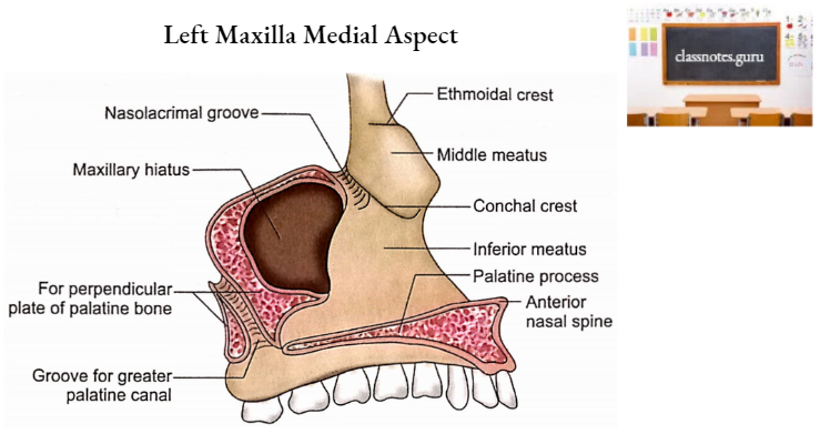
4. Nasal Surface
- It forms the lateral wall of the nasal cavity.
- A large opening (maxillary hiatus) is the most prominent feature of this surface.
- Maxillary hiatus leads into the maxillary sinus, a large air space within the body of the maxilla.
- Maxillary hiatus is greatly reduced in size in the articulated skull by ethmoid (uncinate process) and lacrimal bone above, inferior concha below and perpendicular plate of palatine bone behind.
- Below the hiatus, this surface forms the inferior meatus of the nasal cavity.
- The posterior part of the nasal surface has an oblique groove which is converted into a greater palatine canal by a perpendicular plate of palatine bone. Greater palatine nerves and vessels pass through this canal.
- In front of the hiatus is the nasolacrimal groove. This is converted into the nasolacrimal canal by the lacrimal bone and inferior concha. This canal is meant for the nasolacrimal duct.
- An oblique ridge called the conchal crest is present in front of the nasolacrimal groove. It articulates with inferior concha.
Maxillae Processes
1. Zygomatic Process
It has three surfaces, anterior, posterior and superior. The latter is rough for articulation with the zygomatic bone.
2. Frontal Process
It possesses an upper end, two surfaces (lateral and medial) and two borders (anterior and posterior).
- Upper End
- It articulates with the nasal notch of the frontal bone.
- Surfaces
- Lateral Surface
- It has a vertical ridge (anterior lacrimal crest) in the middle for the attachment of the medial palpebral ligament.
- The area in front of the crest gives attachments to orbicularis oculi and levator labii superioris alaeque nasi.
- The area behind the lacrimal crest contributes to the anterior half of the lacrimal groove.
- Medial Surface
- It has a horizontal ridge (ethmoidal crest) in its middle. It articulates with the middle nasal concha.
- A roughened area above the crest articulates with ethmoid to complete anterior ethmoidal air cells.
- The area below the ethmoidal crest forms the atrium of the middle meatus.
- Borders
- Anterior border
- It articulates with the nasal bone.
- Posterior border
- It articulates with lacrimal bone.
- Anterior border
- Lateral Surface
3. Alveolar Process
- It is an arched lower border of the body.
- It has sockets for the upper teeth.
- Buccinator originates from the posterior part of the outer surface over the sockets for permanent molar roots.
4. Palatine Process
It is a horizontal bracket-like projection from the lower part of the medial surface of the body. It forms the anterior 3/4th of the hard palate. It has two surfaces (superior and inferior) and three borders (medial, posterior and lateral).
- Surfaces
- Superior Surface
- It is concave and smooth.
- It forms the floor of the nasal cavity.
- Inferior Surface
- It has depressions for palatine glands.
- It has several nutrient foramina for nutrient vessels.
- A greater palatine groove for greater palatine nerve and vessels is present in its posterolateral part.
- When two maxillae meet, the incisive fossa is noticed behind the incisor teeth.
- The incisive canal is the communication between the incisive fossa and the nasal cavity. It transmits the greater palatine artery and nasopalatine nerve.
- Superior Surface
- Borders
- Medial Border
- It meets with a similar border of the opposite maxilla to form an intermaxillary suture.
- This border is raised into a ridge called the nasal crest. Nasal crests of two sides enclose a groove to receive the vomer.
- Its anterior end is prolonged and meets with a similar prolongation of the opposite side to form the anterior nasal spine.
- Posterior Border
- It articulates with the anterior border of the horizontal plate of palatine bone to form a palatomaxillary suture.
- Lateral border
- It fuses with the body.
- Medial Border
Maxillae Ossification
- The maxilla is intramembranous in origin.
- It develops in the mesenchyme just superficial to the nasal capsule.
- Three centres of ossification appear:
- One centre appears for the main mass just above the canine fossa at about 6th week of intrauterine life.
- Two centres appear for os incisivum (premaxillary part).
- Note: Remember that the premaxilla is that part of the maxilla which holds incisor teeth and is a separate bone in most mammalian upper jaws.
- The maxillary sinus appears on the nasal aspect as a groove at about 4th month of intrauterine life
Age Changes In Maxilla
1. At birth
- Vertical diameter is lesser than both the transverse and anteroposterior diameters.
- The body is mainly occupied by sockets for the teeth.
- The maxillary sinus is seen as a shallow groove on the nasal aspect.
2. Adult
- Vertical diameter is greater than the transverse and anteroposterior diameters.
- The maxillary sinus has greatly developed within the body.
3. Old Age
- Due to the falling of teeth and resorption of the alveolar margin, the vertical diameter is again greatly reduced.
- The alveolar margin is reduced in thickness at the expense of the labial wall.
Maxillae Applied Anatomy
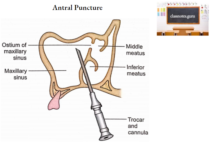
1. Maxillary sinus (antrum of Highmore)
- It is the air space in the body of the maxilla. It is pyramidal in shape with a base towards the nasal cavity and an apex towards the zygomatic process.
- Its height and anteroposterior measurements are 1.5 inches each while width is 1 inch only. It is very important clinically due to the following facts:
- It is the largest paranasal sinus and is commonly involved during the inflammation process (maxillary sinusitis).
- It drains into the middle meatus which is higher than its floor. The latter is about 1.25 cm below the floor of the nasal cavity.
- To facilitate the drainage of pus in the maxillary sinus an opening is made in the inferior meatus by operative procedures like antral puncture or antrostomy.
- Maxillary tumours can produce a bulging in adjacent related surroundings, i.e. superiorly in the floor of the orbit, inferiorly in the roof of the oral cavity.
- Anteriorly in the face, posteriorly in the infratemporal fossa and medially in the lateral wall of the nasal cavity.
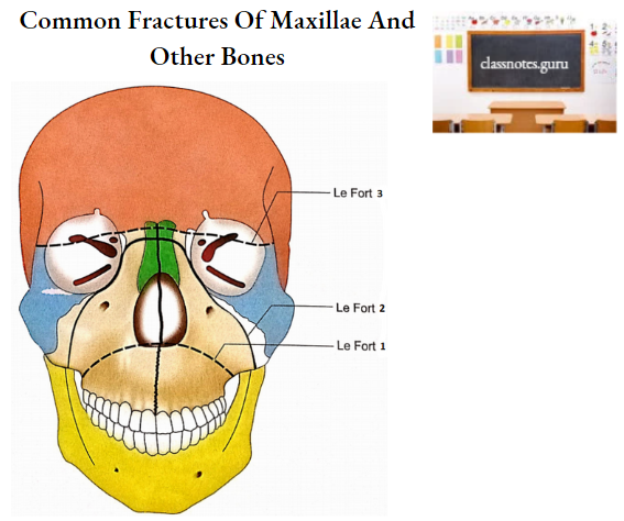
2. Maxillary fractures
- Unilateral fracture of the maxilla usually involves its alveolar process.
- Bilateral maxillary fractures are classified into the following three types:
- Le Fort 1 (Guerin’s fracture)
- It is a horizontal fracture along the floor of the nose and below the zygomatic bone.
- Le Fort 2
- In this fracture line passes through orbits and then runs medial to and below the zygomatic bones towards the alveolar margins.
- Le Fort 3
- In this, the fracture line runs through nasal bones and orbits above the zygomatic bone. This fracture is also called craniofacial disjunction as the face separates from the cranium.
- Le Fort 1 (Guerin’s fracture)