
Kidney Ureter And Suprarenal Gland Question And Answers
Question 1. Describe the anatomical features of kidney.
Answer:
The Anatomical Features Of Kidney
- Bean-shaped excretory organ
- Situated on either side of the vertebral column in the posterior abdominal wall
- Right kidney is slightly lower than the left
- Vertebral Level: T12–L3
- Length: 11 cm
- Breadth: 6 cm
- Thickness: 3 cm
- Weight
- Male: 150 g
- Female: 130 g
- Color: Reddish brown
- Orientation: Directed downwards and laterally
Read And Learn More: Abdomen And Pelvis
Kidney Ureter And Suprarenal Gland Questions And Answers
External Features Of Kidney: It has
- Two Poles:
- Broad upper pole
- Pointed lower pole, which lies 2.5 cm above the iliac crest.
- Two Surfaces:
- Anterior Surface: Irregular, faces anterolaterally
- PosteriorSurface: Flat, faces posteromedially.
- Two Borders:
- Medial Border
- Concave
- The middle part of concavity shows a depression, called hilum
- Lateral Border
- Convex.
- Medial Border
Hilum of Kidney:
- Hilum is present on the central part of the medical border
- Hilum Transmits The Following Structures (From Anterior To Posterior)
- Renal vein
- Renal artery
- Renal pelvis
- Branch of renal artery
- Lymphatics and sympathetic nerves.
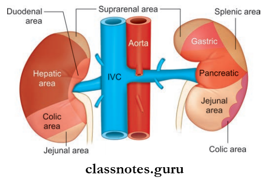
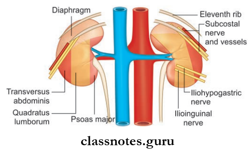
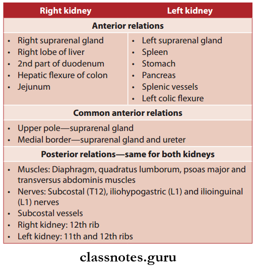
Kidney Relations
Question 2. What are the relations of kidney?
Answer:
The Relations Of Kidney
Kidney And Adrenal Gland Anatomy MCQs
Question 3. Write a note on coverings of kidney.
Answer:
Coverings Of Kidney
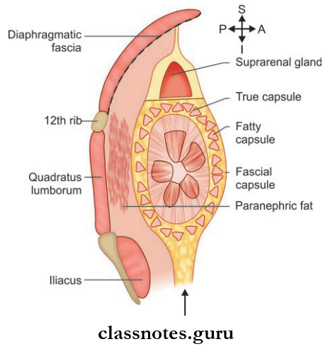
- The kidney has 4 coverings
- They Are (From Inside To Outside):
- Fibrous capsule or true capsule
- Perinephric fat or fatty capsule
- Fascial capsule/false capsule/renal fascia/fascia of Gerota
- Perinephric fat.
- True Capsule Of Kidney
- True Capsule Is Made Up Of:
- Collagen fibers
- Elastic fibers
- Smooth muscle fibers
- True capsule gives a shining appearance to the kidney
- Normally true capsule can be easily stripped of
- But The Capsule Becomes Adherent In Renal Diseases.
- Fatty Capsule Or Perinephric Fat Of Kidney
- It is an adipose tissue layer
- Numerous fibrous strands connecting the true and false capsules pass through the fatty capsule.
- True Capsule Is Made Up Of:
- False Capsule Of Kidney
- False Capsule provides common covering to kidney and suprarenal glands
- False Capsule Consists Of Two Layers:
- Anterior layer: Fascia of Toldt
- Posterior layer: Fascia of Zuckerkandl
- On Horizontal Tracing Of False Capsule:
- Two layers fuse along the lateral border of the kidney and become continuous with transversal fascia
- Medially
- The anterior layer passes anterior to renal vessels and joins with the anterior layer on the opposite side, in front of the abdominal aorta and inferior vena cava
- Posterior layer blends with psoas fascia and gets attached to lumbar vertebral bodies
- On vertical Tracing Of False Capsule:
- Superiorly: Anterior and posterior layers fuse above the suprarenal gland and become continuous with the diaphragmatic fascia
- Inferiorly: Two layers run downwards along the ureter and merge with fascia iliaca and are open inferiorly.
- Perinephric Fat Of Kidney
- Perinephric Fat is a part of retroperitoneal fat
- Lies outside the fascial capsule
- Found more on the posterior aspect of the kidney.
Ureter Anatomy Important Questions
Question 4. What is Morrison’s parallelogram? And how is it drawn?
Answer:
Morrison’s Parallelogram
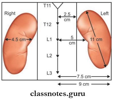
- It is the surface marking of the kidney from the back
- Morris Parallelogram Is Drawn In The Following Way:
- Two horizontal lines are drawn, one at the level of the spine of T11 and the other at the level of the spine of L3
- Then two vertical lines are drawn, one 2.5 cm and the other 9 cm lateral to the median plane
- Hilum lies opposite to the lower border of 1st lumbar spine. Lower on the right side.
Question 5. Describe in detail the blood supply of the kidney. Mention vascular segments of the kidney.
Answer:
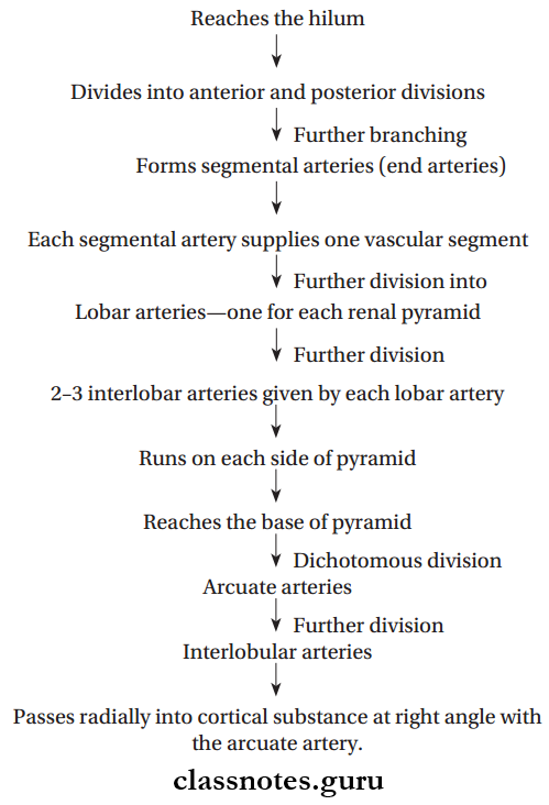
The Blood Supply Of Kidney
- Each kidney is supplied by one renal artery (branch of the abdominal aorta)
- In 30% of individuals accessory renal arteries are also seen
Course Of Kidney:
- Renal arteries arise directly from the abdominal aorta (between L1 and L2)
- The right renal artery passes to the right side and the left renal artery to the left side
- A further course is similar for both
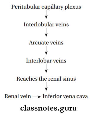
Venous Drainage Of Kidney
- Drained by right and left renal vein into inferior vena cava
- Course Of Venous Blood From Renal Substance Is As Follows:
Kidney And Suprarenal Gland Viva Questions
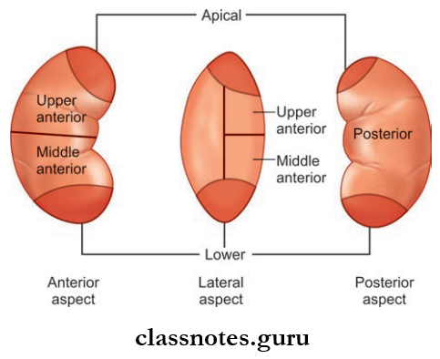
Vascular Segments Of Kidney
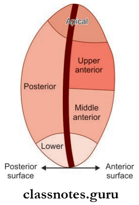
- Based on the distribution of five segmental arteries, each kidney is divided into five vascular segments:
- Apical: It occupies both anterior and posterior surfaces
- Upper Anterior: It occupies the anterior surface only
- Middle Anterior: It occupies the anterior surface only
- Lower: It occupies the entire inferior pole and includes both anterior and inferior surfaces
- Posterior: It occupies the posterior surface only.
Applied Anatomy Of Kidney: Just like the liver, surgical resection of the kidney is done on the basis of vascular segments—which helps to preserve the healthy parenchyma.
Lymphatic Drainage Of Kidney: Drains into para-aortic lymph nodes.
Nerve Supply Of Kidney
- Supplied by renal plexus of nerves
- Renal plexus consists of sympathetic and parasympathetic nerve fibers
- Sympathetic supply from T10–L1 spinal segments
- Parasympathetic supply from vagus nerves.
6. Write a note on the development of the kidney.
Answer:
The Development Of Kidney
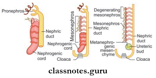
- The kidney develops from the intermediate cell mass
- Intermediate cell mass lies between the paraxial mesoderm and lateral plate mesoderm
- Paraxial mesoderm gives rise to somites
- Intermediate cell masses extend craniocaudally on both sides of the primitive dorsal aorta
- In the cervical and upper thoracic region, it shows segmentation called nephrotomes
- The remaining unsegmented portion below gives rise to the nephrogenic cord
- The nephrogenic cord later divides into 3 parts, from above to below pronephros, mesonephros, metanephros
- Excretory tubules of the kidney are formed from metanephros
- The collecting part of the kidney is formed from a diverticulum called the ureteric bud
- The ureteric bud is derived from the lower part of the mesonephric duct
- Horseshoe kidney—the lower pole of two kidneys fuse together to form an isthmus.
Anatomy Of Kidney Ureter And Adrenal Gland Exam Questions
Question 7. Describe briefly about the external features of suprarenal glands.
Answer:
The External Features Of Suprarenal Glands
- They are endocrine glands present on the upper pole of the kidney enclosed by renal fascia
- Location: Lies in the epigastric region, anterosuperior to the upper part of the kidney, in front of the crus of the diaphragm
Suprarenal Glands Shape:
- Right Suprarenal Gland: Pyramidal in shape
- Left Suprarenal Gland: Semilunar in shape
Suprarenal Glands Size:
- Length: 5 cm
- Breadth: 3 cm
- Thickness: 1 cm
- Weight: 5 g
Suprarenal Glands External Features
- Right Suprarenal Gland: it has
- Apex
- Base
- Two Surfaces: Anterior and posterior
- Three Borders: Anterior, medial, and lateral
- Left Suprarenal Gland: it has:
- Two Ends: Narrow upper end and rounded lower end
- Two Surfaces: Anterior and posterior
- Two Borders:
- Medial Border: Convex
- Lateral Border: Concave.
Kidney Ureter And Suprarenal Gland Short Questions And Answers
Question 8. Write a note on the blood supply of the suprarenal gland. Also, mention its lymphatic drainage and nerve supply.
Answer:
Suprarenal Gland:
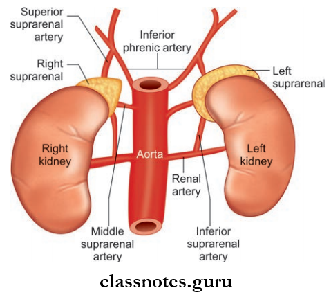
Suprarenal Glands Arterial Supply
- Superior Suprarenal Artery: Branch of the inferior phrenic artery
- Middle Suprarenal Srtery: Branch of the abdominal aorta
- Inferior Suprarenal Srtery: Branch of the renal artery.
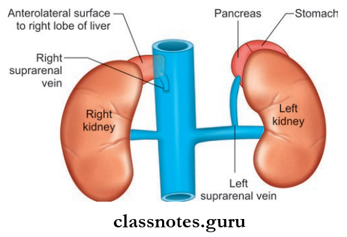
Suprarenal Glands Venous Drainage
- Drained by right and left suprarenal veins:
- Right suprarenal vein drains into the inferior vena cava
- The left suprarenal vein drains into the left renal vein.
Suprarenal Glands Lymphatic Drainage: Drain into lateral aortic nodes.
Suprarenal Glands Nerve Supply: Receive mainly myelinated preganglionic sympathetic fibers derived from splanchnic nerves.
Renal System Clinical Anatomy Questions
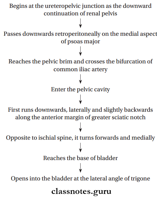
Question 9. Describe in detail the features, division, course, and relations of the ureter.
Answer:
Ureters:
- Ureters are a pair of narrow muscular tubes that transports urine from the kidney to the bladder
- Extent: From the ureteropelvic junction to base of the urinary bladder
- Length: 25 cm, upper half lies in the abdomen and lower half in the pelvis
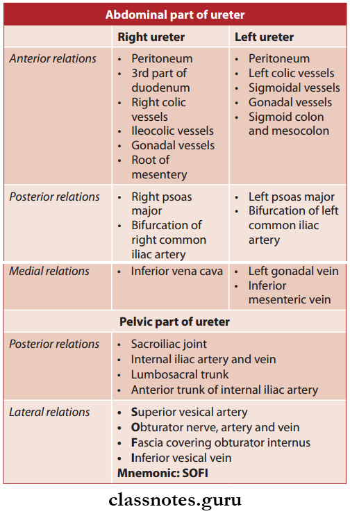
- So, The Ureter Is Divided Into Three Parts Based On Its Location, Namely:
- Abdominal Part: Renal pelvis—pelvic brim
- Pelvic Part: Pelvic brim—base of the urinary bladder
- Intravesical Part.
Ureter Course:
Suprarenal Glands Relations:
Suprarenal Glands Intravesical Part
- Narrowest part
- This part has an oblique course in the wall of the bladder
- This oblique passage acts as a flap valve and prevents the reflux of urine from the urinary bladder back to the ureter
- Ureteric openings are 5 cm and 2.5 cm apart in distended and empty bladders respectively.
Suprarenal Glands Blood Supply
Suprarenal Glands Arterial Supply:
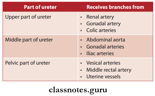
These branches further divide into ascending and descending branches and they form a plexus in the connective tissue sheath on the surface of ureter and then supply it.
Suprarenal Glands Venous Drainage: Drained by veins corresponding to the arteries
Kidney And Suprarenal Gland MBBS Notes
Suprarenal Glands Lymphatic Drainage
- Lateral aortic nodes
- Iliac nodes.
Suprarenal Glands Nerve Supply
- Sympathetic Supply: From T10 to L1 spinal segments through renal, aortic, and hypogastric plexuses
- Parasympathetic Supply: From S2 to S4 through pelvic splanchnic nerves.
Question 10. Name the constrictions of ureter.
Answer:
The Constrictions Of Ureter
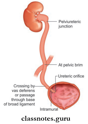
The Ureter Presents With 5 Constrictions:
- At the pelvic ureteric junction at the level of the tip of the transverse process of L2 vertebrae
- At the brim of the true pelvis
- At the point of crossing of the ureter by ductus deferens
- Intravesical or intramural: During its passage through the bladder wall
- At the ureteric orifice in the interior of the urinary bladder.
Ureter Applied Anatomy: These constrictions can act as the site of lodgment of kidney stones.
Kidney And Ureter NEET PG Questions
Kidney Ureter And Suprarenal Gland Multiple Choice Questions And Answers
Question 1. The kidney is related posteriorly to all the structures except:
- Iliohypogastric nerve
- Subcostal nerve
- Small intestine
- Renal fascia
Answer: 3. Small intestine
Question 2. Renal angle lies between:
- 11th rib and lateral border of sacrospinalis
- 12th rib and lateral border of sacrospinalis
- 10th rib and lateral border of sacrospinalis
- 12th rib and lateral border of quadratus lumborum
Answer: 2. 12th rib and lateral border of sacrospinalis
Question 3. The structures in the hilum of the kidney are:
- Renal vein
- Renal artery
- Ureter
- All of the above
Answer: 4. All of the above
Kidney And Suprarenal Gland Labeled Diagram With Questions
Question 4. The suprarenal gland has:
- 1 artery 3 veins
- 2 arteries 2 veins
- 3 arteries 3 veins
- 3 arteries 1 vein
Answer: 4. 3 arteries 1 vein
Question 5. Regarding the relations of the ureter, which is incorrect?
- Cross the vas deferens in males
- Medial to the transverse processes of lumbar spine
- Cross the genitofemoral nerve
- Narrowest at the PUJ
Answer: 1. Cross the vas deferens in males
Question 6. Which of the following structures is normally NOT found at the L1 vertebral level?
- Hilum of kidneys
- Origin of the celiac trunk
- Pylorus of stomach
- 3rd part of duodenum.
Answer: 4. 3rd part of duodenum.