Hypersensitivity
Question 1. Define and classify hypersensitivity reactions. Describe in detail the principle and mechanism of hypersensitivity reaction.
Answer:
Hypersensitivity Reaction Definition:
Hypersensitivity refers to a condition in which an immune response results in excessive reactions leading to tissue damage, diseases, or even death in the sensitized host.
Hypersensitivity Reaction Classification:
- Based on the time required for the immune response.
- Immediate hypersensitivity.
- Delayed hypersensitivity.
- Coomb and gel classification.
- Type 1 – Anaphylactic reaction.
- Type 2 – Cytotoxic reaction.
- Type 3 – Immune complex.
- Type 4 – Delayed or cell-mediated.
Read And Learn More: Microbiology Question and Answers
Hypersensitivity Reaction Principle And Mechanism
1. Type 1 Reaction.
- Cytotropic IgE antibody is the major antibody responsible for it.
- This antibody is induced by an allergen and thus binds to mast cells and basophils.
- When exposed to the allergen again, the bound IgE is cross-linked.
- This induces degranulation and release mediators like histamine.
2. Type 2 – Reaction.
- Antigens on a cell surface combine with antibodies.
- This leads to complement-mediated lysis.
3. Type 3 Reaction.
- Antigen-antibody immune complexes are deposited in tissues.
- By this, the complement is activated and polymorphonuclear.
- As a result, they release lysosomal enzymes. Causing tissue damage.
Short notes on hypersensitivity
4. Type 4 Reaction.
- Helper T lymphocytes sensitized by antigen release lymphokines upon second contact with the same antigen.
- This lymphokine induced inflammation.
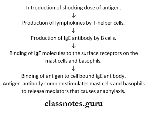
Anaphylaxis
- Anaphylaxis is the typical classical immediate hypersensitivity reaction.
- The term ‘anaphylaxis’ was coined by Richet in 1902.
- Anaphylaxis is a type of IgE-mediated hypersensitivity reaction, which develops quickly after the introduction of a large shocking dose of antigen following one or more small sensitizing doses.
Anaphylaxis Mechanism
Anaphylaxis Types
- Anaphylaxis in vitro.
- Cutaneous anaphylaxis.
Anaphylaxis Features
- The lung is the principal shock organ.
- Bronchospasm.
- Laryngeal edema.
- Respiratory distress.
- Shock
- Death.
Hypersensitivity Definition
Hypersensitivity refers to a condition in which immune response results in excessive reactions leading to tissue damage, diseases, or even death in the sensitized host.
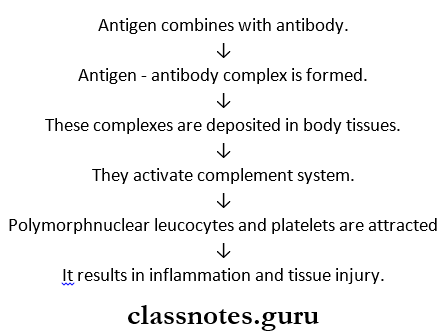
Type 3 – Hypersensitivity:
- Type 3 – Hypersensitivity is also called an immune complex or toxic complex disease.
- Type 3 – Hypersensitivity is a type of humoral antibody-mediated hypersensitivity reaction.
- In it, tissue damage is caused by antigen-antibody complexes.
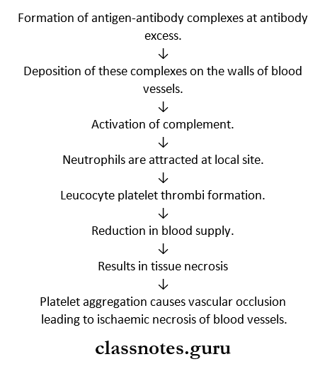
Hypersensitivity Pathogenesis
Hypersensitivity Types
1. Arthus Reactions – Localized.
- Arthus Reactions – Localized results in intense local edema and hemorrhage in persons with repeated administration of the same antigen.
2. Serum Sickness – Generalized.
- Serum Sickness – Generalized appears in 7 – 12 days following administration of a high concentration of antigen.
- Its clinical features include.
- Fever
- Lymphadenopathy.
- Splenomegaly
- Arthritis.
- Glomerulonephritis.
- Endocarditis.
- Vasculitis.
- Urticarial rashes -Abdominal pain.
- Nausea and vomiting.
Type 1 Hypersensitivity Reaction
- An anaphylactic reaction is a type 1 hypersensitivity reaction.
- It is B -a cell-mediated immediate type of hypersensitivity
Type 1 Hypersensitivity Reaction Forms:
1. Anaphylaxis
- Acute
- Potentially fatal
- Systemic form
2. Atopy
- Recurrent
- Non-fata
- Localized form.
Hypersensitivity MCQs
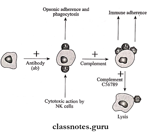
Type 2 Hypersensitivity Reaction
- The cytotoxic reaction is a type 2 hypersensitivity reaction.
- Type 2 Hypersensitivity Reaction is an IgG antibody-mediated reaction.
- Type 2 Hypersensitivity Reaction is intermediate between hypersensitivity and autoimmunity.
Type 2 Hypersensitivity Reaction Pathogenesis
- IgG antibodies bind to an antigen on the cell surface.
- This causes.
- Phagocytosis of the cell.
- Cytotoxicity by natural killer cells.
- Cell lysis.
- In some reactions, the antibody in combination with the cell surface receptors disrupts the normal functioning of the cell.
- Uncontrolled activation.
- Blocking of receptors.
- Examples:
- Autoimmune anemias
- Hemolytic disease
- Drug reactions.
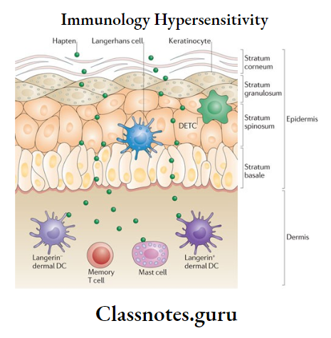
Delayed hypersensitivity or type 4 reaction
Delayed Hypersensitivity
- Delayed hypersensitivity reaction is mediated by sensitized T – T-lymphocytes but not by antibodies.
- Thus, it cannot be passively transferred by serum but can be transferred by lymphocytes or the transfer factor.
Delayed Hypersensitivity Pathogenesis
- Type 4 reaction occurs within 48 – 72 hours of antigen contact.
- Delayed Hypersensitivity Pathogenesis is provoked by intracellular infections or haptens – like simple chemicals applied on the skin.
- These sensitize T-lymphocytes.
- Sensitized T-lymphocytes on contact with specific antigen release lymphokines.
- This causes biological effects on macrophages, leucocytes, and tissue cells.
Delayed Hypersensitivity Types
- Tuberculin type.
- Contact dermatitis type.
- Examples:
- Tuberculosis.
- Typhoid fever
- Allergic contact dermatitis.
- Graft rejection.
Delayed hypersensitivity
Immediate Hypersensitivity Reaction
Based on the time required for a sensitized host to develop clinical reactions on re-exposure to the antigen, hypersensitivity is classified into.
- Immediate hypersensitivity.
- Delayed hypersensitivity.
1. Immediate Hypersensitivity:
- Immediate Hypersensitivity is a B-cell or antibody-mediated reaction.
- Immediate Hypersensitivity appears and recedes rapidly.
- Immediate Hypersensitivity is passively by antigens or haptens.
- Immediate Hypersensitivity is subdivided into the following clinical types.
- Anaphylaxis.
- Atopy
- Antibody-mediated cell damage.
- Arthus phenomenon.
- Serum sickness.
Atopy
Atopy is a form of type 1 hypersensitivity.
- The antigens commonly involved in it are pollens, house dust, and foods.
- These induce IgE antibodies.
Atopy Features:
- Atopy shows marked familial distribution.
- Atopy sensitization is developed spontaneously following natural contact with opens.
- Reactions occur at the site of entry of antigen.
- For example.
- Inhalation of pollens affects the lungs.
- Contact leads to local allergy.
Atopy Manifestations:
- Conjunctivitis.
- Rhinitis.
- Bronchospasm
- GI symptoms.
- Dermatitis.
- Cutaneous eruptions.
Immune system hypersensitivity
Arthus Reaction
Arthus reaction is a localized form of type 3 hypersensitivity reaction.
- When an antigen is injected subcutaneously or intradermally in an animal in whom there are repeated administrations of the same antigen previously, there occurs intense local edema and hemorrhage which reaches a peak in 3 – 6 hours, this is called the Arthus reaction.
Arthus Reaction Mechanism:
Serum Sickness
Serum Sickness is a systemic form of type 3 hypersensitivity that appears 7-12 days following a single injection of a high concentration of foreign serum such as diphtheria anti-toxin.
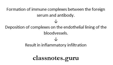
Serum Sickness Pathogenesis:
Tuberculin reaction
Tuberculin reaction is a type of delayed hypersensitivity reaction.
- When a small dose of tuberculin is injected intradermally in an individual sensitized to tuberculoprotein by prior infection or immunization, an indurated inflammatory reaction develops within 48 – 47 hours.
- The injection site is infiltrated by a large number of lymphocytes and 10 – 12% macrophages.
- It occurs in infections caused by bacteria, fungi, and parasites.
Tuberculin Test
- The Tuberculin test is a useful indicator for delayed hypersensitivity.
- In it, purified protein derivative (PPD) is used.
Tuberculin Test Indicates:
- Hypersensitivity to tubercle bacilli.
- Infectious with bacteria, fungi, and parasites.
- Subacute and chronic infections.
- Intracellular pathogen.
Tests Used For Delayed Hhypersensitivity
Tests that are used for delayed hypersensitivity are.
1. Tuberculin Tests.
- Positive for M tuberculosis.
2. Lepromin Test
- Positive in tuberculoid leprosy.
- Negative in lepromatous leprosy.
3. Freitest.
- Positive in lymphogranuloma venereum.
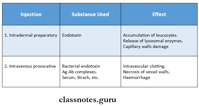
Shwartzman Reaction
Shwartzman in 1928 observed that if a culture of salmonella typhi is injected intradermally in a rabbit, followed by intravenous injection of the same filtrate after 24 hours, a hemorrhagic necrotic lesion develops at the site of in-dermal injection.
- This is called the Shwartzman reaction.
- Because of the absence of specificity and the short interval between the two doses, there is no immunological basis for the reaction.
Hypersensitivity questions and answers
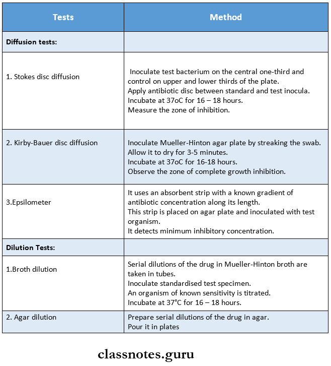
Antibiotic Sensitivity Test