
Gingival Inflammation Important Notes
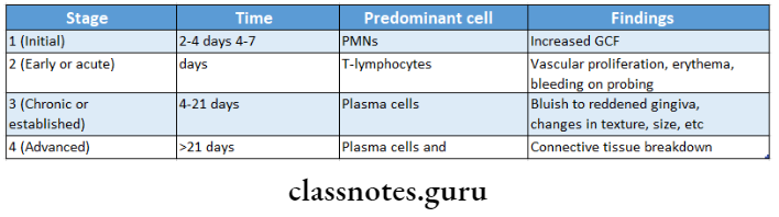
1. Stages Of Gingivitis
2. Stage 3 Gingivitis
Stage 3 Gingivitis Clinical features:
- Engorgement of blood vessels
- The bluish-red appearance of the gingiva
- Changes in size and texture
Read And Learn More: Periodontics Question and Answers
Stage 3 Gingivitis Histological Features:
- Infiltration of connective tissue and junctional epithelium with neutrophils, and lymphocytes with the predominance of plasma cells
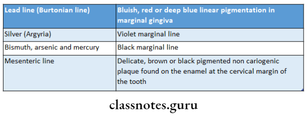
3. Gingival Pigmentation Caused By Heavy Metals
Gingival Inflammation Short Essays
Question 1. Stages of Gingivitis.
Answer:
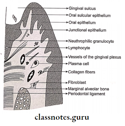
Stage 1 Initial Lesion:
- Classic vasculitis of vessels
- Exudation of fluid from gingival sulcus
- Changes in the coronal portion of JE
- Migration of leukocytes
- Presence of serum proteins
- Loss of collagen
Stage 2-Early Lesion:
- Erythematous Gingiva
- Bleeding on probing
- Development of recipes in JE
- Presence of lymphocytes
- Cytotoxic changes of fibroblasts
- Loss of collagen
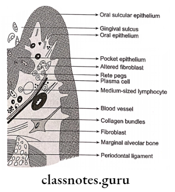
Stage 3-Established Lesion:
- Bluish hue on reddened gingiva
- Moderate to severely inflamed gingiva
- Presence of plasma cells deep into connective tissue
- Apical migration of junctional epithelium
- Collagen destruction
- Continuous loss of ground substance
- Pocket formation
- Elevated levels of acid and alkaline phosphate, B- glucuronidase, aminopeptidase
Stage 4-Advanced Lesion:
- Persistence of infection
- Bone loss
- Loss of collagen
- Presence of all inflammatory cells Formation of pockets
- Leads to periodontics
Question 2. Gingival pigmentation.
Answer:
The color of gingiva is determined by several factors in which pigments within the epithelium are one of the factors
Gingival Pigmentation Types:
- Endogenous pigmentation
- Exogenous pigmentation
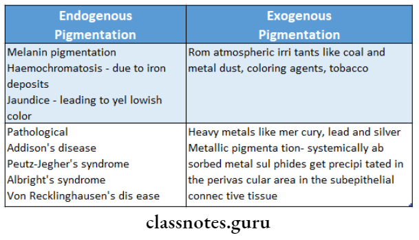
Gingival Pigmentation Causes:
1. Localized:
- Amalgam tattoo
- Graphite
- Nevus
- Melanotic macules
- Malignant melanoma
- Kaposi’s sarcoma
2. Generalized pigmentation:
- Genetics- Peutz-Jegher’s syndrome
- Habits- Smoking, betel chewing
- Drugs
- Antimalarial
- Antimicrobial
- Minocycline
- Ketoconazole
- Contraceptive pills
- Heavy metal exposure
- Endocrine
- Addison’s disease
- Albright’s syndrome
- Pregnancy
- Post-inflammatory
- Periodontal disease
- Post-surgical gingival pigmentation
- Others
- Haemochromatosis
- Generalised neurofibromatosis
- HIV disease
- Thalassaemia
Question 3. Stage 2-Early lesion.
Answer:
Stage 2-Early Lesion:
- Erythematous Gingiva
- Bleeding on probing
- Development of rete pegs in JE
- Presence of lymphocytes
- Cytotoxic changes of fibroblasts
- Loss of collagen
Gingival Inflammation Short Answers
Question 1. Stage III of gingivitis.
Answer:
Stage 3 gingivitis is an established lesion
Stage III of gingivitis Features:
- Bluish hue on reddened gingiva
- Moderate to severely inflamed gingiva
- Presence of plasma cells deep into connective tissue
- Apical migration of junctional epithelium
- Collagen destruction
- Continuous loss of ground substance
- Pocket formation
- Elevated levels of acid and alkaline phosphate, beta-glucuronidase, aminopeptidase
Question 2. Plasma cell gingivitis.
Answer:
- Plasma cell gingivitis is also referred to as atypical and plasma cell gingivostomatitis
- It consists of a mild marginal gingival enlargement that extends to the attached gingiva
- Gingiva appears red, friable and bleeds easily
- Microscopically, connective tissue contains a dense infiltrate of plasma cells that also extends to oral epithelium
Plasma Cell gingivitis Uses:
- Allergy
- Related to components of chewing gums or denitrifies
Gingival Inflammation Viva Voce
- The predominant cell in acute gingivitis is the T lymphocyte
- Gingivitis is the most common form of gingival disease
- Bacteria found in gingivitis are localized in the gingival sulcus
- Extension of inflammation into the supporting structures occurs in stage 4
- Stage I gingivitis is subclinical
- Erythema and bleeding on probing occurs in stage 2
- The established lesion of gingivitis is characterized by the predominance of plasma cells and B lymphocytes
- The B cells found in established lesions are predominantly IgG and IgG, subclasses