Spirochetes Question And Answers

Spirochaetes Long Essays

Question 1. Laboratory diagnosis of syphilis
(or)
Serological tests for syphilis
Answer:

1. Microscopy

  • It is useful in primary and secondary syphilis
  • Direct fluorescent antibody test is used for T. pal¬lidum
  • In this method acetone fixed smear is subjected to fluorescent tagged anti-T. pallidum antiserum

2. Serological tests

  • Tests for antibodies reacting with cardiolipin anti¬gen
    • Standard tests for syphilis
      • Wassermann test
  • It is a complement fixation test

Serological tests for syphilis Method:

  • Patient’s serum is inactivated by heating at 56s C for 30 minutes
  • Incubated with cardiolipin antigen and guinea pig antigen at 37° C for 1 hour
  • Indicator is added to detect presence or absence or complement
  • 0.15 ml of inactivated serum is taken in three test tubes containing 0.05, 0.025 and 0.0125 ml of freshly prepared antigen dilution
  • Tubes are shaken on Kahn’s shaken and examined

Serological tests for syphilis Interpretation:

    • If hemolysis does not occurs, it indicates that the complement is utilized
    • This is positive Wassmann’s reaction
    • It hemolysis takes place, it indicates the comple¬ment is not been utilized in tire primary reaction but utilized by the indicator system
    • Kahn test
    • It is tube flocculation test
    • Negative test shows uniform opalescence
    • Positive test shows floccules
    • VDRL test
    • In this test the inactivated serum is mixed with cardiolipin antigen on a special slide and rotated for 4 minutes
    • Uniform distribution of crystals in the drop indi¬cates tire serum is non reacting
    • Formation of clumps indicates it is reactive
  • Tests for antibodies reacting with group specific treponemal antigen
    • Reiter protein complement fixation test
      • In this method, lipopolysaccharide protein complex antigen derived from the cultivable Reiter’s stain is used
      • It is less sensitive
  • Tests for specific antibodies to pathogenic tre¬ponema
    • Treponema pallidum immobilization test
      • It employs live T. pallidum
      • The test serum is mixed with actively motile Nichol’s strain of T. pallidum and incubated anaerobically
      • If antibodies are present, the treponemas are immobilised

Read And Learn More: Microbiology Question and Answers

    • Fluorescent treponemal antibody absorption
      • Test serum is preabsorbed with an extract of non pathogenic treponemas to remove group specific antigens
      • It detects IgM and IgG
      • It is the earliest test to become positive
    • Microhaemagglutination test for treponema pallidum
      • T. pallidum antigen is coated onto the surface of red cells
      • Tanned sheep RBCs are sensitized with an extract of T. pallidum
      • When these sensitized erythrocytes are mixed with patient’s serum containing anti¬bodies, the erythrocytes clump together
    • Enzyme immunoassay
      • They have been developed using T. pallidum antigens and are available commercially

Spirochaetes Short Question And Answers

Question 1. VDRL test
Answer:

  • Venereal Disease Research Laboratory test is the most widely used serological test for diagnosis of syphilis
  • It is simple and more rapid test which gives more quantitative results
  • It requires only small amount of serum and is sensitive

VDRL test Procedure:

  • In this test the inactivated serum is mixed with cardi- olipin antigen on a special slide and rotated for 4 min¬utes
  • Uniform distribution of crystals in the drop indicates the serum is non reacting
  • Formation of clumps indicates it is reactive
  • By testing serial dilutions, the antibody titre can be determined

Question 2. Vincent’s angina
Answer:

  • Vincent’s angina is a painful condition of the throat characvterized by local ulceration of the tonsils, mouth and pharynx
  • Vincent’s bacilli is the causative organism
  • It may occur as an acute illness with diffuse involve¬ment of tissue or as chronic illness consisting of ulcera¬tion of tonsil
  • It is insidious in onset with less fever and less discom¬fort
  • Membrane which usually forms over the tonsil can be easily removed revealing irregular ulcer on the tonsil

Question 3. Borrelia vincentii
Answer:

  • It is a motile spirochaete which is a normal mouth commensal but a potential pathogen.
  • Under predisposing conditions such as malnutrition (or) viral infections it gives rise to ulcerative gin¬givostomatitis or oropharyngitis (Vincent’s angina).
  • Borrelia vincenti in association with fusiform bacilli causes an infection known as fusospirochetosis.
  • Diagnosis may be made by demonstrating spirochaetes and fusiform bacilli in stained smears of exudates from the lesions.
  • Cultivation is difficult but can be done in enriched me¬dia anaerobically.
  • Penicillin and metronidazole are effective in treatment.

Question 4. Congenital syphilis
Answer:

In congenital syphilis the infection is transmitted from mother to foetus transplacentally.

Congenital syphilis Clinical Features:

  • Hutchinson’s traid
    • Hyperplasia of the incisor and molar teeth
    • Interstitial keratitis
    • Eight nerve deafness.
  • Frontal bossing
  • Saddle nose
  • High palatal arch
  • Poorly developed maxillae
  • Irregular thicknening of clavicle
  • Enlarged liver and spleen
  • Diagnosis may be done by demonstrating spirochetes in lesions under dark ground microscope.
  • IgM FTA – ABS test, the modification of indirect im-mune fluorescence test is useful for diagnosis of con-genital syphilis and distinguishing from seropositivity due to passively transferred maternal antibodies

Spirochaetes Viva Voce

  1. Techoic acid of cell wall of staphylococci facilitates adhesion of cocci to host cell
  2. ASO test is used for diagnosis of rheumatic fever
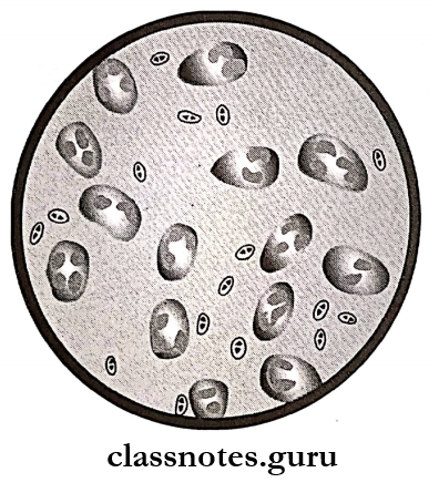
  3. Pneumococci gives draughtsman or carom coin ap-pearance on blood agar
  4. Modified Thayer Martin, selective media is used for meningococci
  5. Corynbacterium diphtheria shows Chinese letter or cuneiform arrangement
  6. DTP vaccine consists of diphtheria toxoid, tetanus toxoid and pertussis vaccine
  7. Bacillus coreus causes food poisoning
  8. Clostridium contains peritrichate flagella for motility
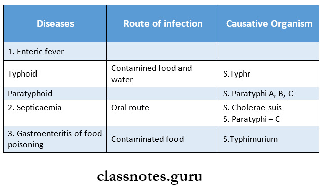
  9. Widal test is used for diagnosis of typhoid fever
  10. Dreyer’s tube and Felix tube are used in widal test
  11. Food poisoning is caused by any salmonella except salmonella typhi
  12. Yersinia pestis is causative agent of plaque or black death
  13. Trepnema pallidum is causative agent for syphilis
  14. VDRL test is used for syphilis
  15. Actinomycetes gives sun ray appearance on culture
  16. H pyroli causes peptic ulcer

Mycobacterium Leparae Question And Answers

Mycobacterium Leparae Long Essays

Question 1. Classify leprosy. Describe morphology, pathogenesis, and laboratory diagnosis of Mycobacterium leprae.
Answer:

Leprosy Classification

Based on the immune status of the host, leprosy is classified into four types

  • Lepramatous leprosy
  • Tuberculoid leprosy
  • Dimorphous leprosy
  • Intermediate leprosy

Mycobacterium Leprae Morphology

  • It is weakly gram-positive and strongly acid-fast bacilli
  • Shape: slender, slightly curved or straight bacilli
  • Size: 1-8 μm * 0.2 * 0.5 pm
  • Arrangement: seen singly and in groups
  • In tissues arranged in clumps resembling cigarette ends
  • Position: intracellularly or lying free outside the cells
  • Inside the cells they are present as bundles of organ¬isms bound together by glia
  • These are known as globe
  • Parallel rows of it appear as cigar bundle

Mycobacterium Leprae Pathogenesis

  • Leprosy is a granulomatous disease of humans
  • It has long incubation period
  • Route of infection: nasal discharge and skin
    • Lepromatous leprosy: features
      • Nodular skin lesions
      • Slow and symmetric thickening of peripheral nerves
      • Anesthesia
      • Loss of sensation
      • Ulceration of nodular lesions
      • Nodules become secondarily infected
    • This leads to distortion and mutilation of extremities
    • Tuberculoid leprosy
      • Few skin lesions occur
      • It consists of elevated hypo or hyper-pigmented macular patches
      • Involvement of peripheral nerves occurs
      • Leads to deformities of hands and feet

Read And Learn More: Microbiology Question and Answers

    • Oral manifestations
      • Oral lesions involve the tongue, lip or hard palate
      • Lesions consist of small tumours called lepromas
      • These tend to ulcerate
      • In the facial region, the following changes occur
      • Atrophy of the anterior nasal spine
      • Saddle nose
      • Premaxillary bone recession

Mycobacterium Leprae Laboratory Diagnosis

  • It includes:
    • Acid-fast staining:
      • Acid-fast staining of smears shows acid-fast bacilli arranged in parallel bundles within macrophages [Lepra-cells) confirm the diagnosis of lepromatous leprosy.
      • The viable bacilli stain uniformly and the dead bacilli are fragmented, irregular or granular.
    • Skin and nerve biopsy:
      • These are required for histological confirmation of tuberculoid leprosy when acid-fast bacilli cannot be demonstrated in direct smear.
      • Skin biopsy is also useful in the diagnosis and accurate classification of leprosy lesions.
    • Animal inoculation:
      • Injection of ground tissue from lepromatous nodules or nasal scrapings from leprosy patients into the foot pad of a mouse produces typical granuloma at the site of inoculation within 6 months.
    • Lepromin test:
      • It is not diagnostic test but is used to assess the resistance of patients of M. leprae infection.
      • It also assesses the prognosis and response to treatment.
    • Serological test:
      • Serodiagnosis of leprosy may be carried out by the detection of anti-phenolic glycolipid -1 antibodies.

The test used are:

  • ELISA
  • Latest agglutination
  • Mycobacterium leprae particles agglutination (MLPA)

Mycobacterium Leparae Short Essays

Question 1. Lepromin test
Answer:

  • It is a delayed type of hypersensitivity reaction
  • It was first described by Mitsuda in 1919

Lepromin Antigen:

  • Lepromin antigens used are
  • Integral lepromin
  • Bacillary lepromin

Lepromin Procedure:

  • 0.1 ml of lepromin is injected intradermally
  • Reaction is observed

Lepromin Reactions:

  • The early reaction of Fernandez
    • Consists of erythema and induration in 24-48 hours
    • It remains for 3-5 days
  • The late reaction of Mitsuda
    • Appears after 1-2 weeks of injection
    • It occurs in the form of nodules that may ulcerate
    • It heals in a few weeks

Lepromin Uses:

  • Classification of leprosy
  • Assessment of prognosis
  • Assessment of resistance

Mycobacterium Tuberculosis Question And Answers

Mycobacterium Tuberculosis Long Essay

Question 1. Describe morphology, cultural characteristics, pathogenicity, and laboratory diagnosis of mycobacterium tuberculosis.
Answer:

Mycobacterium tuberculosis Morphology:

  • M. Tuberculosis is weakly gram-positive, strongly acid- fast bacilli.
  • They are non-sporing, non-capsulated and non-motile.
  • Shape-slender, straight or slightly curved bacilli with rounded ends.
  • Size : 1 – 4 0m * 0.2 – 0.8 0m.
  • Arrangement – singly, in pairs or in small clumps.

Mycobacterium tuberculosis Cultural characteristics:

  • M. Tuberculosis is an obligate aerobe.
  • It grows slowly at a temperature range of 30 – 40°C and pH – 6.4 – 7.0.

1. Lowenstein – Jensen media.

  • It consists of
    • Beaten eggs – Acts as a solidifying agent.
    • Asparagine.
    • Mineral salts.
    • Malachite green – Inhibits the growth of another organism.
    • Glycerol – Improves growth.
    • Colonies are dry, rough, buff-colored, raised with a wrinkled surface.

2. Liquid media.

  • Bacilli grow as a surface pellicle.
  • Virulent strains grow as serpentine cords.
  • Avirulent strains grow in a dispersed fashion.

Mycobacterium tuberculosis Pathogenicity:

  • Route of infection – inhalation, ingestion, and skin inoculation.
  • Tubercle is a characteristic lesion.
  • It is an avascular granuloma composed of a central zone containing giant cells with or without caseation necrosis surrounded by epitheloid cells and a peripheral zone of lymphocytes and fibroblasts.

Mycobacterium tuberculosis Types:

1. Exudative.

  • It is an acute inflammatory reaction with an accumulation of edema fluids, leucocytes, and monocytes.

2. Productive.

  • It is predominantly cellular and composed of tubercles.
  • Tuberculosis can also be classified as.

1. Primary infection.

  • Occurs in young children.
  • Leads to primary complex.
  • Consists of Gohn’s focus.
  • Hilar lymph nodes are involved.

Read And Learn More: Microbiology Question and Answers

2. Postprimaiy infection.

  • Occurs in adults.
  • It heals by resorption, fibrosis and occasionally

Lab Diagnosis of My-cobacterium Tuberculosis

1. Specimen collection

Mycobacterium Tuberculosis Specimen collection

2. Direct microscopy

  • Ziehl Neelsen staining shows acid-fast, bright red tubercle bacilli.

3. Culture.

  • Lowenstein – Jension media is used.

4. Animal inoculation.

  • 0.5 ml of the concentrated specimen is inoculated IM into the thigh of two healthy guinea pigs.
  • Animals are weighed before inoculation and then at weekly intervals.
  • Tested after 3-4 weeks.
  • Auto spy shows a caseous lesion.

5. Molecular method – Includes polymerase chain reaction.

6. Serological test – Includes.

  • ELSI test
  • Radioimmunoassay.
  • Latex agglutination assay.

Mycobacterium Tuberculosis Short Essays

Question 1. Mantoux test.
Answer:

It is routinely used method for tuberculin testing.

Mantoux test Method:

  • 0.1 ml of purified protein derivative, PPD containing 5 TU [tuberculin unit] is injected intradermally into the flexor aspect of the forearm.
  • It is given between layers of the skin.
  • The site is examined after 48 – 72 hours for induration.

Mantoux test Result:

Mycobacterium Mantoux test result

Mantoux test Significance:

Mycobacterium Mantoux test significance

Question 2. BCG vaccine.
Answer:

  • BCG vaccine was prepared by Calmette and Guerin.
  • It is a live attenuated and freeze-dried vaccine.

BCG vaccine Dose and Administration:

  • It is available as a fresh liquid vaccine or in the form of freeze-dried vaccine.
  • It is given intradermally in a dose of 0.1 ml soon after birth.

BCG vaccine Immune response:

  • Induces a self-limited infection with multiplication and dissemination of the bacillus in different organs and production of small tubercles.
  • It gives rise to delayed hypersensitivity.

BCG vaccine Complications:

  • Local abscess, indolent ulcer, keloid, confluent lesion, lipid lesion.
  • Regional enlargement and suppuration of draining lymph nodes.
  • Systemic fever, mediastinal adenitis.
  • Erythema nodosum.

BCG vaccine Contraindications:

  • In patients of AIDS, Eczema, pertussis, measles, and patients on steroids.

Role of BCG:

  • Makes the disease milder.
  • Prevents serious forms of disease.

Mycobacterium Tuberculosis Short Question And Answers

Question 1. Classification of mycobacteria.
Answer:

Classification of mycobacteria is as follows:

1. Cultivable:

Mycobacterium Tuberculosis Cultivable

2. Non-cultivable.

  • M.Leprae

Vibrio Bacteriology Question And Answers

Vibrio Long Essays

Question 1. Describe morphology, cultural characteristics, taxing, and laboratory diagnosis of vibrio cholera.
Answer:

Vibrio cholera Morphology:

  • Vibrio cholera is a gram-negative, non-capsulated, and non-sporing organism.
  • It is motile and possesses a single polar flagellum, thus called darting motility.
  • Size: 1.5 mm x 0.2 – 0.4 mm.
  • Shape: curved or comma-shaped rod.

Vibrio- Vibirio cholerae

Vibrio cholera Cultural Characteristics:

  • Vibrio cholera is strongly aerobe
  • Grows within a temperature range of 16-40°C and alkaline pH of 8.2

Vibrio- Vibirio choleraeCultural characteristics

Vibrio cholera Toxins:

  • Vibrio cholera produces enterotoxin known as cholera toxin (CT)
  • It is heat labile
  • Protein in nature.
  • Molecular weight – 90,000
  • It has two fractions.
    • A (active) subunit – one.
    • B (binding) subunit – five.
  • Its production is determined by phage integrated with bacterial chromosomes.

Vibrio cholera Mechanism:

Vibrio- Vibirio cholerae mechanism

Read And Learn More: Microbiology Question and Answers

Laboratory diagnosis:

1. Direct microscopy.

  • Not reliable.
  • Characteristic motility of the vibrios is demonstrated by dark field or phase contracts microscope.

2. Culture.

  • Nutrient agar.
    • Shows moist, translucent, round disc colonies.
  • Selective media – Monsur’s GTTA media.
    • Shows small, translucent colonies with a greyish-black center and turbid halo.
  • Macconkey’s agar media.
    • Shows initially colorless colonies which later become reddish.

3. Agglutination test

  • Colonies from selective media are picked up with a straight wire and tested by slide agglutination with cholera 0 subgroup I serum.
  • If positive, the agglutination test is repeated using monospecific Ogawa and Inaba sera for serotyping.

4. Serological tests – includes.

  • Agglutination using live or killed vibrio suspension.
  • Indirect haemagglutination.
  • Vibriocidal test.
  • Antitoxin assay.

5. Biochemical test.

  • Fermentation of glucose, mannitol, maltose, sucrose.
  • Indole positive.
  • Reduction of nitrates.
  • Catalase and oxidase positive. Voges – Broskauer negative.

Vibrio Short Essays

Question 1. Cholera.
Answer:

Cholera Etiology:

  • Cholera is caused by vibrio cholera.
  • The route of infection is contaminated food and water.
  • Alkaline pH in the stomach and intestine appears to be more easily infected.

Cholera Pathogenesis:

Vibrio- Cholerae Pathogenesis

Cholera Features:

  • Diarrhea is the major symptom.
  • Feces contain epithelial cells, mucus and large number of V. Cholera.
  • Profuse watery diarrhoea – rice water stools occur.
  • In severe cases, there may be one liter of fluid loss each hour.
  • Abdominal pain.
  • Death due to electrolyte abnormalities and fluid loss.

Treatment, Prevention, and Control:

  • Intravenous administration of fluids.
  • Oral administration of a solution containing glucose and electrolytes.
  • Antibiotics.
    • Doxycycline – In adults.
    • Sulphamethethoxazole – In children.
    • Furazolidone – In pregnant women.
  • Improved – hygiene.
  • Water purification, immunization.

Vibrio Short Question And Answers

Question 1. Whooping cough.
Answer:

Whooping cough is predominantly a childhood disease.

Whooping cough Causative agents:

  • B. Pertusis – 95% cases.
  • B. Parapertusis – 5% cases.
  • B. Bronchiosepetica – 0.1% cases.

Whooping cough Features:

  • Incubation period – 1 – 2 weeks.
  • Infection is transmitted by droplets.
  • Diseases usually last for 6 – 8 weeks.
  • It consists of 3 stages.

1. Catarrhal.

  • Clinical diagnosis is difficult.

2. Paroxysmal.

  • Whooping cough occurs.

3. Convalescent.

  • Frequently and severity of coughing decreases.

Whooping cough Complications:

  • Subconjunctival hemorrhage.
  • Subcutaneous emphysema.
  • Bronchopneumonia.
  • Convulsions, coma.

Whooping cough Prophylaxis:

  • Pertussis vaccine is given.
  • Three injections at intervals of 4 – 6 weeks are given before the age of 6 months.
  • A booster dose is given at the end of the first year.

Question 2. Castaneda’s method of culture.
Answer:

It is used for Brucella.

Culture Composition:

  • It contains:
    • Liquid media – trypticase soy broth.
    • Solid media – trypicase soy agar.

Culture Method:

  • Blood is inoculated into liquid media in a bottle.
  • Incubate it in an upright position.
  • The bottle is tilted and subcultured by solid media.
  • Incubate it again in an upright position.

Brucella Castaneda's medium

Salmonella Bacteriology Question And Answers

Salmonella Long Essays

Question 1. Describe etiopathogenesis and clinical features and lab diagnosis of enteric fever.
Answer:

Enteric fever:

  • Etiopathogenesis:
    • Enteric fever includes:
      • Typhoid fever – caused by salmonella typhi.
      • Paratyphoid fever – caused by salmonella paratyphi.

Virulence factors:

1. Endotoxin.

  • Causes diverse toxic manifestations of enteric fever.

2. Exotoxin – enterotoxin.

3. Capsule.

  • Has anti-phagocytic activity.

Enteric fever Clinical features:

  • Incubation period is 7 – 14 days.
  • Route of infection – contaminated food and water.

1. Typhoid fever – Its manifestations include.

  • Gradual illness
  • Headache
  • Anorexia
  • Congestion of mucous membrane.
  • Hepatosplenomegaly.
  • Step-ladder pyrexia.
  • Relative bradycardia.
  • Leucopenia.
  • Skin rashes called ‘Rose-spots’ appear during the second or third week.

2. Paratyphoid fever.

  • Symptoms are milder than typhoid fever.
  • Septicaemia with suppurative complications occurs.

Read And Learn More: Microbiology Question and Answers

Lab Diagnosis of enteric fever:

1. Isolation of bacilli.

  • For isolation of bacteria specimens are obtained from blood, feces, urine, aspirated duodenal fluid, bile, bone marrow or rose spot.
  • These specimens are then cultured.

Salmonella Isolation of cbacilli culture and features

2. Demonstration of Antibodies.

  • Widal test
    • It is an agglutination for the detection of agglutinins H and 0 in patients with enteric fever

Salmonella Widal test

  • Method:
    • Equal volumes (0.4 ml) of serial dilutions of serum from 1:10 to 1: 640 and H and 0 antigens are mixed.
    • One control tube containing antigen and normal saline is used.
    • All these tubes are incubated in a water bath at 37°C.
  • Results:

Salmonella Lab Diagnosis Result

  • Interpretation:

Salmonella Lab Diagnosis Interpretation

3. Demonstration of circulating antigen.

  • Done by counterimmunoelectrophoresis and ELISA.

Salmonella Short Essays

Question 1. Diseased caused by salmonella.
Answer:

Salmonella Diseased caused by salmonella

Question 2. Antigenic structure of salmonella.
Answer:

Salmonella possesses three types of antigen.

Salmonella Antigenic structure of antigen

Pneumococcus Bacteria Questions and Answers

Pneumococcus Short Essays

Question 1. Morphology and cultural characteristics of pneumococci.
Answer:

Pneumococci Morphology:

  • Pneumococci are gram-positive, alpha-hemolytic organisms.
  • Size – small, 1 micrometer in diameter.
  • Shape – slightly elongated.
  • Each coccus has one end broad or rounded and other pointed.
  • They are arranged in pairs.
  • This shows a flame-shaped or lanceolate appearance.
  • They have large polysaccharide capsules.
  • They are non-motile and non-sporing.

Pneumococcus - Str. Pneumoniae in pus

Pneumococci  Cultural characteristics:

  • They are aerobes and facultative anaerobes.
  • The optimum temperature for growth is 37°C and pH 7.8

Pneumococcus Cultural characteristics

Pneumococcus Short Question And Answers

Question 1. C-reactive proteins (CRP).
Answer:

C-reactive protein is an abnormal protein that precipitates with the somatic C antigen of pneumococci.

  • It is not an antibody produced as a result of pneuma- coccal infection but it is an acute phase substance produced in hepatocytes.
  • It appears in.
    • Acute sera of cases of pneumonia.
    • Some other pathological conditions.
  • It disappears during convalescence.

Read And Learn More: Microbiology Question and Answers

C-reactive proteins Production:

  • Its production is stimulated by.
  • Bacterial infections.
  • Inflammation.
  • Malignancies.
  • Malignancies.
  • Tissue destruction.

C-reactive proteins Uses:

  • As an index of the response of treatment in rheumatic fever.

C-reactive proteins Test:

  • It is tested by passive agglutination using latex particles coated with an anti-CRP antibody.

Question 2. Lab investigations of pneumococci.
Answer:

Pneumococcus Cultural characteristics

Question 3. Mention the difference between pneumococci and streptococcus viridians.
Answer:

Pneumococcus Differences between pneumococci and streptococcus viridians

Question 4. Media used for gonococci.
Answer:

Gonococci are aerobic organisms.

  • They grow best at a temperature of 35 – 36oC in the presence of 5 – 10 % C02 and at pH 7.2 – 7.6
  • The cultural media used are

1. Enriched media.

  • It includes chocolate agar and blood agar media.
  • Colonies obtained are small, round, convex, grey, and translucent.

2. Selective media – Thayer Martin media.

  • It inhibits most contaminants.

Corynebacterium Diphtheriae Question and Answers

Corynebacterium Long Essays

Question 1. Describe the morphology, cultural characteristic, and laboratory diagnosis of Corynebacterium diphtheria.
Answer:

Corynebacterium Diphtheria:

  • Morphology:
    • C. Diphtheria bacillus are thin slender rods.
    • They are gram-positive bacilli.
    • They are pleomorphic, nonsporting, non-motile, and non-capsulated.
    • Size – approx 3-6 pm x 0.6 – 0.8 pm.
    • They show clubbing at one or both ends.
    • They have intracellular polyphosphate granules known as metachromatic or volutin or Babes-Ernst granules usually situated at the poles of bacilli.
    • They are arranged in pairs or small groups.
    • They frequently remains attached after division, which gives them Chinese letter or cuneiform arrangement.
    • The granules represent energy storage depots.

Corynebacterium Morphology of C. diphtheriae

Corynebacterium diphtheria Cultural characteristics:

  • C. Diphtheria grows at a temperature 37°C and pH – 7.3.

Corynebacterium Cultural characteristics of Corynebacterium diphtheria

Corynebacterium Laboratory Diagnosis:

1. Corynebacterium Direct microscopy.

  • Gram staining.
    • Shows gram-positive bacilli.
  • Albert staining.
    • Shows beaded slender green rods in a typical Chinese letter pattern.

2. Corynebacterium Culture.

  • Loeffler’s serum slope.
    • Growth appears within 6.8 hours in it
  • Tellurite blood agar.
    • Incubated at 37°C for at least 48 hours.
  • Blood agar.
    • Differentiate streptococcal or staphylococcal pharyngitis.

Read And Learn More: Microbiology Question and Answers

3. Corynebacterium Biochemical reactions.

  • It ferments carbohydrates like glucose and maltose.
  • It reduces N03.
  • It is catalase-positive and oxidase negative.

4. Corynebacterium Virulence test

  • It demonstrates its toxicity.
  1. In vivo tests:
  • Subcutaneous test:
    • Methods:
      • Growth from an overnight culture on Loeffler’s slope is emulsified in 2 – 4 ml broth.
      • 0.8 ml of it is injected subcutaneously into two guinea pigs.
      • One of the pigs is protected with prior administration of 500 units of diphtheria antitoxin.
    • Result:
      • Virulent strains cause the death of unprotected animals within 4 days.
  • Intracutaneous test:
    • Method:
      • Broth emulsion is injected intracutaneously of about 0.1 ml into two guinea pigs.
      • One of them is protected with 500 units of diphtheria antitoxin – control animal.
      • Test animal is protected with 50 units of diphtheria antitoxin.
  • Result:
      • Virulent strain causes.
      • Inflammatory reaction at the site of injection which progresses to necrosis in 48 – 72 hours in test animals.
      • No change – in control animal.

2. In vitro tests:

  • Eleck’s get precipitation test.
    • It is an in vitro immunodiffusion test.
      • Method:
        • A rectangular strip of filter paper impregnated with diphtheria antitoxin is placed on the surface of a 20% horse serum agar while the medium is still fluid.
        • The surface is dried.
        • Once the agar is set, narrow streaks of the test strains are made at a right angle to the filter paper strip.
        • A positive and negative control is set.
        • The plate is incubated at 37°C for 24 – 48 hours.
      • Result:
        • Virulent strains produces arrow head lines of pre-cipitation where the bacterial toxin meets with the antitoxin in optimum concentration.

Corynebacterium Eleck's gel precipitation test

2. Tissue culture.

  • It demonstrates the toxigenicity of C. diphtheria.
  • The toxins produced diffuse into the cells below and kill them.

Question 2. Describe the morphology, pathogenesis, and immunization of corynebacterium diphtheria.
Answer:

immunization of corynebacterium Pathogenesis:

  • The incubation period in diphtheria is 3 – 4 days or maybe as short as 1 day.
  • It is most commonly seen in children of 2 – 10 years.
  • Site of infection are:
    • Faucial – common.
    • Laryngeal
    • Nasal
    • Conjunctival
    • Otitic
    • Vulvovaginal
    • Cutaneous.
  • It causes local as well as systemic effects.
  • Cutaneous diphtheria may be present as a simple pustule or chronic non-healing ulcer.

1. immunization of corynebacterium Local effects:

CorynebacteriumLocal effects

2. immunization of corynebacterium Systemic effects:

  • Diphtheria toxin diffuses into bloodstream and causes toxemia.
  • It acts systematically on the cells of cardiac tissue, adrenal, and nerve endings.
  • Complications associated with tissue damage at.
    • Heart – Cardiac dysfunction, myocarditis, and circulatory shock.
    • Nervous system – Demyelination, paralysis of throat muscle, and polyneuritis.

Immunization:

1. Active immunization:

  • It is started at 6 weeks of age by toxoid in combination with tetanus toxoid and pertussis vaccine (DPT].
  • It is given by intramuscular route.
    • First dose      – 6 weeks
    • Second dose – 10 weeks
    • Third dose     – 14 weeks
    • Booster dose   – 18 months and 5 years

2. Passive immunization.

  • It is an emergency measure
  • It consists of subcutaneous administration of 500 – 1000 units of antitoxin or antidiphtheric serum (ADS).
  • Used in susceptible exposed to infection.

3. Combined immunization.

  • It should be given in all persons receiving ADS as prophylactic measures.
  • It consists of an alum-containing preparation like an alum-precipitated toxoid.

Corynebacterium Short Question And Answers

Question 1. Name two media used for the cultivation of diphtheria.
Answer:

Media used for the cultivation of diphtheria are.

  1. Loeffler’s serum slope.
  2. Tellurite blood agar.
  3. Hiss’s serum water.

Question 2. Name different specifies of genus corynebacterium.
Answer:

Various species of genus Corynebacterium are:

  1. C. Diphtheria.
  2. C. ulcerans.
  3. C. Minutissimum.
  4. C. Tenuis.
  5. C. Pseudodiphtheriticum.
  6. C. Parvum.

Question 3. Staining of diphtheria.
Answer:

On staining of diphtheria -with Albert’s stain.

  1. Bacilli – looks green.
  2. Granules – Appear bluish-black.

Question 4. Types of C.diphtheria.
Answer:

Based on colony morphology, there are 3 types of C. Diphtheria they are as follows.

Corynebacterium Types of C. diphtheria

Enterobacteriaceae Question And Answers

Enterobacteriaceae Short Question And Answers

Question 1. Name four groups of E-coli causing diarrheal diseases.
Answer:

Groups of E-coli causing diarrhoea:

1. Enteropathogenic E-coli [EPEC]

  • Affects infants.

2. Enterotoxigenic E-coli [ETEC]

  • Causes diarrhea in children.

3. Enteroinvasive E-coli [EIEC]

4. Enterohaemorrhage E-coli [EHEC]

  • Affects infants and young children.

5. Enteroaggregative E-coli [EAEC]

Question 2. Name bacteria causing urinary tract infection and what is significant bacteriuria.
Answer:

Bacteria causing urinary tract infection:

Read And Learn More: Microbiology Question and Answers

1. Gram negative bacilli.

  • E-coli.
  • Klebsiella sp.
  • Pseudomonas aeruginosa.

2. Gram positive cocci.

  • Enterococci.
  • St. Aureus.

3. Miscellaneous:

  • M. Tuberculosis.
  • Salmonellae.
  • St. Pyogenes.

Significant bacteriuria:

  • Kass gave a criterion for active bacteriuria.

Enterobacteriaceae Significant bacteriuria

Question 3. Enterotoxin.
Answer:

They are produced by enterotoxigenic strains of E.coli.

They are:

  1. Heat labile toxin (LT)
  2. Heat-stable toxin (ST)

Enterobacteriaceae Toxin Features andfunctions

Non-Sporing Anaerobes Question And Answers

Non-Sporing Anaerobes Short Essays

Question 1. Lactobacillus.
Answer:

Lactobacilli are non-sporing, anaerobic, Gram-positive bacilli.

  • They are acidophilic and grow best at pH 5 or less.
  • They show bipolar and barred staining.
  • They are normally present in

1. Mouth.

  • Lactobacilli cause dental caries.
  • They form lactic acid by fermentation of sugar which destroys enamel and dentin.

2. Intestine.

  • L-acidophilus synthesize vitamins such as biotin, B12, K

3. Vagina.

  • Lactobacillus is called Doderleins bacilli to ferment the glycogen deposited in the vaginal epithelial cells forming lactic acid.
  • This results in highly acidic pH of the vagina.
  • They protect the adult vagina from infections.

Lactobacillus Culture:

Lactobacillus grow in media enriched with glucose or blood in the presence of 5% CO2 and at pH 6

Lactobacillus Pathogenicity:

  • Lactobacilli are non-pathogenic.
  • Involved in serious infections in immune-compromised individuals.
  • Associated with advanced dental caries.

Question 2. Bacteroides.
Answer:

Bacteroides are non-sporing, non-motile, obligate anaerobes, gram-negative bacilli.

  • They possess capsular polysaccharides
  • They are pleomorphic.
  • They occur normally in the mouth, gastrointestinal, and female genital tracts.

Bacteroides Common species:

  • B. Fragilis – isolated from the large intestine.
  • B. Maleninogenicus – isolated from or pharynx, gut, and vagina.

Read And Learn More: Microbiology Question and Answers

Bacteroides Culture:

  • Bacteroides require enriched media containing blood for growth.
  • They grow readily in brain-heart infusion agar.
  • P. Melaninogenica causes black or brown-coloured colonies.
  • It results in characteristic red fluorescence when exposed to ultraviolet light.

Diseases caused by them:

  • Peritonitis.
    • Occurs following bowel injury and pelvic inflammatory disease.
  • Brain and abdominal abscess.
  • Empyema.
  • Periodontal disease – caused by P. Gingivalis.
  • Dental root canal infections – caused by P. endodon- talis.

Non-Sporing Anaerobes Short Question And Answers

Question 1. Fusobacterium.
Answer:

Fusobacterium Morphology:

  • Gram-negative bacilli.
  • Long slender rods that are wide at the center and taper towards ends.
  • Non-motile.

Fusobacterium Culture:

  • Fusobacterium are strictly anaerobes.
  • They grow on blood agar containing neomycin and vancomycin.

Fusobacterium Pathogenicity:

  • They are commensals in the mouth, gastrointestinal and genitourinary tracts

Fusobacterium Causes:

  • Head and neck infections.
  • Dental and periodontal infections.
  • Cerebral abscess.
  • Intraabdominal infections.
  • Osteomyelitis.
  • Soft tissue infection.

Question 2. Antibiotics are used against anaerobic bacteria.
Answer:

  1. Penicillin.
  2. Tetracycline
  3. Chloramphenicol
  4. Metronidazole.

Question 3. Enumerate four non-sporing anaerobes.
Answer:

1. Cocci.

  • Gram-positive – streptococcus.
  • Gram negative – veillonella.

2. Bacilli.

  • Gram positive – eubacterium, lactobacillus.
  • Gram negative – bacteroides, fusobacterium.

3. Spirochaetes – Treponema, borrelia.

Question 4. Name four anaerobic bacteria.
Answer:

  1. Gram positive cocci – peptococci, peptostreptococci
  2. Gram negative cocci – Veillonella
  3. Gram positive bacilli – Clostridium
  4. Gram negative bacilli – Bacteroides, fusobacterium

Clostridium Question And Answers

Clostridium Long Essays

Question 1. Discuss in detail about organisms causing gas gangrene.
Answer:

Gas gangrene is caused by Clostridium perfringes.

  • It is also known as Clostridium Welchii.

The organism causing gas gangrene Morphology:

  • Cl. Perfringes is Gram-positive, capsulated and non- motile bacilli.
  • Size: large, 4 – 6 pm x 1
  • It has subterminal spores.

The organism causing gas gangrene  Culture:

  • Cl. Perfringes is anerobic.
  • It grows within a temperature range of 20 – 50°C and pH -5.5-8.

Clostridium Organism causing gas gangrene culture

The organism causing gas gangrene Biochemical Reaction:

It undergoes the following biochemical reactions.

  • Ferments glucose, lactose, sucrose and maltose.
  • It is indole negative.
  • It leads to stormy fermentation.

The organism causing gas gangrene Toxins:

Clostridium Organism causing gas gangrene Toxins

Organism causing gas gangrene Pathogenesis:

Clostridium Organism causing gas gangrene Pathogenesis.

Read And Learn More: Microbiology Question and Answers

Clostridium Organism causing gas gangrene Pathogenesis

Organisms causing gas gangrene Diseases caused by them:

  • Gas gangrene
  • Food poisoning.
  • Necrotising enteritis.

The organism causing gas gangrene Complications:

  • Profound toxemia.
  • Prostration
  • Death due to circulatory failure.

The organism causing gas gangrene Laboratory diagnosis.

1. Specimens collected are

  • Muscles – At the edge of the affected area.
  • Tissue – In necrotic area.
  • Exudate – In deeper parts of the wounds – in active regions.

2. Cultures:

  • Aerobic and anaerobic cultures are made on fresh and heated blood agar.
  • A plate of serum or egg yolk agar with C. Perfringes antitoxin spread on one half is used for Nagler re-action.
  • Four tubes of Robortson’s cooked meat broth are inoculated and heated at 100oC for 5, 10, 15, and 20 min and then incubated at 45°C for 4 – 6 hours bacterial isolates are identified.

Clostridium - Clostridium Perfringens

Question 2. Classify Clostridium. Describe lab diagnosis and prophylaxis of gas gangrene.
Answer:

Clostridium Classification:

1. Based on the shape and position of spores.

  • Central or subterminal.
  • C. Perfringes. o C. Botulinum
  • Oval and terminal -C. Tertium.
  • Spherical and terminal – C. Tetani.

2. Based on biochemical properties.

  • Both proteolytic and saccharolytic.
    • Proteolytic – predominating – C. histolyticum, C. Botulinum.
    • Saccharolytic predominating – C. Welchii.
  • (Slighly proteolytic but not saccharolytic – C. Tetani.
  • Saccharolytic but not proteolytic -Botulinum.
  • Neither proteolytic nor sacharolytic – C. Cochlearum.

2. Based on the disease.

  • Gas gangrene – C. Welchii, C. Histolyticum
  • Tetanus – C. Tetani.
  • Food poisoning – C. Botulinum.
  • Acute colitis – C. Difficile.

Clostridium Prophylaxis:

1. Surgical prophylaxis.

  • Prompt removal of all damaged tissue.
  • Irrigation of wound with an antiseptic solution.
  • Uncompromising excision of all affected tissue.

2. Antibiotics – includes.

  • Metronidazole.
  • Penicillin.
  • Sulphonamide.
  • Tetracycline.
  • Amoxycillin.

3. Antitoxin.

  • Anti-gas gangrene serum provides passive immunization.

4. Introduction of hyperbaric oxygen.

Question 3. Describe morphology, cultural characteristics, toxins liberated, and lesions produced by clostridial stains:
Answer:

Some of the clostridial strains are as follows:

Clostridium Organism causing gas gangrene Clostridial strains

Question 4. Enumerate the various pathogenic Clostridia. Describe morphology, cultural characteristics and laboratory diagnosis of Clostridium tetani.
Answer:

Pathogenic Clostridia:

  • C. Welchii.
  • C. Tetani.
  • C. Botulinum.
  • C. Septicum.
  • C. histolyticum.
  • C. Bifermentans.
  • C. Difficle.

Pathogenic Clostridia Morphology:

  • Cl. Tetani is gram-positive, slender bacilli.
  • Size – 4 – 8 pm x 0.5 pm.
  • It has a straignt axis, parallel sides, and rounded ends.
  • It is non-capsulated.
  • It has spherical terminal spores which gives it a drum-stick appearance.
  • It is motile and possess peritrichate flagella.
  • It occurs singly and occasionally in chains.

Pathogenic Clostridia Cultural characteristics:

  • Cl. Tetani is strictly an anaerobe.
  • It grows at 37°C and pH. 7.4.

1. Robertson cooked meat broth.

  • Growth occurs as turbidity.
  • Some gas formation occurs.
  • Meat is not digested but turns black on prolonged incubation.

2. Blood agar media.

  • Produces swarming growth.

3. Horse blood agar media.

  • Produces alpha-hemolytic colonies.
  • These develop into beta-hemolytic due to the production of hemolysin.

Pathogenic Clostridia Laboratory diagnosis:

1. Direct microscopy.

  • Gram staining shows gram-positive bacilli with drums tick appearance.

2. Culture.

  • Blood agar media.
    • The specimen is inoculated on one-half of blood agar plate at 37oC for 24 – 48 hours anaerobically.
    • It shows swarming growth.
  • Cooked meat broth (CMB)
    • The specimen is inoculated in three tubes of CMB.

Clostridium Three tubes of CMB

    • Heating kills vegetative bacteria.
    • These tubes are incubated at 37°C and subcultured on blood agar plates for 4 days.

3. Pathogenicity test.

  • Blood agar plates are used.
  • Tetanus antitoxin – 1500 units/ml is spread over one-half of the plate.
  • The suspected C. Tetani strains are stab-inoculated on each half of the plate.
  • This is incubated anaerobically for 2 days.

Pathogenicity test Result:

  • Toxigenic strains shows hemolysis around colonies in one half of the plate which does not contain antitoxin.

4. Animal inoculation.

  • 0.2 ml of 2 – 4 days old cooked meat culture is injected into the tail of 2 mice.
  • One of them that has received tetanus antitoxin – 1000 units one hour before acting as a control.

Animal inoculation Result:

  • Test animal (without antitoxin)
    • Symptoms begin within 12 – 24 hours.
    • Stiffness of tail occurs.
    • Rigidity then proceeds to one leg, another leg, the trunk, and the forelimb.
    • Death occurs within 2 days.
  • Control animal (with antitoxin)
    • No change occurs due to neutralization of toxin with antitoxin.

Clostridium - Clostridium tetani

Clostridium Short Essays

Question 1. Immunization against tetanus
(or)
Prophylaxis of tetanus.
Answer:

1. Surgical prophylaxis.

  • Aims at the removal of foreign bodies, blood clot, etc.
  • It involves procedures like simple cleansing to radical excision.

2. Antibiotic prophylaxis.

  • It destroys or inhibits tetanus bacilli.
  • By this production of toxins is prevented.
  • It involves use of long-acting penicillin or erythromycin.

3. Immunization.

  • Active immunization.
    • It is achieved by tetanus toxoid which is available as a plain toxoid or adsorbed on aluminum hydroxide or phosphate (APT),
    • Three doses of 0.5 ml are given intramuscularly.

Clostridium Three doses of 0.5 mi are intramuscularly

    • Booster dose is given after 10 years.
    • Tetanus toxoid is given along with diphtheria toxoid and pertussis vaccine (DPT).
      • First dose       – 6 weeks
      • Second dose  – 10 weeks
      • Third dose      – 14 weeks
      • Booster dose  – 18 months or 5 years
  • Passive immunization.
    • It is achieved by antitetanus serum (ATS) prepared from hyperimmune horses.
    • 1500 IU of it is given intramuscularly immediately after wounding.
    • It may cause hypersensitivity, to avoid it human antitetanus immunoglobulin (HTIG) is given as 250 units.
  • Combined immunization.
    • It involves the first dose of
      • Tetanus toxoid – on one arm.
      • ATS or HTIG – on another arm.
    • Second and third doses of tetanus toxoid are given at monthly intervals.

Question 2. Tetanus OR lockjaw.
Answer:

Tetanus is an acute infection of the nervous system characterized by intense activity of motor neurons and resulting in severe muscle spasms.

Tetanus Etiopathogenesis:

  • It is caused by an exotoxin produced by Clostridium tetani bacilli.
  • This acts at the synapse of the interneurons of inhibitory pathways and motor neurons to produce a blockade of spinal inhibition.

Tetanus Clinical features:

Incubation period – 6 – 10 days.

Clostridium Tetanus clinical features

Tetanus Treatment:

1. General measures.

  • Cardiopulmonary monitoring.
  • Sedation.
  • Airway maintenance.

2. Antibiotics.

  • Includes antibiotics like metronidazole.

3. Antitoxin.

  • HTIG is given, 3000 – 6000 units 1M

4. Prophylaxis – includes.

  • Wound debridement.
  • Booster doses of tetanus toxoid.

5. Unimmunised individuals are given.

  • ATS – 1500 units or
  • HTIG – 250 units.

Clostridium Short Question And Answers

Question 1. Prophylaxis and treatment of gas gangrene.
Answer:

Clostridium Prophylaxis and Treatment of gangrene

Question 2. Toxins of Cl. Tetani.
Answer:

1. Tetanolysis.

  • Heat labile
  • Oxygen labile
  • Causes hemolysis on blood agar.
  • May act as leukotoxin.

2. Tetanospasmin.

  • Heat labile.
  • Oxygen stable.
  • Gets rapidly destroyed by proteolytic enzymes.
  • Blocks release of neurotransmitters.
  • Neurotoxin.
  • Responsible for manifestations of tetanus.

Question 3. Nagler’s reaction.
Answer:

It is a cultural characteristic of C. Welchii.

Nagler’s reaction Method:

  • Cl. Welchoi is grown on a media containing.
    • 6 % agar.
    • 5 % hide’s peptic digest of sheep blood.
    • 20% human serum or 5% egg yolk.
    • Neomycin sulphate
    • It is collected in a plate, half of which contains antitoxin.
    • It is incubated at 37oC for 24 hours.

Nagler’s reaction Result:

1. Colonies without antitoxin.

  • Surrounded by opacity.

2. Colonies with antitoxin.

  • Do not show any opacity.

Nagler’s reaction Mechanism:

  • Alpha toxin splits lecithin into phosphorylcholine and diglyceride.
  • This lipid deposit results in opacity.

Question 4. Prevention and treatment of botulism.
Answer:

1. Spore germination prevented by

  • Maintaining food in an acid pH, by use of fruit preservatives.
  • Storage of food at 4°C or colder.

2. Prevention of infant botulism.

  • Preventing consumption of honey or food containing it in infants younger than 1 year.

Botulism Treatment:

  • Administration of metronidazole or penicillin
  • Trivalent botulinium antitoxin.
  • Ventilatory support.