Gingival Inflammation Question and Answers

Gingival Inflammation Important Notes

1. Stages of gingivitis

Gingival Inflammation Stages of gingivitis

2. Stage 3 gingivitis

Clinical features:

  • Engorgement of blood vessels
  • Bluish red appearance of the gingiva
  • Changes in size and texture

Histological features:

  • Infiltration of connective tissue and junctional epithelium with neutrophils, and lymphocytes with the predominance of plasma cells

3. Gingival pigmentation caused by heavy metals

Gingival Inflammation Gingival pigmentation caused by heavy metals

Gingival Inflammation Short Essays

Question 1. Stages of Gingivitis.
Answer:

Stage 1 Initial Lesion:

  • Classic vasculitis of vessels
  • Exudation of fluid from gingival sulcus
  • Changes in the coronal portion of JE
  • Migration of leukocytes
  • Presence of serum proteins
  • Loss of collagen

Gingival Inflammation Initial lesion

Stage 2-Early Lesion:

  • Erythematous Gingiva
  • Bleeding on probing
  • Development of recipes in JE
  • Presence of lymphocytes
  • Cytotoxic changes of fibroblasts
  • Loss of collagen

Stage 3-Established Lesion:

  • Bluish hue on reddened gingiva
  • Moderate to severely inflamed gingiva
  • Presence of plasma cells deep into connective tissue

Read And Learn More: Periodontics Question and Answers

  • Apical migration of junctional epithelium
  • Collagen destruction
  • Continuous loss of ground substance
  • Pocket formation
  • Elevated levels of acid and alkaline phosphate, B- glucuronidase, aminopeptidase

Stage 4-Advanced Lesion:

  • Persistence of infection
  • Bone loss
  • Loss of collagen
  • Presence of all inflammatory cells Formation of pockets
  • Leads to periodontics

Gingival Inflammation Early lesion

Question 2. Gingival pigmentation.
Answer:

  • The color of gingiva is determined by several factors in which pigments within the epithelium are one of the factors

Types:

  1. Endogenous pigmentation
  2. Exogenous pigmentation

Gingival Inflammation Endogenous pigmentation and Exogenous pigmentation

Causes:

1. Localized:

  • Amalgam tattoo
  • Graphite
  • Nevus
  • Melanotic macules
  • Malignant melanoma
  • Kaposi’s sarcoma

2. Generalized pigmentation:

  • Genetics- Peutz-Jegher’s syndrome
  • Habits- Smoking, betel chewing
  • Drugs
    • Antimalarial
    • Antimicrobial
    • Minocycline
    • Ketoconazole
    • Contraceptive pills
    • Heavy metal exposure
  • Endocrine
    • Addison’s disease
    • Albright’s syndrome
    • Pregnancy
  • Post-inflammatory
    • Periodontal disease
    • Post-surgical gingival pigmentation
  • Others
    • Haemochromatosis
    • Generalised neurofibromatosis
    • HIV disease
    • Thalassaemia

Question 3. Stage 2-Early lesion.
Answer:

Stage 2-Early Lesion:

  • Erythematous Gingiva
  • Bleeding on probing
  • Development of rete pegs in JE
  • Presence of lymphocytes
  • Cytotoxic changes of fibroblasts
  • Loss of collagen

Gingival Inflammation Short Answers

Question 1. Stage III of gingivitis.
Answer:

  • Stage 3 gingivitis is an established lesion

Features:

  • Bluish hue on reddened gingiva
  • Moderate to severely inflamed gingiva
  • Presence of plasma cells deep into connective tissue
  • Apical migration of junctional epithelium
  • Collagen destruction
  • Continuous loss of ground substance
  • Pocket formation
  • Elevated levels of acid and alkaline phosphate, beta-glucuronidase, aminopeptidase

Q. 2. Plasma cell gingivitis.
Answer:

  • Plasma cell gingivitis is also referred to as atypical and plasma cell gingivostomatitis
  • It consists of a mild marginal gingival enlargement that extends to the attached gingiva
  • Gingiva appears red, friable, and bleeds easily
  • Microscopically, connective tissue contains a dense infiltrate of plasma cells that also extends to oral epithelium

Auses:

  • Allergy
  • Related to components of chewing gums or denitrifies

Gingival Inflammation Viva Voce

  1. The predominant cell in acute gingivitis is the T lymphocyte
  2. Gingivitis is the most common form of gingival disease
  3. Bacteria found in gingivitis are localized in the gingival sulcus
  4. Extension of inflammation into the supporting structures occurs in stage 4
  5. Stage I gingivitis is subclinical
  6. Erythema and bleeding on probing occurs in stage 2
  7. The established lesion of gingivitis is characterized by the predominance of plasma cells and B lymphocytes
  8. The B cells found in established lesions are predominantly IgG and IgG, subclasses


Epidemiology Of Gingival And Periodontal Diseases

Epidemiology Of Gingival And Periodontal Diseases Definitions

1. Epidemiology

  • It is defined as the study of the distribution and determinants of health-related states or events in specified populations and the application of this study to the control of health problems

2. Index

  • Numerical values describing the relative status of the population on a graduated scale with definite upper and lower limits designated to permit and facilitate comparisons with other populations that are classified by the same criteria and methods are referred to as index

3. Incidence

  • It is defined as the number of new cases of a specific disease occurring in a defined population during a specified time

4. Prevalence

  • The total number of all individuals who have an attribute or disease at a particular time divided by the population at risk of having the attribute or disease at this point in time or midway through the period

Epidemiology Of Gingival And Periodontal Diseases Long Essays

Question 1. Define epidemiology. Write in detail about indices used in assessing gingival inflammation.
Answer:

Epidemiology

It is defined as the study of the distribution and determinants of health-related states or events in specified populations and the application of this study to the control of health problems

Indices Used To Assess Gingival Inflammation:

Epidemiology Of Gingival And Periodontal Diseases Of Epidimology

1. Papillary marginal attachment index by Schour and Massler:

  • The gingival unit is divided into three parts
    • Papillary gingiva
    • Marginal gingiva
    • Attached gingiva
  • The presence or absence of inflammation in each unit is recorded and scored
    • -0- Absence of inflammation
    • -1- Presence of inflammaion
  • The severity of gingivitis is assessed by the following scoring
    • Papillary gingiva-0-5
    • Marginal gingiva-0-3
    • Attached gingiva- 0-3

2. Gingivitis component of the periodontal disease:

  • Teeth selected- 3, 9, 12,19, 25, 28
  • Sulcus depth is examined in these teeth and scored

3. Gingival index by Loe and Silliness:

  • All teeth are examined
  • The surfaces examined are:
    • Distal facial papillaae
    • Facial margin
    • Mesial facial papillae
    • Lingual margin
  • Scoring is done

Epidemiology Of Gingival And Periodontal Diseases Score and Interpretation

Interpretation:

Epidemiology Of Gingival And Periodontal Diseases Interpretation

4. Indices of gingival bleeding:

  • Sulcular bleeding index by Muhlemann and Son
    • All teeth are examined
    • Four gingival units are scored
      • Labial and lingual marginal gingiva
      • Mesial and distal papillary gingiva
    • The probe is helped parallel with the long axis of the tooth and scoring is done after 30 seconds

Scoring:

Epidemiology Of Gingival And Periodontal Diseases Sulcular bleeding index by muhlemann and son

  • Papillary bleeding index by Muhlemann
    • A blunt periodontal probe is inserted into the gingival sulcus, bleeding is examined and scored

Scoring:

Epidemiology Of Gingival And Periodontal Diseases Papillary bleeding index by muhlemann

  • Bleeding point index by Lennox and Kopczy
    • It determines the presence or absence of gingival bleeding interproximal and on facial and lingual surfaces of each tooth
  • Interdental bleeding index by Caton and person
    • A triangular-shaped interproximal cleaner is inserted to depress the interproximal papillae up to 2 mm
    • The presence or absence of bleeding within 15 sec is noted

Read And Learn More: Periodontics Question and Answers

  • Gingival bleeding index by Ainamo and Bay
    • The presence or absence of gingival bleeding is determined by gentle probing of gingival cer-vice

Epidemiology Of Gingival And Periodontal Diseases Short Essays

Question 1. Plaque Index.
Answer:

  • Selected tooth – entire dentition

1. Selected teeth

  • Surfaces: Distofacial, facial, mesiofacial and lingual
  • Site: Cervical third of the tooth

Plaque Index Scoring:

Epidemiology Of Gingival And Periodontal Diseases Plaque index

Plaque Index Calculation:

  • Per tooth-Score/4
  • Per person – Score per tooth/No. of teeth

Question 2. OHI-S by Greene and Vermillion.
Answer:

  • The oral hygiene index-simplified was described by John C. Greene and Jack R. Vermillion in 1964

Tooth Examined:

  • 16/17, 11, 26/27
  • 46/47,31 36/37

Surfaces: Facial of 16/17, 11, 26/27, 36/37 Lingual of 31

Sites:

  • D1-S – Incisal third to gingival third
  • C1-S Distal gingival crevice subgingivally from dis- tal to medial contact

Scoring: For D1-S:

  • 0-No debris
  • 1-Debris covering 1/3rd of tooth surface
  • 2-Debris covering more than 1/3rd and less than 2/3rd of tooth surface
  • 3-Debris covering more than 2/3rd of tooth surface

For CI-S:

  • O-No calculus
  • 1-Supragingival calculus covering 1/3rd of tooth surface
  • 2-Supragingival calculus covering more than 1/3rd but less than 2/3rd of tooth surface
  • 3-Supragingival calculus covering more than 2/3rd of the tooth surface, heavy bands of subgingival
    calculus also present

Calculation:

  • OH1 – S = D1-S+C1-S

Interpretation:

  • Good – 0.0 to 1.2
  • Fair – 1.3 to 3.0
  • Poor – 3.1 to 6.0

Question 3. Russel’s Periodontal Index.
Answer:

By Russell Al.

Teeth examined:

All

Surfaces:

Epidemiology Of Gingival And Periodontal Diseases Clinically and Radiographically

Interpretation:

  • 0-0.2 – Normal
  • 0.3-0.9 – Simple Gingivitis
  • 0.7-1.9 – Beginning of destructive disease
  • 1.6-5.0 – Established destructive disease
  • 3.8-8.0 – Terminal disease

Question 4. Gingival Index – by Loe and Stillness.
Answer:

  • Teeth Examined: All or selected

Surfaces:

  • Distal facial papillae
  • Facial margin
  • Mesial facial papillae
  • Lingual margin

Scoring:

  • 0-No inflammation
  • 1-Mild inflammation, no bleeding on probing
  • 2-Moderate inflammation, and bleeding on probing
  • 3-Severe inflammation

Calculation:

  • Per tooth-Score/4
  • Per person\( -\frac{\text { TotalScore }}{\text { Total teeth examined }}\)

Interpretation:

  • 0.1-1.0 – Mild gingivitis
  • 1.1-2.0 – Moderate gingivitis
  • 2.1-3.0 – Sever gingivitis

Question 5. CPITN.
Answer:

Objectives:

  • Mild gingivitis Moderate gingivitis Severe gingivitis
  • To survey and evaluate periodontal treatment needs Identify actual and potential problems posed by periodontal diseases both in the community and in the individual

Teeth examined: Ten specified index teeth are

  • 17-16  11  26-27
  • 47-46  31  36-37

Scoring:

  • Code X – When only one tooth or no teeth are present in the sextant
  • Code 0 – Healthy periodontium
  • Code 1 – Bleeding on probing
  • Code 2 – Calculus present
  • Code 3 – Pocket of 4-5 mm
  • Code 4 – Pocket of more than 6 mm

Treatment Needs:

  • TN-0 -No treatment
  • TN-1 -Improvement of personal oral hygiene
  • TN-2 -Professional scaling
  • TN-3 Complex treatment involving deep scaling, root planning, and complex procedures

Question 6. Incidence and prevalence.
Answer:

Incidence:

  • It is defined as the number of new cases of a specific disease occurring in a defined population during a specified period

Uses:

  • Provide a clue for the etiology and pathogenesis of the disease
  • Study the distribution of the study
  • Control the disease

Types:

  1. Episode incidence
    • It is the rate of occurrence of new episodes of a dis- case arising in the population
  2. Cumulative incidence
    • Similar to incidence but the time interval is ex-presses as a fixed period

Prevalence:

  • The total number of all individuals who have an attribute or disease at a particular time divided by the population at risk of having the attribute or disease at this point or midway through the period

Uses:

  • Estimate the magnitude of disease or health problems in the community
  • Identify the potential high-risk population
  • Useful in administrative and planning purposes like assessing manpower needs in health services

Types:

1. Point prevalence:

  • It is the number of all current cases of a specific disease at one point in time about a defined population

2. Period prevalence:

  • It is defined as the total number of existing cases of a specific disease during a defined period of time ex- pressed concerning a defined population

Question 7. Describe the possible causes as to why the incidence, and prevalence of periodontal diseases are very high in India.
Answer:

  • The prevalence of periodontal disease in India is high due to:
  • Low socioeconomic group
  • Poor oral hygiene practice
  • Greater prevalence in mentally retarded children due to
    • Lack of awareness of oral hygiene
    • Nutritional deficiency
    • Malocclusion
    • Oral health habits like bruxism, tongue thrusting, mouth breathing
    • Low power of concentration
    • Low neuromuscular coordination
  • Vegetarian diet
  • Hereditary
  • Presence of habits like smoking and betel nut chewing
  • Evaluate the efficacy of preventive and therapeutic
  • Malnutrition
  • measures
  • Presence of systemic disease

Question 8. Define index, uses and ideal requirements of an index.
Answer:

Index Definition:

  • Numerical values describing the relative status of the population on a graduated scale with definite upper and lower limits designated to permit and facilitate comparisons with other populations that are classified by the same criteria and methods are referred to as index

Index Uses:

  1. In the case of individual patients
    • Provides individual assessment
    • Reveals the degree of effectiveness
    • Motivates the patient
    • Evaluates the progress of treatment
  2. In the case of research studies
    • Determines the baseline data before the introduction of experimental factors
    • Measures effectiveness of specific agents for pre-version control and treatment of oral conditions
    • Measures effectiveness of devices for personal care
  3. In community health
    • Provides baseline data
    • Assesses the needs of a community
    • Evaluates the results

Index Ideal Requirements:

  • Clarity simplicity
  • The examiner should remember the rules of the index clearly
  • The index should be simple & easy to apply
  • The criteria should be objective
  • Validity
  • The index should measure what it is intended to measure
  • It should correspond to the clinical stages of the disease under study
  • Reliability
  • The index should be measured consistently at different times & under a variety of conditions
  • Quantifiability
  • The index should be amenable to statistical analysis so that the status can be expressed by a number
  • Sensitivity
  • The index should be able to detect small shifts in either direction
  • Acceptability
  • The use of an index should not be painful or demeaning to the subject

Epidemiology Of Gingival And Periodontal Diseases Short Answers

Question 1. Index.
Answer:

Numerical values describing the relative status of the population on a graduated scale with definite upper and lower limits designated to permit and facilitate comparisons with other populations that are classified by the same criteria and methods

Index Ideal Requisites:

  • Clarity, simplicity, and objectivity
  • Validity Reliability
  • Quantifiability
  • Sensitivity
  • Acceptability

Question 2. Epidemiology.
Answer:

The study of the distribution and determinants of health-related states or events in specific populations and the application of this study to control health problems.

Epidemiology Principles:

  • Exact observation
  • Correct interpretation
  • Rationale explanation
  • Scientific construction

Epidemiology Types:

  • Descriptive epidemiology
  • Analytical epidemiology

Question 3. Incidence and prevalence.
Answer:

Incidence:

  • It is defined as the number of new cases of a specific disease occurring in a defined population during a specified period of time

Incidence Types:

  • Point prevalence
  • Period prevalence

Question 4. Indices used to measure calculus.
Answer:

  • The following are the various indices used to measure calculus
  • Calculus surface index
  • Calculus surface severity index
  • Marginal line calculus index by Muhlemann and Villa
  • Volpe-Manhold index
  • PDI
  • Calculus component of OHI-S
  • Calclus component of PDI by Ramfjord
  • Probe method of calculus assessment by Volpe and as- sociates
  • Calculus surface index by Ennever and co-workers

Halitosis Treatment Question and Answers

Oral Malodor

Definitions

1. Halitosis Definition: Unpleasant odor exhaled in breathing

Oral Malodor Important Notes

1. Bana test

  • It is a chairside test
  • It exploits unusual trypsin enzymes found in Treponema denticola, P.gingivalis, and Bacteroides
    Forsyth

Bana Test Procedure:

  • Subgingival plaque is obtained with a curette
  • The samples are placed on the BANA strip, which is then inserted into a slot on a small toaster-sized incubator
  • The incubator automatically heats the sample to 55 degrees for 5 minutes
  • If P.gingivalis, T.denticola, and B.forsythus is present the test strip turns blue
  • The bluer it turns, the higher the concentration and greater the number of organisms

2. A trained judge sniffs the expired air and tests whether it is unpleasant or not using an intensity rating normally from 0 to 5

Oral Malodor Whether it is unpleasant or unpleasant or not using an intensity rating normally from 0 to 5

Oral Malodor Short Essays

Question 1. Diagnosis of halitosis.
Answer:

Halitosis Diagnosis:

1. Medical history:

  • Evaluates the cause
  • The patient is asked about
    • Relevant pathologies
    • Frequency
    • Time of appearance
    • A time when the problem first appeared
    • Presence of contributing factors like dry mouth, allergies
    • Medications taken

2. Clinical examination:

  • Self-examination is done by
    • Smelling metallic/plastic spoon after scraping the back of the tongue
    • Smelling toothpick after introducing it in the inter-dental area
    • Spitting in a small cup

3. Measurement approach:

  • Subjective organoleptic approach:
    • The subject is instructed not to eat, chew, rinse, or smoke before 2 hours
    • Trained clinician sniffs the expired air and rates it from 0-5
    • 0- No odor present
    • 1- Barely noticeable odor
    • 2-Slight but clearly noticeable odor
    • 3-Moderate odor
    • 4- Strong offensive odor
    • 5- Extremely foul odor
  • Gas chromatography:
    • Consists of a monitor which measures levels of Hydrogen Sulphide, methyl mercaptan, and dimethyl sulfide and displays in the form of a graph
  • Halimeter:
    • Measures sulfide levels in healthy persons
  • BANA test:
    • BANA – Benzoyl-d, L-arginine-naphthylamide
    • Bacteria like P-gingivalis, T denticola, and B- forsythias degrade the BANA compound
    • This changes its color
    • Thus, used to measure bad breath
  • Chemiluminescence:
    • Step Mixing of the sulfur compound and mercury compound
    • Resultant product results in fluorescence

Question 2. Halitosis.
Answer:

Halitosis Definition:

  • Unpleasant odor exhaled in breathing

Read And Learn More: Periodontics Question and Answers

Halitosis Classification:

  • Physiologic
  • Pathologic
  • Oral
  • Extraoral

Halitosis Causes:

1. Physiologic:

  • Mouth breathing
  • Medication
  • Fasting
  • Aging
  • Tobacco
  • Food

2. Pathologic:

  • Periodontal infection
  • Tongue coating
  • Stomatitis, Xerostomia
  • Faulty restoration
  • Unclean denture
  • Ulcers, Abscess
  • Systemic diseases

Halitosis Pathogenesis:

Microbial putrefaction

Proteolysis of protein

Rises to volatile sulfur compounds

Halitosis Diagnosis:

  • Clinical examination
  • Subjective organoleptic
  • Halimeters
  • BANA test
  • Chemiluminescence
  • Gas chromatography

Halitosis Treatment:

  • Scaling
  • Irrigation
  • Tongue brushing
  • Use of mouth rinse

Recent: use of Halita

Oral Malodor Short Answers

Question 1. Halimeter.
Answer:

  • It is an electronic device that analyzes the concentration of hydrogen sulfide and methyl mercaptan
  • Mouth air is aspirated by inserting a drinking straw fixed on the flexible tube of the instrumental
  • Straw is kept about 2 cm behind the lips while the per- son keeps the mouth slightly open and breathes through the nose
  • It uses a voltammetric sensor that generates a signal when exposed to sulfur-containing gases

Halimeter Result:

  • 150 ppb or lower-indicates absence of oral malodor
  • 300-400 ppb- Indicates elevated concentration of volatile sulfur compounds

Halimeter Advantages:

  • Easy to use
  • Inexpensive
  • Less embarrassing for patients

Halimeter Disadvantages:

  • Lack specificity
  • Detects only sulfur compounds
  • Used only for intraoral causes

Question 2. BANA test.
Answer:

  • BANA – Benzoyl-d, L-arginine-naphthylamide
  • Bacteria like P-gingivalis, T denticola, and B-forsythias degrade the BANA compound
  • This changes its color
  • Thus, used to measure bad breath

Question 3. Halita.
Answer:

  • Used for treatment of Halitosis

Halita Composition:

  • 0.05% Chlorhexidine
  • 0.05% Cetyl Pyridium chloride
  • 0.14% zinc lactate

Halita Mechanism:

  • Bonding of zinc ion with twice negatively charged sulfur radicals
  • Results in a reduction in the expression of volatile sulfur compounds
  • Halita also has antimicrobial action

Question 4. Causes of halitosis
Answer:

Physiologic

  • Mouth breathing
  • Medication
  • Fasting
  • Aging
  • Tobacco
  • Food

Pathologic

  • Periodontal infection
  • Tongue coating
  • Stomatitis, Xerostomia
  • Faulty restoration
  • Unclean denture
  • Ulcers, Abscess
  • Systemic diseases

Oral Malodor Viva Voce

  1. Organoleptic assessment by a trained judge is the gold standard in the examination of breath malodor
  2. 0.05% chlorhexidine is used in halitosis
  3. Mouth spray reduces oral malodor by means of masking the oral malodor
  4. Skatole has the highest volatility

Macrophage Phagocytosis Question and Answers

Host Response – Basic Concepts Important Notes

1. Leucocytes

  • They constitute a major protective mechanism against the extension of plaque into the sulcus
  • They are attracted by plaque bacteria

2. Leukotoxin

  • It is an exotoxin produced by A.a. contains which has a toxic effect on PMNs
  • This enables these microorganisms to evade the host defense of phagocytosis

3. Interleukin-1-includes

  • Osteoclast activating factor – causes bone resorption
  • Lymphocyte activating factor – has the ability to stimulate the proliferation of T cells
  • Interleukin-1 and TNF are key cytokines in the pathogenesis of periodontitis

4. Prostaglandin Ez

  • The cells that produce it in periodontium are macrophages and fibroblasts
  • It induces the secretion of metal matrix proteins and osteoclastic bone resorption
  • Contributes to the loss of alveolar bone as seen in periodontitis

Host Response – Basic Concepts Short Essays

Question 1. Role of saliva in oral defense mechanism.
Answer:

  • Swallows bacteria
  • Inhibits bacterial attachment
  • Bacteriocidal action

Peroxidase System:

Peroxidase (Synthesize by ductal cells)

Bound to bacteria (or) Hydrogen peroxide (secreted by bacteria, neutrophils & host cells)

This combines with thiocyanate secreted by ductal cells
↓Oxidation

This leads to hypothiocyanous acid

Causes the death of bacteria

Lactoferrin:

Secreted by serous salivary gland

Binds to iron

Cut off nutrition to bacteria

Results in bacteriostatic action

Lysozyme:

Secreted by mucous salivary gland

Degrades cell wall

Lysis of cell

Question 2. Phagocytosis.
Answer:

Phagocytes reach the site of inflammation

Opsonization recognizes micro-organisms coated by Cзb

Attach to them

Extends pseudopodia and engulfs microorganisms

Fusion of lysozymes and phagosomes occurs

Resulting in phagolysosome

Kill the infectious agent by following the mechanism

1. Oxidative:

  • Stimulation of Phagocytes
  • This leads to increased O2 consumption
  • Formation of O2 metabolite
  • Conversion of the phagocyte to superoxide anion
  • Conversation to H2O2 [microbicidal]

Read And Learn More: Periodontics Question and Answers

2. Non-Oxidative:

  • Granules involved
    • Primary granules
    • Secondary granules
    • Tertiary granules

Host Response - Basic Concepts Neutrophil Oxidative and non oxidative

Host Response - Basic Concepts Functions of macrophage in periodontal tissues

Question 3. Lymphocytes.
Answer:

Lymphocytes Types:

1. T-cells:

  • Derived from thymus
  • Secrete prostaglandins

Helper T-cell (TH):

  • CD4
  • Releases IL2 and Interferon
  • In adult periodontitis, TH increased

Suppressor T-cell (TS):

  • CDa
  • Releases IL4 and IL5

2. B-Cells:

  • Derived from the liver, spleen, and bone marrow
  • Help in humoral immunity

3. Natural Killer Cells:

Host Response - Basic Concepts Natural killer cell

  1. Plaque antigens diffuse through the junctional epithelium
  2. Langerhans cells within the epithelium capture and process the antigens
  3. Antigen-presenting cells (macrophages & Langerhans cells) leave the gingiva in the lymphatics
  4. Antigen-presenting cells reach the lymph node and begin to stimulate lymphocytes to produce a specific immune response
  5. Periodontal microbe-specific antibodies are produced by plasma cells within the lymph nodes and travel back to the gingiva via blood vessels
  6. Antibodies leave the circulation and are carried to the crevice in the transudate from the inflamed and dilated blood vessels
  7. Antibody action on microbes in the crevice can result in killing, aggregation, precipitation, detoxification, opsonization, and phagocytosis of bacteria

Lymphocytes Functions:

Recognizes antigen:

Divides and provides a large no. of cells called “clonal expansion”

Differentiate into

Humoral responses
Different lymphocytes into plasma cells Secret es antibody

Question 4. Inflammatory cells.
Answer:

1. Neutrophils:

  • The first cell of defense
  • Exit circulation and reaches the site

Inflammatory cells Functions:

  • Emigration:
    • Neutrophils adhere to endothelial cells
    • These, then migrate across the endothelium
  • Chemotaxis:
    • Attracted by chemical signals from multiple sources
  • Phagocytosis:
    • Attaches to micro-organisms and engulfs it

2. Macrophages:

  • Develop from blood monocytes
  • Migrate to the site of inflammation
  • Triggered by cytokines, inflammatory mediators, and bacterial products

Inflammatory cells Functions:

  • Phagocytose kill bacteria
  • Remove damaged host tissue
  • Present antigens to lymphocytes
  • Secretes inflammatory mediators
  • Secretes tissue-damaging enzymes
  • Secretes complement components

Host Response - Basic Concepts Derivation and response of Band T lymphocytes

Host Response – Basic Concepts Short Answers

Question 1. Complement.
Answer:

Components – C1-C9:

Effect: Cytolytic and cytotoxic damage to cell

Functions:

Question 2. Neutrophils.
Answer:

  • The first cell of defense
  • Exit circulation and reaches the site

Functions:

1. Emigration:

  • Neutrophils adhere to endothelial cells
  • These, then migrate across the endothelium

2. Chemotaxis:

  • Attracted by chemical signals from multiple

3. Phagocytosis:

  • Attaches to micro-organisms and engulfs it

Question 3. Antibacterial factors in saliva.
Answer:

  • The antibacterial action of saliva is through

1. Perioxidase System:

Peroxidase (Synthesize by ductal cells)

Bound to bacteria (or) Hydrogen peroxide (secreted by bacteria, neutrophils & host cells)

This combines with thiocyanate created by ductal cell

Oxidation
This leads to hypothiocyanous acid

This leads to hypo thiocyanic acid

Causes the death of bacteria

2. Lactoferrin:

Secreted by serous salivary gland

Binds to iron

Cut off nutrition to bacteria

Results in bacteriostatic action

3. Lysozyme:

Secreted by mucous salivary gland

Degrades cell wall

Lysis of cell

Question 4. Any 5 Neutrophil disorders causing periodontitis.
Answer:

  • Papillon Lefevre syndrome
  • Down’s syndrome
  • Chediak-Higashi syndrome
  • Agranulocytosis
  • Cyclic Neutropenia

Question 5. Functions of leukocytes
Answer:

1. Phagocytosis:

  • Leukocytes engulf bacteria and foreign material

2. Chemotaxis:

  • Enables leukocytes to locate their target

3. Antiallergic effect:

  • Eosinophil inhibits histamine release during allergic conditions

4. Antibody formation:

  • Lymphocytes are responsible for antibody formation

5. Heparin production:

  • Basophils produce heparin which prevents in-intravascular clotting

6. Trephone formation:

  • Leukocytes help in the formation of the telephone from plasma proteins

Question 6. Functions of IgG.
Answer:

Functions of IgG:

  • Complement fixation Delayed antibody response
  • Opsonization
  • Cross placental barrier
  • Increased concentration in GCF

Question 7. Name functional defects of leukocytes.
Answer:

Host Response - Basic Concepts Name functional defects of leukocytes

Host Response – Basic Concepts Viva Voce

  1. Predominant immunoglobulin in saliva is IgA
  2. Orogranulocytes are PMNs that reach the oral cavity


Dental Calculus and Other Local Predisposing Factors Question and Answers

Dental Calculus And Other Etiological Factors

Definitions

1. Calculus: Calculus is an adherent, calcified, or calcifying mass that forms on the surfaces of teeth and dental appliances

2. Food impaction: Forceful wedging of the food into the periodontium by occlusal forces

Dental Calculus And Other Etiological Factors Important Notes

1. Composition of calculus

Calculus And Other Etiological Factors Inorganic constituents and Organic constituents

2. Theories of calculus formation

  • Booster mechanism
  • Colloidal proteins in saliva
  • Liberation of phosphate
  • Epideictic concept
  • Inhibition theory

3. Differences between supra gingival and subgingival calculus

Calculus And Other Etiological Factors differences between supragingival and subgingival calculus

4. Modes of attachment of calculus

  • By means of an organic pellicle
  • By mechanical interlocking into lacunae and caries
  • Penetration into cementum
  • Close adaptation to cementum surface

5. Sequele of food impaction

  • Gingivitis
  • Periodontal pocket
  • Bone loss
  • Tooth mobility

Read And Learn More: Periodontics Question and Answers

6. Factors causing vertical food impaction

  • Uneven occlusal wear
  • Opening of the contact point
  • improper restoration

7. Stains and their causes

Calculus And Other Etiological Factors Brown and Lack of oral hygiene

8. Features of different terms

Calculus And Other Etiological Factors Features of different terms

Dental Calculus And Other Etiological Factors Long Essays

Question 1. Define and classify calculus. Describe its structure, composition, and etiology.
Answer:

Calculus Definition:

  • Calculus is an adherent, calcified, or calcifying mass that forms on the surfaces of teeth and dental
    appliances

Calculus Types:

1. Supra gingival:

  • Location – Above gingival margin
  • Formation – From salivary secretion

2. Subgingival:

  • Location – Below free gingiva
  • Formation – Gingival exudates

Calculus Structure

  1. Supra gingival: Whitish yellow in color
  2. Subgingival: Dark brown or greenish black

Calculus Forms:

  • Ring like
  • Ledge like
  • Crusty
  • Spiny
  • Nodular
  • Finger-like
  • Fern-like

Calculus Composition:

Calculus Inorganic constituents:

  • Calcium
  • Phosphorous
  • Carbonate
  • Sodium
  • Magnesium
  • Fluoride
  • Hydroxyapatite
  • Whitlock lite
  • octacalcium phosphate
  • Brush ite

Calculus Organic constituents:

  • Carbohydrate
  • Proteins
  • Lipids
  • Leukocytes
  • Micro-organisms
  • Desquamated cells

Calculus  Etiology:

  • Bacterial deposits
  • Poor oral hygiene
  • Iatrogenic factors
    • Faulty restoration
    • Overhanging
  • Plunger cusps
  • Orthodontic appliances
  • Unreplaced missing teeth
  • Malocclusion
  • Habits

Question 2. Discuss the role of iatrogenic factors in the etiology of periodontal diseases.
Answer:

Calculus And Other Etiological Factors latrogenic factors and effects
Calculus And Other Etiological Factors latrogenic factors and effects.

Dental Calculus And Other Etiological Factors Short Essays

Question 1. Theories regarding mineralization of Dental calculus. (or) Theories of Calculus Formation.
Answer:

  • Precipitation of minerals due to a local rise in Calcium and Phosphate

1. Booster mechanism:

Dental Calculus and Other Local Predisposing Factors Booster mechanism

2. Colloidal proteins in saliva:

Dental Calculus and Other Local Predisposing Factors Colloidal proteins

3. Liberation of phosphatize:

  • By precipitation of calcium phosphate

4. Epideictic concept:

  1. Seeding agents (intercellular matrix of plaque, carbohydrate, protein complexes, and plaque bacteria)
  2. Induce small foci of calcification
  3. These enlarge and form calcification

5. Inhibition theory:

  • States that calcification occurs at specific sites because of inhibiting mechanism at non-calcifying sites
  • At the calcifying site, the inhibiting mechanism is removed

Example: Pyrophosphate

Question 2. Supra and Subgingival Calculus/Difference between supra gingival and subgingival Calculus. Answer:

Calculus And Other Etiological Factors difference between supregingival and subgingival calculus

Calculus And Other Etiological Factors Subgingival deposits on the root surface of extracted lower anterior tooth

Calculus And Other Etiological Factors Supra gingival calculus on the lingual surfaces of lower anterors

Question 3. Modes of attachment of calculus.
Answer:

  • By means of an organic pellicle
  • By mechanical interlocking into lacunae and caries
  • Penetration into cementum
  • Close adaptation to cementum surface

Dental Calculus And Other Etiological Factors Short Answers

Question 1. Pathologic migration of tooth/ Causes of Patho- Faulty restoration logic migration.
Answer:

Refers to tooth displacement that results when the balance among the factors that maintain physiologic tooth position is disturbed by periodontal disease. Teeth affected: Common in the anterior region

Pathologic Migration of Tooth Symptoms:

  • Mobility of teeth
  • Rotation of teeth
  • Extrusion

Pathologic Migration of Tooth Causes:

  • Weakened periodontal support
  • Changes in forces exerted on teeth
  • Unreplaced missing teeth
  • Failure to replace the first molar

Pathologic Migration of Tooth Other Causes:

  • Pressure from tongue
  • Pressure from granulation tissue

Question 2. Epideictic concept of calculus formation.
Answer:

  1. Seeding agents (intercellular matrix of plaque, carbohydrate, protein complexes, and plaque bacteria)‎
  2. Induce small foci of calcification
  3. These enlarge and form calcification

Question 3. Modes of Attachment of Calculus.
Answer:

  • By means of an organic pellicle
  • By mechanical interlocking into lacunae and caries
  • Penetration into cementum
  • Close adaptation to cementum surface

Question 4. Etiological Significance of Calculus.
Answer:

  • Initially, calculus causes damage to the gingival margin
  • Provides retention of more microorganisms
  • Brings microorganisms close to supporting structures
  • Interferes with a self-cleansing mechanism
  • Initiates periodontal destruction
  • Results in periodontal diseases

Question 5. Bruxism.
Answer:

It is unconscious grinding of teeth, usually at nighttime.

Bruxism Types:

  • Nocturnal
  • Diurnal

Bruxism Features:

  • Presence of Occlusive factors
  • No association with periodontal disease
  • Lead to tooth fracture
  • Tooth wear
  • Muscle hypertrophy

Bruxism Etiology:

  • Occlusive disharmony
  • Psychological

Bruxism Treatment:

  • Maxillary stabilization

Question 6. The sequence of food impaction.
Answer:

  • Gingivitis
  • Periodontal pocket
  • Bone loss
  • Tooth mobility

Question 7. Etiological significance of calculus in periodontitis (or) What is the role of calculus in periodontitis?
Answer:

Role Of Calculus In Periodontitis:

  • Calculus provides a fixed nidus for continuous plaque accumulation
  • It brings plaque bacteria close to the supporting tissues • Interferes with a local self-cleansing defense mechanism
  • Acts as a reservoir for irritating substances such as en- endotoxins, antigenic material, and bone-resorbing factors
  • Makes plaque removal more difficult for the patient

Question 8. Define Food Impaction.
Answer:

Forceful wedging of the food into the periodontics by occlusive forces

Food Impaction Causes:

  • Uneven occlusal wear
  • Loss of proximal contact
  • Congenital abnormalities
  • Faulty restoration

Dental Calculus And Other Etiological Factors Viva Voce

  1. Anti-tartar agents are triclosan, pyrophosphate, and zinc salts
  2. Anti-tartar agents inhibit mineralization
  3. Calculus embedded deeply into the cementum is called calculocementum
  4. Calculus provides a fixed nidus for the continued accumulation of plaque
  5. The irritating effect of material alba on the gingival is caused by bacteria and their products
  6. Distolingual cusp of upper second molar is common- est plunger cusp
  7. Subgingival calculus is the dark green or dark brown because of the presence of blood products
  8. A. viscous possess fimbriae that help bond to the dental pellicle
  9. The location and extent of the subgingival calculus may be evaluated clinically by the explorer
  10. Calculus attaches to the tooth surface through chemical bonding to the hydroxyapatite
  11. In the pathogenesis of periodontal disease, calculus acts as a contributing factor.

Periodontics Miscellaneous Short Essay Question And Answers

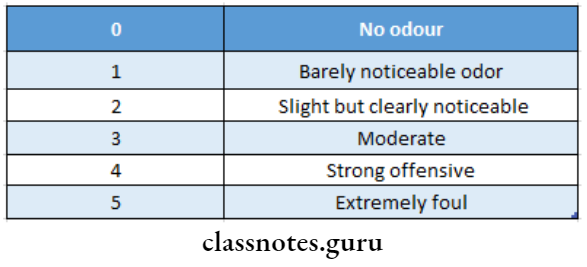
Miscellaneous Short Essays

Question 1. Oral Candidiasis.
Answer:

  • It is a fungal infection occurring in the oral cavity

Oral Candidiasis Causative Organisms:

  • Candida albicans
  • C. Krusei
  • C. pseudotropicalis

Oral Candidiasis Features:

  • Oral thrush
  • Mucosa appears as a thick, white soft plaque
  • However, it can be wiped off easily
  • Method Of Preparation:

Oral Candidiasis Treatment:

  • Topical and systemic administration of nystatin.

Question 2. Night guards
Answer:

  • A night guard is a removable appliance that is worn by the patient during sleep, to protect his teeth and jaws from the damage that is caused by the habit.
  • It is not necessarily a treatment procedure because the grinding habit is not exactly resolved, but the wearing of the appliance reduces the harmful effects of teeth grinding.

Night guards Types:

  1. A ready-made appliance that comes in standard sizes;
  2. A boil-and-bite appliance that offers some customization; and
  3. Custom-made appliances that dentists prefer and commonly prescribe. Custom dental night guards are different and rather effective,

Night guards Method of preparation:

  • Step 1: Impression Taking.
  • Step 2: Night Guard Fabrication.
  • Step 3: Delivery. As soon as the night guards have been fabricated, they are delivered to the patients along with proper instructions for wear and care.

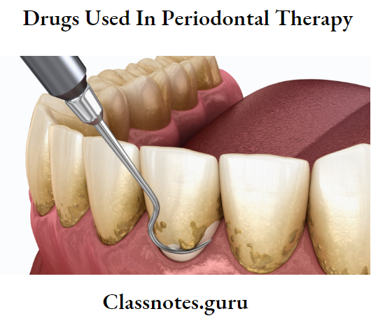
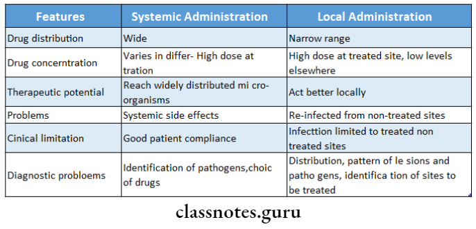
Question 3. Full mouth disinfection
Answer:

  • Full mouth disinfection refers to an intense course of treatment for periodontitis typically involving scaling and root planing in combination with adjunctive use of local antimicrobial adjuncts to periodontal treatment such as chlorhexidine mouthwash
  • It consists of organisms, keratotic debris, and inflammation. It is a technique to eliminate or reduce the pathogenic cells, desquamated cells bacteria from the buccal habitats which can provoke the reinfection of the places already treated

Full mouth disinfection Aims

  • Complete and simultaneous elimination of all the bacteria in the periodontal sacs within less than 24 hours

Full mouth disinfection Steps

  • Periodontal probing and diagnosis
  • Hygiene instructions
  • Removal of supra and subgingival plaque with ultrasound
  • Radicular scraping and smoothing with Gracey curettes

Read And Learn More: Periodontics Question and Answers

  • Radicular scaling with preset
  • Brushing of the back of the tongue for 60 seconds with 0.12% chlorhexidine gel
  • Mouthwashes with 0.12% chlorhexidine
  • Subgingival irrigation of periodontal pockets above 5 mm with chlorhexidine gel

Question 4. Subepithelial connective tissue graft.
Answer:

  • As described by Langer and Langer

Tissue graft Indications:

  • Larger and multiple defects
  • Defects with good vestibular depth
  • Defects with an adequate gingival thickness

Tissue graft Surgical procedure:

  • Raise a partial thickness flap with a horizontal incision 2 mm away from the tip of the papilla and 2 vertical incisions 1-2 mm away from the gingival margin
  • Extend the flap to the mesiobuccal fold
  • Thorough root planning is done
  • Obtain connective tissue graft from the palate through a horizontal incision from the gingival margin of molars and premolars
  • A palatal wound is sutured by primary closure
  • Place the connective tissue graft over a denuded root and suture it
  • The graft is covered by the partial thickness flap and sutured interdentally
  • The graft is protected by surgical dressing
  • After 7 days sutures and dressing is removed

Miscellaneous Short Question And Answers

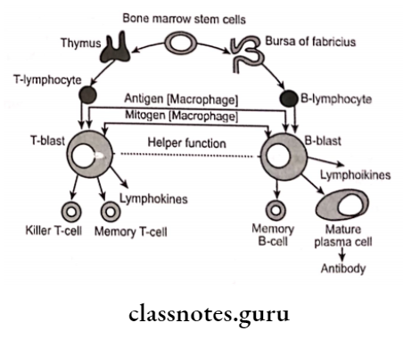
Question 1. Active Immunity.
Answer:

  • It is the immunity that an individual develops as a result of infection or by specific immunization
  • It may be acquired in 3 ways
    • Following clinical infection – Ex. Chickenpox
    • Following subclinical infection – Ex. Polio
    • The following immunization
  • It is superior to passive immunity

Question 2. Define.
Answer:

  1. Neoplasia: A mass of tissue formed as a result of the abnormal, excessive, uncoordinated, autonomous, and purposeless proliferation of cells.
  2. Hypertrophy: It is an increase in the size of cells re- resulting in the enlargement of the organ or tissue, without any change in the number of cells.
  3. Hyperplasia: It is an increase in the number of parenchyma cells resulting in the enlargement of the organ or tissue.

Question 3. Stillman’s Clefts.
Answer:

  • Apostrophe shaped indentation

Extend of Stillman’s Clefts: Marginal gingiva to varying depth

Clefts Site of Stillman’s Clefts: Facial surface

Clefts Margins of Stillman’s Clefts: Rolled underneath linear gap in the gingiva

Clefts Size of Stillman’s Clefts: Blunt over remaining gingiva

  • Slight to 5-6 mm.

Cause of Stillman’s Clefts: Occlusal trauma

  • Pathological pockets

Stillman’s Clefts Treatment: Repair spontaneously

Stillman’s Clefts Types:

  1. Simple: Occurs in a single direction
  2. Complex: Occurs in more than one direction

Question 4. McCall’s Festoons.
Answer:

  • Life-preserver-shaped enlargements of the marginal gingiva.
  • Site: Canine and premolar areas on the facial surface

McCall’s Festoons Features:

  • Initially – Normal gingiva
  • Accumulation of food debris leads to secondary in-inflammatory changes

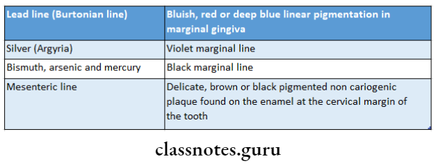
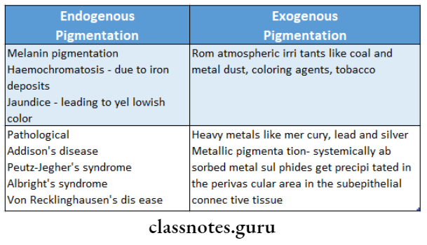
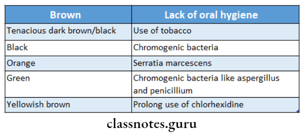
Question 5. Gingival Pigmentations.
Answer:

  • By therapeutic use or occupational environment
  • Various metal causes different pigmentation.
    • Lead Bluish red linear pigmentation of the marginal gingiva
    • Silver – Violet marginal line
    • Results from perivascular precipitation of metallic sul- fides in the subepithelial connective tissue.

Gingival Pigmentations Sites:

  • Areas of inflammation
  • Irritated mucosal areas

Gingival Pigmentations Treatment:

  • Elimination of local irritating factors
  • Restoration of tissue health
  • Topical application of concentrated peroxide
  • Insufflation of the gingiva with oxygen to oxidize the dark metallic sulfides

Question 6. Dentoalveolar Ablation.
Answer:

  • Forceful frictional action between the oral soft tissue tissues.

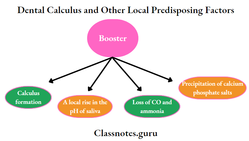
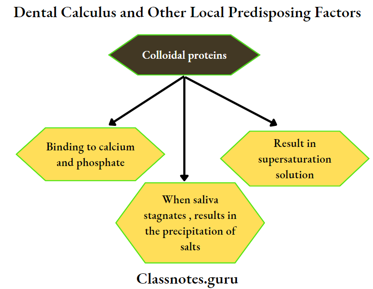
Normal – Salivary pH, buffering capacity, and calcium and phosphorous content

Elevated – mucin level

Feature Sharply defined wedge-shaped depression in the cervical area of the facial tooth surface

Cause – Decalcification by acid beverages.

  • The combined effect of salivary secretion and friction

Question 7. Fremitus test and tension test.
Answer:

Tension test Fremitus Test:

  • Diagnostic for TFO cases

Tension test Procedure:

Miscellaneous Fremitus test and tension test

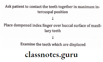
Tension test result:

  • Class 1 – Mild vibration
  • Class 2 – Easily palpable but no visible movements
  • Class 3 Movements visible with the naked eye

Tension Test:

  • Diagnostic to measure the width of attached gingiva

tension test Procedure:

Miscellaneous Procedure

tension test Result:

  • If the free gingival margin moves then the attached gingiva is considered inadequate.

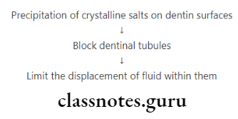
Question 8. Name 2 desensitizing agents. Write about the mode of action of anyone.
Answer:

Agents:

  • Dentrifices
  • Varnishes
  • Fluoride compounds

Mode Of Active

Miscellaneous mode of action

Question 9. Osteoplasty and Osteotomy.
Answer:

Osteotomy Osteoplasty:

  • Reshaping of the alveolar process to achieve a more physiologic form without the removal of supporting bone
  • Reshaping the bone without removing tooth-supporting bone

Osteotomy:

  • Includes removal of tooth-supporting bone
  • Place dampened index finger over the buccal surface of the maxilla
  • It is defined as the excision of bone/portion of bone
  • It is done to correct/reduce deformities caused by pe- periodontitis and includes the removal of the supporting bone

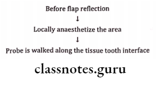
Question 10. Transgingival Probing.
Answer:

Transgingival Probing Use:

  • To detect alveolar bone los
  • Done under local anesthesia
  • Confirms the extent and configuration of infrabony component of the pocket and of furcation defects

Transgingival Probing How To Use:

Miscellaneous How to use

Question 11. Attrition
Answer:

Attrition Definition:

  • Loss by wear of the surface of tooth or restoration caused by tooth to tooth contact during mastication or para-function

Attrition Clinical Features:

  • Wear on occluding surfaces
  • Shiny facets on amalgam contacts
  • Fractures of cusps or restorations

Attrition Types:

  1. Physiological-Causes
    • Old age- increasing age causes attrition
  2. Pathological-causes
    • Abnormal occlusion
    • Premature extraction of teeth
    • Abnormal chewing habits
    • Structural defects in teeth

Question 12. Hydroxyapatite
Answer:

  • It is calcium phosphate biomaterials used for grafting

Hydroxyapatite Advantages:

  • Excellent tissue compatibility
  • Doesn’t cause any inflammation or foreign body re- response
  • Act as a scaffold for blood clots
  • Allows bone formation

Hydroxyapatite Disadvantage:

  • It is encapsulated by collagen

Question 13. Supracontacts
Answer:

  • The gradual movement of teeth as a result of changes, such as a loss of a tooth or wear, can cause “supra contacts”.
  • This is where the upper and lower teeth meet at this point first and causes the opposing teeth not to meet evenly.
  • The jaw tries to grind away this contact that is in the way and may lead to excessive muscle activity and jaw joint dysfunction.
  • Very thin marking paper, called articulating paper, can highlight the contacts of the teeth and an adjustment may be made to allow a more even bite or allow the jaw to move more freely.
  • Three separate assessments are made:
  1. Occlusal assessment:
  2. Muscle assessment:
    • Within the mouth
    • Outside the mouth
    • Neck muscles
    • Measuring the amount of opening of the jaw and the various movements of the jaw and neck.
  3. Joint assessment

Question 14. Measurements of tooth mobility
Answer:

1. Miller mobility index:

  • Grade 0- Normal tooth mobility
  • Grade 1- slightly more than normal
  • Grade 2- Moderately more than normal
  • Grade 3- Severe mobility faciolingually

2. Grace and Smales mobility index:

  • Grade 1- Mobility <1 mm buccolingually
  • Grade 2- Mobility 1-2 mm Buccolingually
  • Grade 3- Mobility >2 mm buccolingually and/or vertical tooth mobility

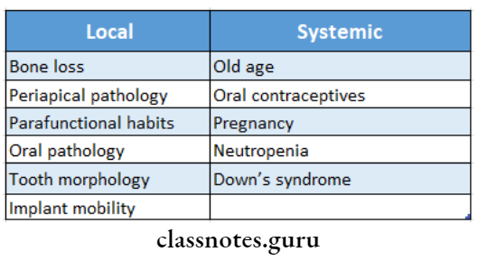
Question 15. Causes of tooth mobility
Answer:

  • Alveolar bone loss
  • Extension of inflammation to periodontal region
  • Trauma from occlusion
  • Hormonal changes
  • Cysts and tumors involving the maxilla and mandible
  • Periodontal surgery

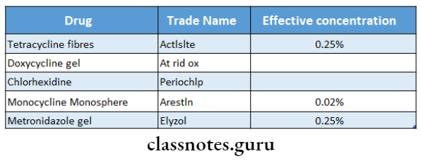
Question 16. Priceline
Answer:

  • Periocline contains a therapeutic agent which acts as a first-line defense in the treatment of moderate to severe chronic adult periodontal disease
  • The active ingredient in it is minocycline
  • It binds to the surface of the tooth and is gradually re- leased over time to provide simple adjunctive treatment for periodontitis

Question 17. Bicuspidization
Answer:

  • Section of the root along with the respective crown portion

Bicuspidization Indications:

  • Periodontal disease
  • Severe bone loss
  • Untreatable roots
  • Root fracture

Bicuspidization Contra-Indications:

  • Fused roots
  • Lack of Osseous support
  • Uncooperative patients

Dental Implants Periodontics Short And Long Essay Question And Answers

Dental Implants Definitions

Dental Implants

  • It is an integral component of the oral implant complex, which also consists of supportive bone, interposed keratinized and mucosal oral soft tissues, and prosthetic superstructure.

Structure of dental Implants:

Dental Implants Periodontics Structure Of Dental Implants

Peri-implantitis

  • It is a progressive peri-implant bone loss in conjunction with soft tissue inflammatory lesions.

Osseointegration

  • It is defined as the direct structural and functional connection between ordered, living bone and the surface of a load-bearing implant without intervening soft tissues

Osteoconduction.

  • It is an effect by which the matrix of the graft forms a scaffold that favors outside cells to penetrate the graft and form new bone

Osteoinduction

  • A process by which graft material is capable of promoting cementogenesis, osteogenesis, and new periodontal ligament

Dental Implants Important Notes

1. Classification of implants

  • Based on shape and forms
    • Endosteal
    • Subperiosteal
    • Transosteal
    • Intramucosal
    • Endodontic

Dental Implants Solid tapering

  • Based on Surface characteristic
    • Titanium plasma sprayed coating
    • Sand blasting-surface etching
    • Laser-induced surface roughening
    • Hydroxyapatite coating

Dental Implants Long Essays

Question 1. Define Osseointegration and classify dental. implants. Add a note on the maintenance of im- plants.
Answer:

Classify dental Osseointegration

classify dental Definition:

  • It is defined as the direct structural and functional connection between ordered, living bone and the surface of a load-bearing implant without intervening soft tissue
  • It is a fundamental requirement and essential component for implant success

classify dental Maintenance Of Implants

1. At the office

  • The patient is recalled at regular intervals to provide optimal preventive services
  • Probing pocket depth and mucosal margins are notes
  • Radiographic crestal bone levels are established
  • Evaluation is done initially after 1 day, 1 month, 3 months, 6 months, and then at yearly intervals
  • At every visit following evaluation is done
    • Evaluation of soft tissue around the implant
    • Implant mobility
    • Prosthesis

Read And Learn More: Periodontics Question and Answers

    • Radiograph
    • Plaque and calculus scores
  • Debridement of the implant should be carried out with plastic instruments that do not damage the implant surface
  • A rubber cup can be used to polish the implant surface with a non-abrasive toothpaste, fine polishing paste, or tin oxide

2. Home care methods

  • A soft circular toothbrush is used around implant restoration
  • Powered and sonic toothbrushes are used around titanium implants
  • Foam tips, interproximal brushes, and disposable wooden picks are used for plaque removal from embrasures
  • Chemical antiplaque agents can be delivered with interdental aids
  • Every implant patient requires lifetime careful maintenance to ensure implant health and longevity

Question 2. Dental implants.
Answer:

  • It is an integral component of the oral implant complex, which also consists of supportive bone, interposed keratinized and mucosal oral soft tissues, and prosthetic superstructure.

Dental implants Material Used:

Dental implants Metals:

  • Stainless steel
  • Gold
  • Titanium
  • Tantalum
  • Zirconium

Dental implants Ceramics:

  • Aluminium oxide
  • Titanium oxide

Dental implants Calcium Phosphate:

Bioactive and Biodegradable Ceramic:

  • Hydroxyapatite
  • Bioglass
  • Carbon

Dental implants Polymers:

  • Polymethyl methacrylate
  • Polytetrafluoroethylene
  • Polyethylene
  • Polypropylene

Dental implants Indications:

1. Edentulous patient:

  • Edentulous mandible
  • Edentulous maxilla

2. Partially edentulous:

  • Free end edentulous
  • Multiple missing

3. Single tooth loss:

Dental implants Contraindications:

  • Diseases
  • Uncontrolled DM
  • Malignancy
  • Disease of CT
  • Blood dyscrasia
  • Malignancy
  • Psychologic disorders
  • Iatrogenic
    • Immunosuppressant therapy
    • Drug addiction
    • Radiation to jaw

Dental implants Procedure:

Dental Implants Dental Implants Procedure

Dental Implants Short Essays

Question 1. Perl-Implantis.
Answer:

  • It is a progressive peri-implant bone loss in conjunction with soft tissue inflammatory lesions.
  • It begins at the coronal portion of the implant.

Perl-Implantis Clinical Features:

  • Erythema over the area
  • Bleeding on probing
  • Pocket formation
  • Bone destruction
  • Suppuration
  • Presence of calculus
  • Tooth mobility present

Perl-Implantis Diagnosis:

  • Bone loss
  • Clinical attachment loss

Perl-Implantis Management:

  • Occlusal therapy – Occlusal correction
  • Anti-infective therapy
  • Scaling and root planning
  • 0.12% chlorhexidine

Perl-Implantis Surgical Techniques:

  • Correction of bone defects
  • Re-osseointegration
  • Maintenance
  • Recall visits planned for at least every 3 months

Question 2. Osseointegration
Answer:

Osseointegration Definition:

  • It is defined as the direct structural and functional connection between ordered, living bone and the surface of a load-bearing implant without intervening soft tissues
  • It is a fundamental requirement and essential component for implant success

Osseointegration Process:

Dental Implants Osseintegration

Osseointegration Requirements:

  • Immobility of implant relative to the bone
  • Avoid excessive occlusal forces
  • Proper vascular supply and oxygen tension
  • A strict aseptic technique should be maintained
  • Profuse irrigation during drilling

Osseointegration Significance:

  • Once osseointegration is achieved, implants can resist and function under occlusal forces for many years

Question 3. Failures of implants
Answer:

 Failures Of Implants Are Due To:

1. Improper patient selection:

  • Patients who are unmotivated to control plaque
  • Chronic smokers
  • Patients with systemic conditions like uncontrolled diabetes
  • Patients with insufficient quality and quantity of A process by which graft material is capable of promoting- bone to support the implant fixture

2. Surgical complications:

  • Oversized osteotomy site preparation
  • Broken burs
  • Improper angulation
  • Inappropriate instrumentation
  • Perforation
  • Hemorrhage
  • Inadequate amount of soft tissue

3. Complication in early stages:

  • Postoperative infections
  • Dysaesthesia
  • Dehiscence
  • Sinusitis
  • Radiolucencies
  • Mobility

4. Late failures:

  1. Mechanical complications
    • Occlusal overloading
    • As osseointegrated implants have no periodontal ligament, the adverse forces generated by occlusal activity may lead to high stress and microfractures in the coronal bone to im- plant contact
    • Abutment screw fracture
  2. Biological complication
    • They are bacterial in origin
    • Characterized by bone loss combined with soft tissue inflammatory response that- demonstrates suppuration with probing depth greater than 6 mm
    • Presence of pockets, bleeding on probing, and purulence

Question 4. Osteoinduction, osteoconduction, osseointe-gradation
Answer:

Osteoconduction:

  • It is an effect by which the matrix of the graft forms a scaffold that favors outside cells to penetrate the graft and form new bone

Osteoinduction:

  • A process by which graft material is capable of promoting cementogenesis, osteogenesis, and new periodontal ligament

Osseointegration:

  • It is defined as the direct structural and functional connection between ordered, living bone and the surface of a load-bearing implant without intervening soft tissues
  • It is a fundamental requirement and essential component for implant success

Question 5. Maintenance of dental implants
Answer:

1. At the office:

  • The patient is recalled at regular intervals to provide optimal preventive services
  • Probing pocket depth and mucosal margins are notes
  • Radiographic crestal bone levels are established
  • Evaluation is done initially after 1 day, 1 month, 3 months, 6 months, and then at yearly intervals At every visit following evaluation is done
    • Evaluation of soft tissue around the implant
    • Implant mobility
    • Prosthesis
    • Radiograph
    • Plaque and calculus scores
  • Debridement of the implant should be carried out with plastic instruments that do not damage the implant surface
  • A rubber cup can be used to polish the implant sur- face with a non-abrasive toothpaste, fine polishing paste, or tin oxide

2. Home care methods:

  • A soft circular toothbrush is used around implant restoration
  • Powered and sonic toothbrushes are used around
  • Foam tips, interproximal brushes, and disposable wooden picks are used for plaque removal from embrasures
  • Chemical antiplaque agents can be delivered with interdental aids
  • Every implant patient requires lifetime careful maintenance to ensure implant health and longevity

Question 6. Biology of soft tissue around an implant.
Answer:

  • Mucosal tissues around intraosseous implants form a tightly adherent band consisting of dense collagenous lamina propria
  • It is covered by keratinized stratified squamous epithelium
  • Implant epithelium junction is similar to junctional epi-thelium
  • The epithelial cells are attached to the titanium implant employing hemidesmosomes and basal lamina
  • A biologic seal exists between epithelial cells and implant
  • Sulcus forms around implant lined with sulcular epithelium that is continuous apically
  • Collagen fibers are non-attached and run parallel to the implant surface

Dental Implants Short Answers

Question 1. Home care methods for implant maintenance.
Answer:

  • A soft circular toothbrush is used around implant restoration
  • Powered and sonic toothbrushes are used around titanium implants
  • Foam tips, interproximal brushes, and disposable wooden picks are used for plaque removal from embrasures
  • Chemical antiplaque agents can be delivered with inter-dental aids
  • Every implant patient requires lifetime careful maintenance to ensure implant health and longevity

Question 2. Implant-bone interface
Answer:

  • The relationship between implant and bone involves mechanisms like

1. Fibro-osseous integration:

  • When soft tissues are interposed between surface and bone, it is known as fibro-osseous integration.

2. Osseointegration:

  • It is defined as the direct structural and functional connection between ordered, living bone and the surface of a load-bearing implant without intervening soft tissues
  • It is a fundamental requirement and essential component for implant success

3. Biointegration:

  • It is achieved in cases where the implant is coated with bioactive materials like hydroxyapatite
  • These materials stimulate bone formation

Dental Implantsl Implant Bone Interface

Dental Implants Viva Voce

  1. The epithelial cell attaches with the implant surface by hemidesmosomes
  2. The probing depth of 3 mm with no bleeding around 15. The minimum amount of bone required all around an implant is presumed to be healthy
  3. The arrangement of collagen fibers around an implant is parallel and unattached
  4. Functional ankylosis is a synonym for osseointegration
  5. The critical temperature of bone cells is 47°C for 1 min
  6. Osteoclasts are derived from monocytes
  7. The oxide layer on the titanium surface mainly contains titanium dioxide
  8. The probable reason for poorer clinical outcomes with implants in the posterior maxilla is the deficiency in primary stability
  9. Periotest device is used for detecting the mobility of both the implants and teeth
  10. The vascular supply of the peri-implant gingival tissue is less than that of gingival tissue around teeth
  11. The inflammatory reaction in the soft tissues around implants is regarded as periimplantitis
  12. The term osseoperception refers to tactile sensitivity at the bone-implant surface
  13. The noninvasive method to assess the implant mobility is the resonance frequency analyzer
  14. An absolute contraindication for implant therapy is radiotherapy of the head and neck
  15. The minimum amount of bone required all around an implant after its placement is 1-1.5 mm
  16. Modification of implant surfaces using the additive technique includes HA coating and oxidative process
  17. The subtractive implant surface modifications include acid itching and sandblasting
  18. The recommended speed for drilling the surgical site for implant placement is 800-12

Questionnaire Periodontics Short And Long Essay Question And Answers

Questionnaire Short Question and Answers

Question 1. Tooth mobility.
Answer:

Tooth mobility Causes:

Questionaire Tooth mobility

Tooth mobility Grading:

Grade 1: Horizontal mobility of about 0.2-1mm

Grade 2: Horizontal mobility of about more than 1 mm

Grade 3: Horizontal and vertical mobility of teeth.

Question 2. Inter-dental Alds.
Answer:

Type 1:

  • Interdental papilla completely fills the embrasure space
  • Dental floss can be used

Type 2:

  • Mild loss of inter-dental papilla
  • Miniature bottle brushes are advised

Type 3:

  • No proximal contact present
  • Unitufted brushes are used

Question 3. Retrograde Periodontitis.
Answer:

  • Periodontitis can also be caused by pulpal infections that have entered the periodontal ligament either through the apical foramen or through the lateral canal.
  • Such a lesion is termed retrograde periodontitis

Question 4. Wasting Diseases.
Answer:

1. Attrition:

  • It is the occlusal wear resulting from functional contact with opposing teeth

Questionaire Attrition of mandibular teeth

2. Abrasion:

  • It refers to the loss of tooth substance induced by mechanical wear other than that of mastication.

Questionaire Abrasion

Read And Learn More: Periodontics Question and Answers

3. Erosion:

  • It is a sharply defined wedge-shaped depression in the cervical area of the facial tooth surface.

Questionaire Erosion of maxillary lateral incisor

Drugs Used In Periodontal Therapy Short Essay Question And Answers

Drugs Used In Periodontal Therapy Short Essays

Question 1. Why antibiotics are not routinely used in periodontal therapy?
Answer:

Periodontal therapy:

Drugs Used In Periodontal Therapy Perodontal therpy image

Periodontal Therapy Uses Of Antibiotics:

  • Reduce/eliminate bacteria
  • Retards bone loss
  • Reduce/Eliminate the need for surgery
  • Useful in aggressive periodontitis

periodontal therapy Not Used Routinely:

  • Despite the above use, systemic administration is not recommended routinely as
    • It produces systemic effects
    • Disturbs the functioning of various systems of the body, such as GIT
    • Certain drugs are contraindicated in certain conditions like pregnancy
    • Besides, this systemic administration is useless unless there is plaque and calculus removal.
    • The presence of a thick band of calculus prevents the penetration of the drug into the site
  • Thus, it is used only as an adjunctive.

Question 2. Tetracycline in periodontics
Answer:

  • Tetracycline are widely used drugs in the treatment of periodontal diseases

Tetracycline in Periodontics Clinical Use:

  • It is used as an adjunct in the treatment of localized aggressive periodontitis
  • A contains is a frequent microorganism associated with localized aggressive periodontitis and is tissue invasive, Therefore mechanical removal of calculus and plaque from root surfaces may not eliminate this bacterium from periodontal tissues
  • Systemic tetracycline in conjunction with scaling and root planning can
    • Eliminate tissue bacteria
    • Arrest bone loss
    • Suppresses A.a. comitans
    • Allows mechanical removal of root surface deposits and elimination of pathogenic bacteria from within tissues

Read And Learn More: Periodontics Question and Answers

Tetracycline in periodontics Actions:

  • Has the ability to concentrate in periodontal tissues
  • Inhibits growth of A.a. contains
  • Exerts anti-collagenase effect
  • Inhibits tissue destruction
  • Aids in bone Regeneration

Tetracycline in periodontics Dose:

  • 250 mg Qid

Tetracycline in Periodontics Side Effects:

  • GI disturbances
  • Photosensitivity
  • Hypersensitivity
  • Increased blood urea nitrogen
  • Dizziness, headache
  • Blood dysplasias
  • Tooth discoloration in children

Question 3. LDD (Local Drug Delivery).
Answer:

LDD Advantages:

  • Greater concentrations of drug at the site
  • Slow release of drug
  • Direct effect on the area
  • Reduced systemic effects

LDD Contraindications:

  • Allergic to drug
  • Children below 10 years

LDD Drugs Used:

Drugs Used In Periodontal Therapy Drugs used

Question4. Methods of Delivery.
Answer:

1. Keye’s technique:

  • Apply slurry of sodium bicarbonate and hydrogen peroxide over tooth brush
  • Tooth brushing

Limitation: Does not reach periodontal pocket

2. Root Bio-modification:

  • Application of root conditioner during surgery

Methods of Delivery Effects:

  • Prevents long junctional epithelium
  • Improves healing

Methods of Delivery Agents Used:

  • Tetracycline
  • Citric acid pH1
  • Fibronectin

3. Irrigation:

Methods of Delivery Types:

  • Home Irrigation
    • Supra gingival
    • Subgingival
    • Marginal
  • Professional Irrigation
    • It delivers medicament into the periodontal pockets via irrigation devices

Question 5. Compare local and systemic drug delivery systems.
Answer:

Drugs Used In Periodontal Therapy Compare local and systemic drug dellivery system

Question 6. Metronidazole in periodontal therapy
Answer:

  • Metronidazole is a nitroimidazole compound used to treat protozoal infections

Metronidazole  Spectrum Of Activity:

  • Effective against
    • A. contains
    • P. gingivalis
    • P. intermedia

Metronidazole Uses In Periodontics:

  • To treat
    • Gingivitis
    • Acute necrotizing ulcerative gingivitis
    • Chronic periodontitis
    • Aggressive periodontitis
  • A single dose of metronidazole appears in both serum and GCF
  • When administered systemically, it reduces the growth of anaerobic flora
  • Used as a supplement to rigorous scaling and root planning Subgingival use
    • A dental gel containing metronidazole benzoate is used
    • It gets converted into an active substance by esterases in GCF

Adverse Effects:

  • GIT effects
    • Nausea, anorexia, abdominal pain, metallic taste in the mouth, looseness of stool
    • Headache, stomatitis, glossitis, dryness of mouth, furry tongue, dizziness, rashes, neutropenia, insomnia
    • Prolonged use causes peripheral neuropathy High doses cause convulsions

Drugs Used In Periodontal Therapy Short Question And Answers

Question 1. Advantages of LDD
Answer:

  • Greater concentration of drug at the site
  • Slow release of drug
  • Direct effect on the area
  • Reduced systemic effects

Question 2. Periochip
Answer:

  • It is a small chip composed of a biodegradable hydrosol-lazed gelatin matrix cross-linked with glyceraldehyde
  • It also contains glycerin and water
  • 2.5 mg of chlorhexidine is incorporated into it
  • It slowly releases chlorhexidine and maintains drug concentration in gingival crevicular fluid for at least 7 days
  • Size of chip: 4*5*0.35 mm

Question 3. Keye’s technique
Answer:

  • It refers to the application of a slurry of sodium bicarbonate and hydrogen peroxide over the toothbrush
  • Tooth brushing of it is done

Keye’s technique Limitation:

It does not reach the periodontal pocket

Question 4. Activity
Answer:

  • Among tetracycline-releasing devices, the most widely. It should be selective and effective against micro- used is activity periodontal fiber
  • It is a monolithic thread of a biologically inert, non-than retard resorbable plastic copolymer containing 25% tetracycline hydrochloride powder
  • The fiber is packed into a periodontal pocket secured with a thin layer of cyanoacrylate adhesive and left in place for 7–12 days
  • Due to the continuous delivery of tetracycline, a local concentration of active drug in excess of 1000 mg/l can be. Maintained throughout the period

Activity Effects:

  • Decreases pocket depth
  • Increases attachment levels
  • Decreases bleeding tendency

Question 5. Define antiseptic and antibiotics
Answer:

Antiseptic:

  • Antiseptic is an agent that destroys microorganisms and can be used on living tissues

Antibiotics:

  • An antibiotic is a chemical substance produced by microorganisms that have the capacity to inhibit the growth or kill another organism in a dilute solution

Question 6. Arestin
Answer:

  • Ares tin is a locally delivered, sustained-release form of minocycline microsphere
  • It is used for subgingival placement as an adjunct to scaling and root planning
  • 2% minocycline is encapsulated into bioresorbable mi- mi-microspheres in a gel carrier

Ares tin Effects:

  • Increase in clinical attachment level in patients with pockets of 6 mm or greater
  • Reduction in probing depth
  • It should destroy microorganisms rather

Question 7. Properties of ideal antibiotics
Answer:

  • It should be selective and effective against microorganisms without injuring the host
  • It should destroy microorganisms rather than retard their growth
  • It should not become ineffective as a result of bacterial resistance
  • It should not be inactivated by enzymes, plasma pro- teens or body fluids
  • It should quickly reach bactericidal levels in the entire body and be maintained for long periods
  • It should have minimal side effects

Drugs Used In Periodontal Therapy Viva Voce

  1. Metronidazole belongs to nitroimidazole
  2. The minimum effective concentration of tetracycline needed in GCF is 2-4 μg/m
  3. The mechanism of action of metronidazole is to disrupt bacterial DNA synthesis
  4. The mechanism of action of penicillin is it inhibits bacterial cell wall production
  5. Penicillin is bactericidal
  6. Pseudomembranous colitis with diarrhea or cramping is a side effect of clindamycin
  7. All strains of A.a.comitans are susceptible to ciprofloxacin
  8. The mechanism of action of erythromycin is it inhibits protein synthesis by binding to the 50S ribosomal subunit
  9. Atridox is used for subgingival delivery of doxycycline

Occlusal Evaluation And Therapy In The Management Of Periodontal Disease Short Essay Question And Answers

Occlusal Evaluation And Therapy In The Management Of Periodontal Disease Long Essays

Question 1. Enumerate occlusal evaluation procedures.
Answer:

Clinical Occlusal Evaluation Procedures:

1. TMJ screening examination

  • TMJ screening evaluation includes
    1. Interincisal opening
      • Interincisal distance is recorded in millimeters
    2. Opening/closing pathway
      • Any deviations from the midline path are noted
    3. Temporomandibular joint sounds
      • Clicking or crepitus is noted
    4. Temporomandibular joint tenderness
      • Bilateral palpation over condyles is examined
    5. Muscle tenderness
      • Masseter, pterygoid and temporal muscles are examined

2. Intraoral evaluation of occlusion

  • It includes
    • Identification of occlusion in maximum inter-occupation or intercuspal position
      • The patient is asked to close into the maximum inter-cuspal position
      • The presence or absence of contacts is examined
    • Excursive movement
      • The patient is asked to move into right and left excur- sions and observe tooth contact patterns
    • Initial contact in centric relation
      • Guide patient’s mandible in centric relation
      • Record any deflection present
    • Tooth mobility
      • Mobility of the tooth is recorded
    • Attrition
      • It is defined as wear caused by tooth-to-tooth contact
      • Significant attrition of teeth indicates bruxism

3. Role of articulated casts

  • Identifies occlusal contacts that can deflect mandible, deflect mobile teeth or cause trauma to teeth
  • Localizes wear facets, trial occlusal adjustment

Read And Learn More: Periodontics Question and Answers

Question 2. Describe the steps of occlusal adjustments.
Answer:

  • Occlusal adjustments also called occlusal equilibration or coroplast is the selective reshaping of occlusal surfaces with the goal of establishing a stable, non-traumatic occlusion

Steps:

1. Removal of retrusive prematurities

  • Remove retrusive prematurities and eliminate the deflective shift from retruded contact position to in a tra-occlusal position
  • Retrusive prematurities are located on the mesial inclines of maxillary cusps and distal inclines of mandibular cusps

2. Adjustments of the intercuspal position

  • Adjustment of intra-occlusal position relieves su- pra contacts and achieves occlusal stability
  • It can be achieved by reducing in size of the cusp and deepening the fossa

3. Test for excessive contact on incisive teeth in intra- cuspal position

  • Contact relationship may be tested with Mylar strips and checking for fremitus
  • Supracontacts are marked and reduced

4. Remove posterior protrusive supra contacts

  • Obtain bilateral protrusive movement
  • Prematurities are corrected by grinding maxillary teeth

5. Correct prematurities on the balancing side

  • Prematurities on the balancing side are corrected next
  • It is present on the inner inclines of mandibular buccal cusps and the inner inclines of maxillary lingual cusps of the first and second molars

6. Reduce supra-contacts on the working side

  • Reduce supra contacts on laterotrusive side
  • They are reduced by reducing inclines of buccal up-per and lingual lower cusps (BULL)

7. Elimination of undesirable gross occlusal features

  • Extruded teeth, plunger cusp, uneven marginal ridges of adjacent teeth, rotated or malposed teeth, occlusal wear facets, etc are corrected

8. Recheck the occlusal contact relationship in all positions

  • Recheck occlusal contact relationship in all positions

9. Finishing and polishing

  • Adjusted surfaces of the teeth are smoothened and polished

Occlusal Evaluation And Therapy In The Management Of Periodontal Disease Short Essays

Question 1. Indications and steps of coroplast
Answer:

Indications:

  • Trauma-occlusal trauma
  • TMJ problems
  • After the elimination of gingival and infra bony pockets

Steps:

  1. Removal of retrusive prematurities
  2. Adjustments of the intercuspal position
  3. Test for excessive contact on incisive teeth in intra- cuspal position
  4. Remove posterior protrusive supra contacts
  5. Correct prematurities on the balancing side
  6. Reduce supra contacts on the working side
  7. Elimination of undesirable gross occlusal features
  8. Recheck the occlusal contact relationship in all positions
  9. Finishing and polishing

Question 2. Effects of orthodontic treatment on periodontal tissues
Answer:

Effects Of Orthodontic Treatment On Perio- Dental Tissues:

1. Iatrogenic effects associated with orthodontic treatment

  • Orthodontic treatment may cause injuries to the teeth and periodontium in most of the cases
  • Usually, these changes are reversible, and regeneration and repair of the tooth structures and period-dental tissues can occur while in some cases the changes may result in irreparable damage

2. Root resorption

  • During orthodontic therapy some amount of root resorption is unavoidable

3. Effects of orthodontic bands on the periodontium

  • Gingivitis and gingival hyperplasia are short-term effects
  • Long-term effects are loss of attachment, root resorption, or no effects

4. Effects of orthodontics on dentition with normal height of attachment apparatus

  • Orthodontic forces cause no damage to the supra- Time of treatment: alveolar connective tissue

Question 3. The rationale for orthodontic tooth movement in periodontal therapy
Answer:

Rationale For Orthodontic Treatment:

1. Reducing plaque retention:

  • Crowded teeth and mesially inclined teeth create plaque accumulation sites that are difficult to clean
  • Crowding creates enlarged contact surfaces and al-tiered embrasure spaces that are displaced apically

2. Improving gingival and osseous form:

  • There is an interrelation between the position of the tooth, the shape of the gingiva, and the bone that surrounds it
  • Orthodontic treatment may improve the shape of the periodontium and reduces the need for bone surgery

3. Facilitating prosthetic replacements:

  • The uprighting of tilted abutment teeth may be im- important for a better-contoured crown which will benefit the surrounding periodontal condition

4. Improving esthetics:

  • Correction of pathologic tooth migration and di-asthma between anterior teeth results in improved esthetics

Occlusal Evaluation And Therapy In The Management Of Periodontal Disease Short Answers

Question 1. Coronoplasty.
Answer:

  • The procedure of selective reshaping of the occlusal surface with the goal of establishing a stable – nontraumatic occlusion.

It Is an Invasive Procedure:

Goals:

  • Reshaping crown surface
  • Elimination of supra contacts
  • Creation of stable occlusion

Indications:

  • Trauma-occlusal trauma
  • TMJ problems

Time of treatment:

  • After the elimination of gingival inflammation and periodontal pockets

Question 2. Forced eruption
Answer:

  • Forced eruption applies to procedures that involve orthodontic movement with gentle forces
    The purpose is the coronal shift of the bone at the base of infrabony defects, thus reducing the depth of the de-fact
  • The elongated tooth thereafter can be reduced in height by grinding and elimination of the infra-bony pocket Forced eruption can also be done to manage teeth that have fractures to make possible the restoration of the tooth
  • To erupt the tooth forcefully, either the adjacent teeth must be bracketed and a wire placed or a wire must be bonded directly to the adjacent teeth and an elastic trac- tion applied from the wire to the tooth

Occlusal Evaluation And Therapy In The Management Of Periodontal Disease Viva Voce

  1. The location of bands and brackets determines the outcome of orthodontic therapy
  2. Orthodontic brackets on the posterior teeth are positioned relative to the marginal ridges and cusps
  3. Orthodontic brackets on the anterior teeth are positioned relative to the incisal edges
  4. The tooth should erupt 4 mm orthodontically for the purpose of restoration if a tooth fracture extends to the level of alveolar bone
  5. To avoid relapse and intrusion of an orthodontically erupted tooth 6 months time period is necessary for stabilization