Question 1. What is shoulder joint complex?
Answer:
Shoulder joint complex
- The upper limb is attached to the axial skeleton by the shoulder girdle which consists of clavicle and scapula.
- This is achieved by 4 articulations. They are:
- Sternoclavicular joint
- Acromioclavicular joint
- Scapulothoracic articulation
- Glenohumeral joint (shoulder joint)
- This is called shoulder joint complex.
Question 2. Write in detail about the shoulder joint or glenohumeral joint under the headings—type, articular surfaces, ligaments, related bursae, relations, blood supply, nerve supply, movements, and muscles involved in it.
Answer:
Glenohumeral joint:
Shoulder Joint Type
- It is a ball and socket variety of synovial joint.
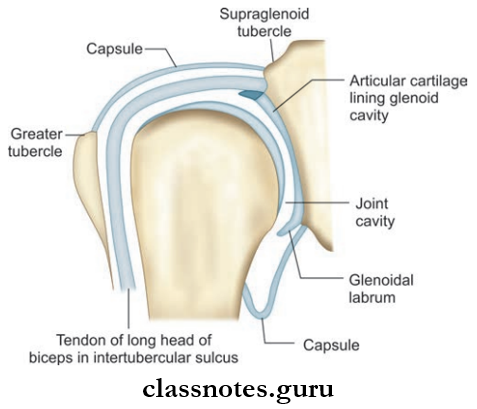
Shoulder Joint Articular Surfaces
- It is formed between the large head of the humerus and the relatively shallow glenoid cavity of the scapula this difference reduces the stability of the joint.
- But there are structures that contribute to the stability. They are:
- Glenoid labrum which deepens the glenoid fossa.
- Rotator cuff of the shoulder.
- Coracoacromial arch/secondary socket for the head of humerus.
- Muscles attaching the humerus to the shoulder joint.
Upper Limb Bones Viva Questions
Shoulder Joint Ligaments
1. Capsular Ligament
- It is loose and allows free movement.
- The capsule is lined inside with a synovial membrane.
Attachments of capsular ligament
- Medially
- To the scapula beyond the supraglenoid tubercle and margin of the glenoid labrum
- Laterally
- To the anatomical neck of the humerus except at:
- Inferiorly extends upto anatomical neck of humerus
- Superiorly, it is deficient for the passage of the tendon of the long head of the biceps brachii
- Anteriorly
- Capsule is strengthened by superior, middle, and inferior glenohumeral ligaments
2. Coracohumeral Ligament
- It strengthens the capsule.
3. Transverse Humeral Ligament
- It bridges the greater and lesser tubercle of the head of the humerus giving a tunnel for the long head of the biceps.
4. Glenoid Labrum
- It is a fibrocartilaginous rim around the margin of the glenoid cavity
- It deepens the glenoid fossa
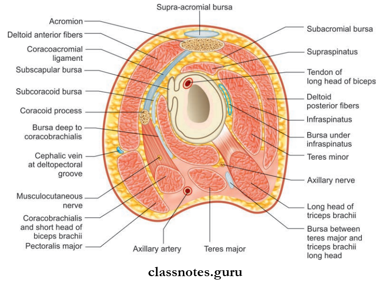
Shoulder Joint Relations
- Superiorly
- Coracoacromial arch
- Subacromial bursa
- Supraspinatus muscle
- Deltoid muscle
- Inferiorly
- Long head of triceps
- Axillary nerve
- Posterior circumflex artery and nerve
- Anteriorly
- Subscapularis
- Subscapular bursa
- Coracobrachialis
- The short head of biceps brachii
- Deltoid
- Posteriorly
- Infraspinatus
- Teres minor
- Deltoid
Bursae Related to the Joint
- Subacromial (subdeltoid) bursa
- Subscapular bursa
- Infraspinatus bursa
Shoulder Joint Arterial Supply
- Anterior circumflx humoral artery
- Posterior circumflx humoral artery
- Suprascapular artery
- Subscapular artery
Shoulder Joint Nerve Supply
- Axillary nerve
- Suprascapular nerve
- Lateral pectoral nerve
Human Upper Limb Bones Important Questions
Movements of Shoulder Joint
- Shoulder joint is the most mobile joint in the body.
- It is having wide range of mobility at the cost of its stability.
- Loose fibrous capsule and a relatively larger humeral head contribute to this.
- The basic groups of movements and the muscles involved are:
Question 3. Why abduction at the shoulder joint is considered as more complex compared to other movements occurring at the shoulder joint?
Answer:
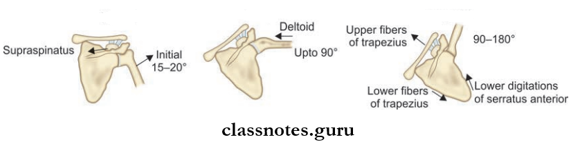
- Complete abduction of the shoulder joint occurs through 180 degree.
- Abduction up to 90 degrees occurs at the glenohumeral
- joint by the action of the deltoid.
- Further movement is only possible if the humerus rotates laterally.
- Therefore, the arm rotates laterally and carries abduction up to 120 degree.
- Abduction from 120 to 180 degrees occurs with the forward rotation of scapula on the chest wall by the action of trapezius and serratus anterior.
- In short, for every 15 degree of abduction, 10 degrees is contributed by the movement of the humerus and 5 degrees from the anterior rotation of the scapula.
Anatomy Of Upper Limb Bones Exam Questions
Question 4. Write in detail about the elbow joint under the headings—type, articular surfaces, ligaments, relations, blood supply, nerve supply, movements, and muscles involved in it.
Answer:
Elbow Joint Type
- It is a hinge variety of synovial joint.
Elbow Joint Articular Surfaces: Elbow joint consists of two articulations:
- Humeroulnar: Between the trochlea of the humerus and the trochlear notch of the ulna
- Humeroradial: Between capitulum of humerus and head of radius.

Elbow Joint Ligaments
- Joint capsule
- Attachments:
- Superiorly
- The attachment makes the trochlea, capitulum, radial fossa, coronoid fossa, and olecranon fossa intracapsular.
- Inferomedially
- The margin of trochlear notch of ulna except laterally
- Inferolaterally
- Annular ligament of superior radioulnar joint
- Synovial membrane lines inside the capsule and fossae
- Anterior ligament
- Posterior ligament
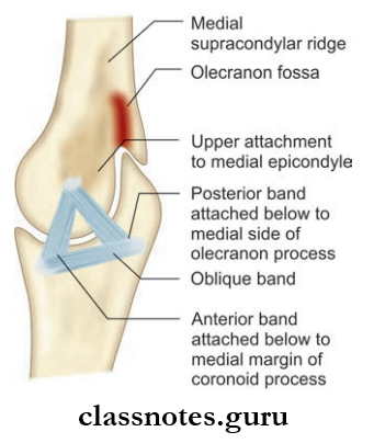
- Ulnar collateral ligament
- It is triangular in shape.
- Its apex is attached to medial epicondyle superiorly and base to the ulna inferiorly.
- The ulnar nerve crosses this ligament and flexor digitorum superficial takes its origin from it.
- Radial collateral ligament
- It is a fan-shaped band extending from the lateral epicondyle to the annular ligament.
Bones Of Arm And Shoulder Question Answers
Elbow Joint Relations
- Anteriorly
- Brachialis
- Median nerve
- Brachial artery
- Tendon of biceps brachii
- Posteriorly
- Medially
- Ulnar nerve
- Flexor carpi ulnaris
- Common flexors
- Laterally
- Supinator
- Extensor carpi radialis brevis
- Remaining common extensors
Elbow Joint Blood Supply
- From the anastomosis around elbow joint.
Elbow Joint Nerve Supply
- Radial nerve
- Median nerve
- Ulnar nerve
- Musculocutaneous nerve.
Elbow Joint Movements and Muscles involved
Elbow Joint Movement
Elbow Joint Muscles involved
- Brachialis
- Biceps brachii
- Brachioradialis
Elbow Joint Movement
Elbow Joint Muscles involved
Question 5. What is the carrying angle?
Answer:
Carrying angle
- Since the transverse axis in the elbow joint is directed medially and downwards, a fully extended forearm will not make a line with the arm.
- The angle made by the forearm with the arm in full extension is called the carrying angle and will be around 13 degrees.
- The carrying angle disappears during flexion at the elbow joint or pronation in the forearm.
Carrying Angle Clinical Anatomy
- If any effusion in the elbow joint occurs, distension, occurs posteriorly. This is due to the fact that the capsule is weaker posteriorly and deep fascia covering is thin. In such cases, aspiration of the joint is also done posteriorly.
- Fracture dislocation of the elbow joint occurs most commonly posteriorly. In such cases, the triangular relationship between the lateral, medial epicondyles of the humerus and the olecranon process of the ulna will be lost.
- When the carrying angle exceeds 13 degrees (cubitus valgus), such as in malunion of the supracondylar fracture of the humerus, stretching of the ulnar nerve occurs leading to weakening of the intrinsic muscles of the hand.
- Students’ elbow or miner’s elbow is the inflammation of the bursa over the olecranon process.
Upper Limb Osteology Questions
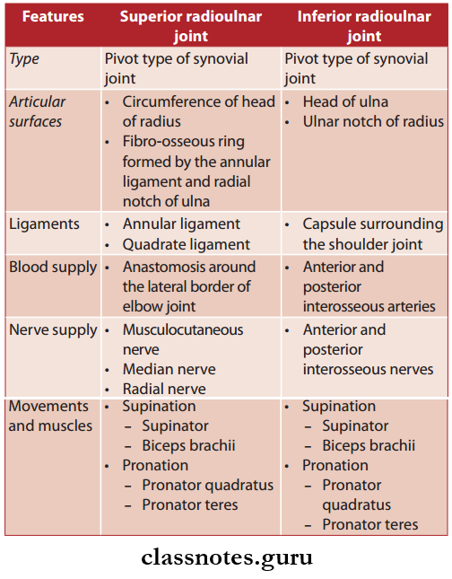
Question 6. Write a note on superior and inferior radioulnar joints.
Answer:
Superior and inferior radioulnar joints
- Radius and ulna are joined to each other by radioulnar joints.
- The superior radioulnar joint is formed between the upper end of the radius and ulna.
- Likewise, the inferior radioulnar joint is formed between the lower end of the radius and ulna.
- The shaft of these two bones are attached together by interosseous membranes which is considered as middle radioulnar joint.
Comparison between superior and inferior radioulnar joints

Superior and Inferior radioulnar joints Clinical Anatomy
- In synostosis (fusion) of upper end of the radius and ulna, pronation is not possible.
Question 7. Explain in detail about the wrist joint or radiocarpal joint under the headings—type, articular surfaces, ligaments, relations, blood supply, nerve supply, movements, and muscles involved in it.
Answer:
Wrist Joint or Radiocarpal Joint Type
- Wrist joint is the synovial joint of the ellipsoid variety.
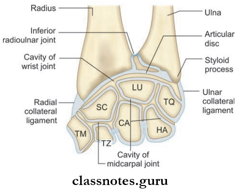
Joint or Radiocarpal Joint Articular Surfaces
- Upper End
- The inferior surface of lower end of radius
- The articular disk which separates the ulna from articulation
- Lower End
- The lateral bones of the proximal row of the carpus
- They are scaphoid, lunate, and triquetral
- But triquetral bone comes into contact with the radius only when wrist is fully adducted, otherwise, it is always in contact with articular disk
Joint or Radiocarpal Joint Ligaments
- Articular capsule: Attached superiorly to the distal end of radius and ulna and inferiorly to the proximal row of carpal bones.
- Palmar radiocarpal ligament
- Palmar ulnocarpal ligament
- Dorsal radiocarpal ligament
- Radial collateral ligament
- Ulnar collateral ligament.
Joint or Radiocarpal Joint Relations
- Anteriorly
- Long flxor tendons with their sheaths
- Median nerve
- Posteriorly
- Extensor tendons of the wrist and their fingers
- Laterally
Joint or Radiocarpal Joint Blood Supply
- Anterior and posterior carpal arteries.
Joint or Radiocarpal Joint Nerve Supply
- Anterior and posterior interosseous nerves.
Movements and Muscles Involved
- Flexion
- Flexor carpi ulnaris
- Flexor carpi radialis
- Palmaris longus
- Extension
- Extensor carpi radialis longus
- Extensor carpi radialis brevis
- Extensor carpi ulnaris
- Abduction
- Flexor carpi radialis
- Extensor carpi radialis longus
- Extensor carpi radialis brevis
- Abductor pollicis longus
- Adduction
- Flexor carpi ulnaris
- Extensor carpi ulnaris
Joint or Radiocarpal Joint Clinical Anatomy
- Nerves, vessels, and tendons pass superficial to this joint, hence, are more vulnerable for injuries.
- The wrist joint and interphalangeal joints are commonly involved in rheumatoid arthritis.
- The Dorsum of the joint is a common site for ganglion (non-tender cystic swelling).
- Fractures can occur at the distal end of radius about one inch proximal to the wrist joint. If this fragment is displaced posteriorly, it causes dinner fork deformity of the wrist and is called Colles fracture. If the displacement is occurring anteriorly, It is called Smith’s fracture.
Mnemonic: Carpal Bones
- ‘She Looks Too Pretty, Try To Catch Her
- Proximal row, lateral-to-medial:
- Scaphoid
- Lunate
- Triquetrum
- Pisiform
- Distal row, lateral-to-medial:
- Trapezium
- Trapezoid
- Capitate
- Hamate
Upper Limb Bones Short Questions And Answers
Question 8. Explain briefly about first carpometacarpal joint under the headings—type, articular surfaces, ligaments, related bursae, relations, blood supply, nerve supply, movements, and muscles involved in it.
Answer:
This joint has a separate joint cavity, so movements are much easier than other carpometacarpal joints.
Joints Of Upper Limb Multiple Choice Questions
Question 1. In which of the following at the elbow region, does the secondary center of ossifiation appear first?
- Head of radius
- Capitulum
- Medial epicondyle
- Olecranon process
Answer: 2. Capitulum
Question 2. Which of the following does not connects radius and ulna?
- Annular ligament
- Interosseous membrane
- Oblique cord
- Quadrate ligament
Answer: 1. Annular ligament
Upper Limb Bones Anatomy MCQs
Question 3. The ulnar collateral ligament of the elbow joint is related to
- Median nerve
- Basilic vein
- Ulnar nerve
- Ulnar artery
Answer: 3. Ulnar nerve
Question 4. The artery supplying the scaphoid usually enters the bone at?
- Waist
- Distal half
- Proximal half
- Proximal end
Answer: 2. Distal half
Question 5. Which movement is impossible at the shoulder joint?
- Medial and lateral rotation
- Adduction and abduction
- Circumduction
- All the above are possible
Answer: 4. All the above are possible

