
Blood Supply Of Brain And Spinal Cord Question And Answers
Question 1. Write a short note on the circle of Willis.
Answer:
Circle of Willis
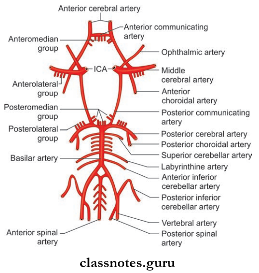
Abbreviation: ICA = Internal carotid artery
At the base of the brain around interpeduncular fossa the branches of basilar artery and internal carotid artery (ICA) anastomose form a six-sided polygon known as the circle of Willis or Circulus arteriosus.
Willis Formation
- Anteriorly by the anterior communicating artery and anterior cerebral artery
- Posteriorly by the basilar artery dividing into two posterior cerebral arteries
- Laterally by posterior communicating artery connecting the internal carotid artery with the posterior cerebral artery.
Willis Functional Significance
- Normally, there is no mixing of blood of two vertebral arteries in basilar artery, two anterior cerebral arteries in anterior communicating artery, and internal carotid and posterior cerebral arteries in posterior communicating arteries
- As a result, the right half of brain is supplied by right vertebral and right internal carotid arteries, and left half of brain is supplied by left vertebral and left internal carotid arteries
- In case of blockade of any major arteries, the collateral circulation will be provided and thus act as an arterial traff circle.
Willis Applied
Berry Aneurysms: These are congenital aneurysms due to the deficiency of tunica media layer of the arterial wall.
Question 2. Write a short note on blood supply of spinal cord.
Answer:
Blood Supply Of Spinal Cord
The Blood Supply Of The Spinal Cord Can Be Studied Under Two Headings:
- Arterial supply
- Venous drainage.
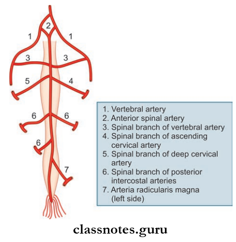
1. Arterial Supply: The spinal cord is mainly supplied by the following arteries
- Anterior Spinal Srteries
- Posterior Spinal Arteries
- Segmental Arteries.
Anterior Spinal Arteries
- Formed by the union of spinal branches of the vertebral artery
- It runs along the anterior median fissure and terminates along the filum terminale.
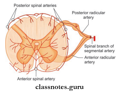
Posterior Spinal Arteries
- There are two posterior spinal arteries arising from vertebral arteries or posterior inferior cerebellar artery
- Each artery runs along the posterolateral sulcus and divides into two collaterals along the medial and lateral sides of the posterior nerve roots
- These arteries are reinforced by segmental arteries which communicate around the cord to form a plexus called vasocorona and supply superficial regions of the cord.
Segmental Arteries
- These are spinal branches of deep cervical, ascending cervical, posterior intercostal, lumbar, and lateral sacral arteries
- They reach the spinal cord, as the anterior and posterior radicular arteries.
2. Venous Drainage: The veins form six longitudinal venous channels which are as below
- Two median longitudinal veins: one in the anterior median sulcus and the other in the posterior median sulcus
- Two anterolateral veins: one on either side posterior to anterior nerve roots
- Two posterolateral veins: one on either side posterior to posterior nerve roots.
Spinal Cord Applied: Anterior spinal artery syndrome occurs due to occlusion of the anterior spinal artery.
Question 3. Write a note on the blood supply of the brain.
Answer:
Blood Supply Of Brain
The blood supply of the brain can be divided into:
- Arterial supply
- Venous drainage.
1. Arterial Supply: The arterial supply of the brain can be studied under the arterial supply of cerebral surfaces and arterial supply of other parts of the brain.
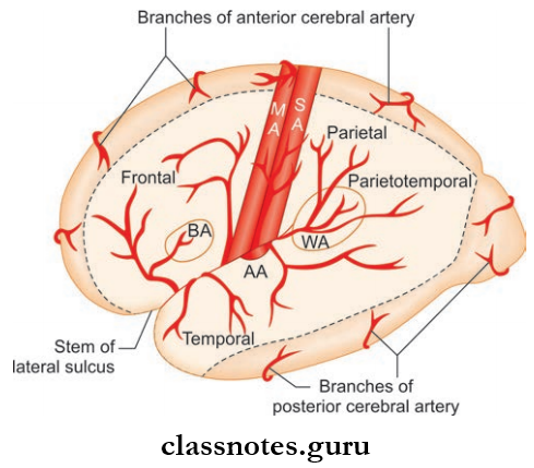
- Arterial Supply of Cerebral Surfaces
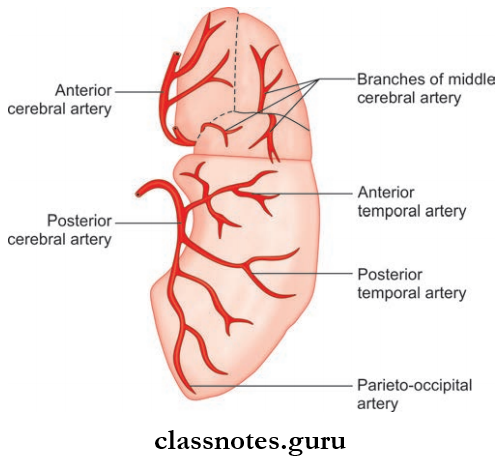
- Superolateral Surface: It is mainly supplied by the following arteries
- Middle Cerebral Artery: About two-thirds of the superolateral surface is supplied. It supplies the primary sensory and motor area, frontal eye field, Broca and Wernicke’s area
- Anterior Cerebral Artery: It supplies the narrow strip of cortex adjoining the superomedial border up to the parieto-occipital sulcus
- Posterior Cerebral Artery: It supplies a narrow strip of cerebral cortex along with temporal lobe and occipital lobe.
- Medial Surface: It is mainly supplied by the following arteries
- Anterior Cerebral Artery: ThE anterior 2/3rd of the medial surface is supplied by the anterior cerebral artery. It includes mainly the paracentral lobule
- Middle Cerebral Artery: It supplies the temporal pole of the temporal lobe
- Posterior Cerebral Artery: It supplies the occipital lobe including the visual cortex.
- Inferior Surface: It is mainly supplied by the following arteries
- Posterior Cerebral Artery: It supplies almost the entire inferior surface except for the temporal pole
- Middle Cerebral Artery: It supplies the lateral part of the orbital surface of frontal lobe and temporal pole of temporal lobe
- Anterior Cerebral Artery: It supplies the medial part of orbital part of frontal lobe.
- Superolateral Surface: It is mainly supplied by the following arteries
- Arterial Supply Of Other Parts Of Brain: The arterial supply of other parts of the brain includes the following
- The corpus striatum and internal capsule: By central branches of the middle cerebral artery and anterior cerebral artery
- Thalamus: By central branches of posterior communicating, posterior cerebral and basilar arteries
- Midbrain: By posterior cerebral, superior cerebellar and basilar arteries
- Pons: By basilar, superior cerebellar and anterior inferior cerebellar arteries
- Medulla Oblongata: By vertebral, anterior spinal, posterior spinal, posterior inferior cerebellar and basilar arteries
- Cerebellum: By superior, anterior inferior and posterior inferior cerebellar arteries.
2. Venous Drainage of Brain: The venous drainage of the brain can be studied under the following headings
- Venous Drainage of Cerebral Surfaces: The venous drainage of the cerebral surfaces is as follows:
- Superolateral Surface: The superolateral surface is drained by the following veins
- Superior cerebral veins drain the upper part of the hemisphere into the superior sagittal sinus
- Inferior cerebral veins drain the lower part of the cortex into a superficial middle cerebral vein and into the transverse sinus.
- Medial Surface: The medial surface is drained by the following veins
- Superior cerebral veins drain the upper part into the superior sagittal sinus
- Inferior cerebral veins drain the lower part into the inferior sagittal sinus
- Anterior cerebral vein drains the anterior part.
- Inferior Surface
- The inferior surface is drained by the following veins:
- Inferior cerebral veins from the orbital part drain into superficial, middle cerebral, and anterior cerebral veins
- Inferior cerebral veins from the tentorial part drain into venous sinuses at base of skull and superficial middle cerebral vein which ultimately drains to straight sinus.
- Superolateral Surface: The superolateral surface is drained by the following veins
- Venous Drainage Of Other Parts Of The Brain
- The corpus striatum and internal capsule are drained by two sets of striate veins and ultimately drains into basal vein
- The thalamus is drained by the internal cerebral vein into the cavernous sinus
- The midbrain is drained by veins into the great cerebral or basal vein
- The pons and medulla drain into superior and inferior petrosal sinuses, transverse and occipital sinuses
- The cerebellum is drained into the straight, transverse, and superior petrosal sinus.
Blood Supply Of Brain Applied: Subdural hemorrhage occurs due to rupture of cerebral veins in subdural space.
Blood Supply Of Brain And Spinal Cord Multiple Choice Question And Answers
Question 1. All of the following arteries take part in the formation of a circle of Willis except:
- Anterior communicating
- Anterior cerebral artery
- Middle cerebral artery
- Posterior cerebral artery
Answer: 3. Middle cerebral artery
Question 2. All of the following arteries arise from the basilar artery except:
- Posterior cerebral
- Posterior inferior cerebellar
- Superior cerebellar
- Anterior inferior cerebellar
Answer: 2. Posterior inferior cerebellar
Question 3. Anterior choroid artery is a branch of:
- Anterior cerebral artery
- Middle cerebral artery
- Internal carotid artery
- Posterior cerebral artery
Answer: 3. Internal carotid artery