Identification Of Bacteria Question And Answers

Identification Of Bacteria Important Notes

1. Types Of Microscopy Used For The Identification Of Bacteria

  • Optical or light microscopy
  • Phase contrast microscopy
  • Darkfield microscopy
  • Interference microscope
  • Polarization microscope
  • Electronic microscope

2. Staining Of Bacteria

Identification Of Bacteria Staining of bacteria

Identification Of Bacteria Short Answers

Question 1. Indole Test?
Answer:

Indole Test Principle: To determine the ability of an organism to decompose amino acid tryptophan into indole.

Indole Test Procedure:

  • Inoculate the test bacterium into tryptophan-rich peptone water and incubate at 37°C for 48-96 hours.
  • 0.5 ml of Kovac’s reagent is added and gently shaken.

Indole Test Interpretation:

  • Indole positive – A red-coloured ring near the surface of the medium
  • Indole negative – A yellow-coloured ring near the surface of the medium.

Indole Test Use:

  • To identify and classify bacteria.
    • Indole positive: E.Coli, Proteus species
    • Indole negative: Klebsiella species

Question 2. Urease test?
Answer:

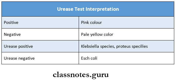
Urease Test Principle: To determine the ability of an organism to produce an enzyme urease which splits urea to ammonia.

Urease Test Procedure:

  • The test is done is Christensen’s urease medium
  • Test organism was inoculated on the entire slope of the medium and incubated at 37°C.
  • It is examined after 4 hours and overnight incubation.

Read And Learn More: Microbiology Question and Answers

Urease Test Interpretation:

Identification Of Bacteria Urease test interpretation

Question 3. Voges – proskouev test:
Answer:

Voges – Proskauer Test Principle: This test depends on the production of acetyl methyl-carbinol from pyruvic acid as an intermediate stage in its conversion to 2:3 butylene glycol.

Voges – Proskouev Test Procedure:

  • 1 ml of glucose phosphate medium culture of the organisms incubated at 30°C for 5 days (or) 37°C for 48 hours is taken
  • Then 1 volume (0.2 ml) of 40% KOH and 3 volumes (0.6 ml) of 5% a-naphthol solution in ethanol are added.

Voges – proskouev Test Interpretation:

  • Positive- Pink colour appears in 2 – Minutes
  • Negative- No colour change for 30 minutes it remains colourless.

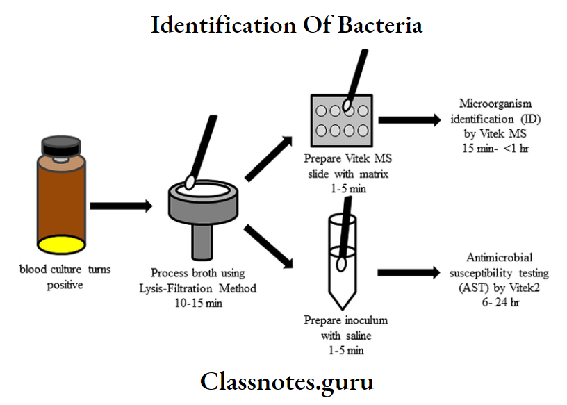
Identification Of Bacteria Identification And Antimicrobial Susceptibility Testing Of Bacteria Directly From Positive Blood Culture

Identification Of Bacteria Viva Voce

  1. Casoni test is used to diagnose hydatid cyst
  2. DNA in bacteria is composed of two strands of complementary nucleotides wound together
  3. Weil Felix reaction is used for typhus fever
  4. Paul Bunnel test is used for infectious mononucleosis
  5. Montoux test is used for tuberculosis
  6. Naggler’s reaction is used for Clostridium

General Microbiology Culture Media And Methods Question And Answers

Culture Media And Culture Methods Important Notes

1. Culture Media

Culture Media And Culture Methods Culture media

Read And Learn More: Microbiology Question and Answers

2. Common Culture Media Used In Mycology

  • Sabouraud’s agar
  • Cornmeal agar

3. Czapek – Dox

  • Anaerobic culture method
    • Obtained by McIntosh and Flide’s anaerobic jar
    • It is the most reliable and widely used method

Culture Media And Culture Methods Long Essays

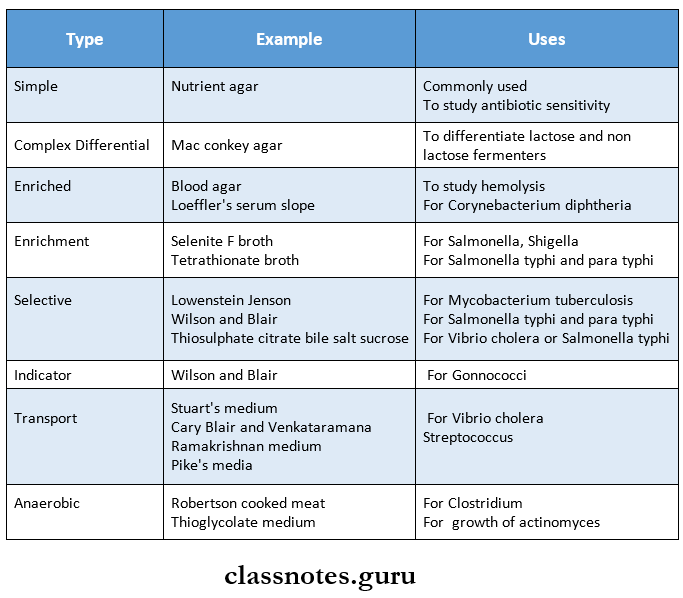
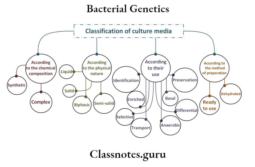
Question 1. Define and classify cultural media with examples. Write briefly on selective and anaerobic media.
Answer:

Culture Media

The culture media are the bacteriological media used for isolation and characterization of various bacterial pathogens

Cultural Media Classification: Cultural media are classified as:

Selective Media:

  • They are solid media which contain a substance incorporated into it
  • It has a selective stimulating effect on the bacteria to be grown and an inhibitory effect on the growth of the unwanted bacteria
  • They enable a greater number of the required bacteria to form colonies
  • They are used to isolate a particular bacteria from specimens

Culture Media And Culture Methods Selective media

Anaerobic Media:

  • These media are used to grow the anaerobic organism
  • They contain a reducing agent that absorbs all the oxygen and creates an anaerobic condition favouring the growth of anaerobes

Culture Media And Culture Methods Anaerobic Media

Question 2. Classify culture media. Describe anaerobic culture methods.
(or)

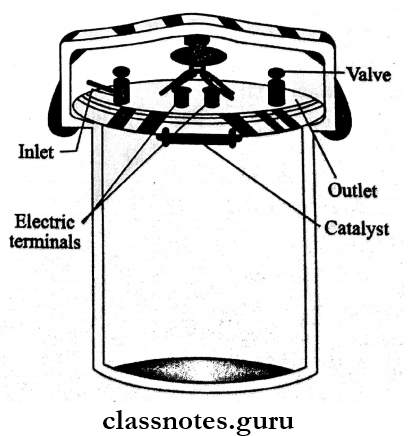
McIntosh and Filde’s anaerobic jar
Answer:

Anaerobic Culture Methods:

1. Cultivation In Vacuum.

  • Anaerobic chamber (or) cabinets (or) glove boxes
  • Anaerobic bags (or) pouches

2. Displacement Of Oxygen With Gases Like Hydrogen, Nitrogen, And Helium. Candle jar method.

3. Absorption Of Oxygen.

  • Chemical Methods
    • Alkaline pyrogallol method.
      • Spray anaerobic dish
      • Ordinary petri dish.
    • Buchner’s tube
    • Rosenthal method
    • Phosphoprous stick method.
  • Biological Methods.

4. Me Intosh Filde Anaerobic Jar

  • It is the most widely used anaerobic method
  • It consists of a glass or metal jar with a metal lid that can be clamped down on a gasket to make it airtight with a screw clamp
  • The lid has two tubes with taps, one is a gas inlet and the other is an outlet
  • One or more capsules containing palladiumized alumina pellets are suspended under the lid with wires and connected to electric terminals on the lid for heating
  • Alternatively, a temperature catalyst called de-oxo pellets can be used
  • Inoculated culture plates are placed in the jar with the medium in the bottom half of the plates and lids are clamped tight
  • The production of anaerobic conditions is brought about by
    • Displacement of oxygen
    • Displacement and combustion of oxygen

General Microbiology Culture Media And Methods Classification Of Culture Media

5. Gaspak System

6. Reduction Of Oxygen.

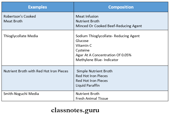
  • An aerobic media
    • Example: Robertson’s cooked meat broth
  • Thioglycollate medium
  • Smith Noguchi medium.

Culture Media And Culture Methods Mcintosh Filde's anaerobic jar

Culture Media And Culture Methods Short Essays

Question 1. Enriched media
Answer:

Enriched Media

  • This is a special medium,
  • When a basal medium is added with some nutrients such as blood, serum (or) egg it is called an enriched medium.
  • Enriched media = Basal media + some nutrients [blood, serum, egg]

Enriched Media Examples:

  • Blood Agar:
    • Blood agar – nutrient agar + blood
    • Used for routine culture
    • Example: streptococcus
    • Mostly sheep blood 5 – 10% is used.
  • Chocolate Agar:
    • It is prepared by heating 10% sterile blood in sterile nutrient agar at 55oC for 2 hours.
    • Used for the cultivation of Neisse via, Haemophilus influenza.
  • Loeffler’s Serum Slope:
    • It is composed of nutrient broth, glucose, and horse serum.
    • Used for the cultivation of diphtheria, the core bacterium
    • Dorset egg medium is used for the cultivation of diphtheria and tubercle bacilli.

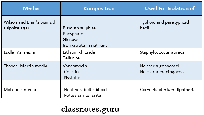
Question 2. Mention three selective media with their uses
Answer:

Three Selective Media With Their Uses

Culture Media And Culture Methods Three selective media with their uses

Question 3. Robertson’s cooked meat media
Answer:

Robertson’s Cooked Meat Media It is an anaerobic culture media

Robertson’s Cooked Meat Media Composition: Composed of Nutrient broth, Predigested cooked meat of ox heart

Robertson’s Cooked Meat Media Uses:

  • It is used for anaerobic culture
  • For the preservation of the stock culture of the aerobic organism
  • Valuable medium for preserving cultures of the delicate organism
  • May be used as a recovery medium for spores

Culture Media And Culture Methods Short Answers

1. Blood agar
Answer:

Blood Agar

  • Blood agar is an enriched media
  • In it blood is added as a nutritive substance to the basal media
  • Though the blood agar is enriched media, the bacteria lysing red cells show a dearing around the colonies
  • It helps in the growth of bacteria

Question 2. Blood Culture
Answer:

Blood Culture

Blood culture is one of the most important investigations in clinical microbiology

Blood Culture Indications:

  • Where there is a possibility of septicaemia or bacteriemia
  • For diagnosis of pyrexia of unknown origin

Question 3. Transport media
Answer:

Transport Media

For the transport of specimens to the laboratory, special media are devised and these are termed as transport media.

Transport Media Examples:

1. Stuart’s Transport Medium:

  • It is a non-nutrient soft agar gel containing a reducing agent to prevent oxidation and charcoal to neutralise bacterial inhibitors.
  • It is used in organisms such as gonococci.

2. Buffered Glycerol Saline Transport Medium: Used for enteric bacilli.

Question 4. Enrichment media
Answer:

Enrichment Media

  • Some substances are incorporated in the liquid medium which has a stimulating effect on the bacteria to be grown (or) inhibits its competitors.
  • This results in an absolute increase in the number of wanted bacteria related to other bacteria. Such media are called enrichment media.
  • They are very useful for the culture of faeces where foe non – pathogenic tend to overgrow than pathogenic ones.
  • Example: Salmonella being overgrown by E. Coli.

Enrichment Media Examples:

1. Tetrathionate Broth:

  • Composed of nutrient broth, Sodium thiosulphate, Calcium carbonate, and Iodine solution.
  • Used for the culture of faeces, especially for salmonellae.

2. Selenite F Broth

  • Composed of Peptone water and sodium selenite.
  • For culture of Salmonellae, shigellae.

3. Alkaline Peptone Water: Peptone water at pH 9.0 was used for the culture of vibrio.

4. Robertson’s Cooked Meat Broth:

  • Composed of Nutrient broth, Predigested cooked meat of ox heart.
  • Used for the culture of anaerobic bacteria.

Question 5. Preparation of blood agar media
Answer:

Preparation Of Blood Agar Media

  • Blood agar is prepared by adding 10 ml of defibrinated sheep blood to 100 ml of nutrient agar
  • The sterile nutrient agar is melted by steaming and then the temperature is brought down to 45° C
  • The required quantity of sheep blood is added aseptically with constant gentle shaking without froth formation
  • The medium is immediately poured into the plates and allowed to set

Culture Media And Culture Methods Viva Voce

  1. Robertson cooked meat media is an anaerobic media
  2. Blood agar is an enriched media
  3. Loeffler serum slope is composed of nutrient broth, glucose and horse serum


Growth And Nutrition Of Bacteria Question And Answers

Growth And Nutrition Of Bacteria Important Notes

1. Bacterial Growth Curve

Growth And Nutrition Of Bacteria - Bacteria growth and curve

Read And Learn More: Microbiology Question and Answers

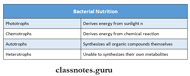
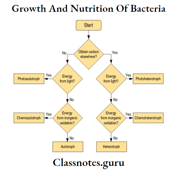
2. Bacterial Nutrition

Growth And Nutrition Of Bacteria - Bacterial Nutrition

Growth And Nutrition Of Bacteria Short Essays

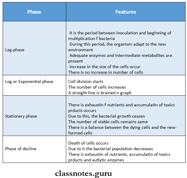
Question 1. Growth curve
Answer:

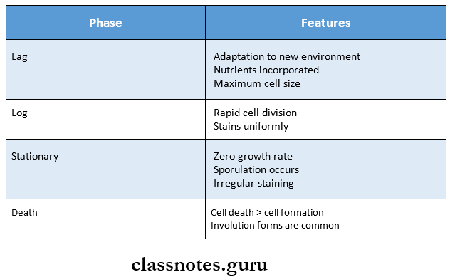
Growth Curve: The bacterial growth curve has four phases

Growth And Nutrition Of Bacteria Four phases of Growth Curve

Growth And Nutrition Of Bacteria Short Answers

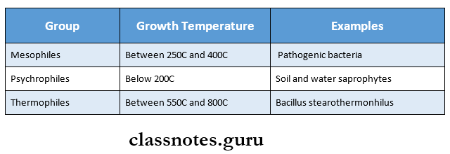
Question 1. Classify bacteria based on growth temperature.
Answer:

Based On The Growth Temperature, Bacteria Are Grouped Into The Following:

Growth And Nutrition Of Bacteria Classify bacteria based on growth temperature

Growth And Nutrition Of Bacteria Viva Voce

  1. Moisture is an essential ingredient of protoplasm
  2. The thermal point is the lowest temperature that kills a bacterium under standard conditions in a given time
  3. Lactobacillus is acidophilic bacteria
  4. Vibrio cholera is a basophilic bacteria

Growth And Nutrition Of Bacteria Autotrophic Bacteria

Herpangina In Adults Long Eassay

Viral Infections Important Notes

1. Herpangina

  • Caused by coxsackie virus
  • The prodromal phase consists of fever, chills, anorexia, sore throat, and dysphagia
  • Oral lesions start as punctuate macules and quickly evolve into papules and vesicles on the posterior pharynx, tonsils, faucial pillars, and soft palate

2. Herpes Zoster

Common sites involved are the areas innervated by spinal cord segments D3 to L2 and the ophthalmic branch of the trigeminal nerve

3. Measles

  • It is an acute, contagious, hepatotropic viral infection
  • Characterized by fever, malaise, Koplik’s spots, cough, maculopapular rash
  • Koplik spots are prodromal and disappear after the onset of the rash
  • They usually occur on the buccal mucosa as small, irregular bluish-white flecks surrounded by a red margin
  • Histologically it shows multinucleated giant cells

Viral Infections Long Essays

Question 1. Define vesicle. Write pathogenesis, clinical features, investigations, and management of primary herpetic infection.
Answer:

Vesicle:

Vesicles are elevated blisters containing clear fluid that are under 1 cm in diameter

Read And Learn More: Oral Medicine Question and Answers

Primary Herpetic Infection

  • Pathogenesis: Herpes simplex virus infects skin and neurons of the dorsal root ganglia

Primary Herpetic Infection Clinical Features:

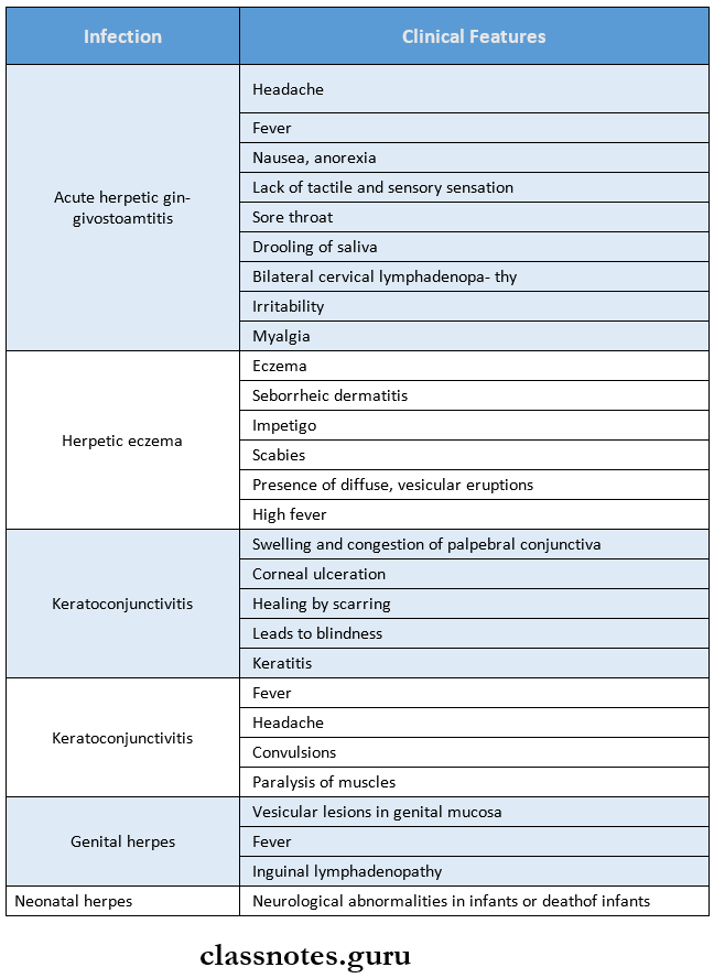
Oral Medicine Viral Infections Primary herpetic infection clinical features

Primary Herpetic Infection Oral Manifestations:

  • Site involved
  • Gingiva
  • Hard palate
  • Dorsum of tongue
  • Lips
  • Vermillion border
  • Perioral skin
  • Nasopharynx
  • Reddening of the oral mucosa
  • Formation of numerous small, dome-shaped, or pin-head type vesicle
  • Size-2-3 mm in diameter
  • Vesicles contain clear fluid and rupture to form ulcers
  • Ulcers are multiple, small, circular, punctuate, shallow, and painful

Primary Herpetic Infection Treatment:

  • Fluid administration
  • Acetaminophen- to reduce fever
  • Topical anesthesia- to decrease oral pain
  • Have red margins and yellowish or greyish floor
  • Small ulcers fuse to form diffuse, large, whitish ulcers
  • They are surrounded by a red halo
  • Gingival margins are red, swollen, and painful and have punched-out erosions
  • Difficulty in taking food
  • Difficulty in mastication
  • Difficulty in swallowing
  • Numerous vesicle formations over the tonsillar area and posterior pharynx

Primary Herpetic Infection Investigations:

  • Lesions show features of epithelial hyperplasia with acanthosis and hyperkeratosis
  • Superficial epithelial cells show dense aggregates of nuclear chromatin
  • Minor atypical changes include basal cell hyperchromatic and an increased number of mitoses

Primary Herpetic Infection Treatment:

  • Fluid administration
  • Acetaminophen- to reduce fever
  • Topical anesthesia- to reduce oral pain
  • Acyclovir- to reduce symptoms of infection

Viral Infections Short Essays

Question 1. Herpangina.
Answer:

Herpangina Clinical Features:

  • Age – Young children of the aged group 3 to 10 years
  • Incubation period – 2 – 10 days
  • Site – Commonly occurs over posterior pharynx, tonsil, faucial pillars, and soft palate

Herpangina Prodromal Symptoms:

  • Fever, chills
  • Headache
  • Anorexia, vomiting
  • Abdominal pain
  • Sore throat, dysphagia

Herpangina Ulceration:

  • The lesion initially appears as a punctuate macule
  • This turns into papules and vesicles
  • Within 24 – 48 hours, vesicles rupture to form a 1-2 mm ulcer
  • They show a grey base
  • They generally heal without treatment

Herpangina Differential Diagnosis:

1. Primary Herpes Simplex Infection: Herpangina occurs in epidemic

2. Herpes Zoster: Segmental distribution of vesicles occurs

Herpangina Treatment – Only palliative treatment is done

Question 2. Infectious Mononucleosis.
Answer:

Infectious Mononucleosis Synonym – Glandular fever

  • Infectious Mononucleosis is a benign acute infectious disease caused due to the Epstein-Barr virus, a herpes virus that infects the B- B-lymphocytes

Infectious Mononucleosis Clinical Features:

  • Incubation period – 10 – 40 days
  • Age-Young age

Infectious Mononucleosis Features:

  • Sore throat
  • Fever -101oF – 103oF
  • Headache
  • Photophobia
  • Nausea, vomiting, diarrhea
  • Erythematous macular rash
  • Splenomegaly, hepatomegaly
  • Lymphadenopathy
  • Myalgia, arthralgia
  • Depression and cognitive defect

Infectious Mononucleosis Oral Manifestations: Site affected – soft palate, labial and buccal mucosa

Infectious Mononucleosis Features:

  • Petechiae over the soft palate
  • Ulcerative gingivitis, periodontitis
  • Stomatitis
  • Inflamed and enlarged tonsils
  • Tonsils are covered by pseudo-membrane
  • Sore throat
  • Dysphagia
  • Bleeding occurs from the oro-nasopharyngeal region and the gingiva

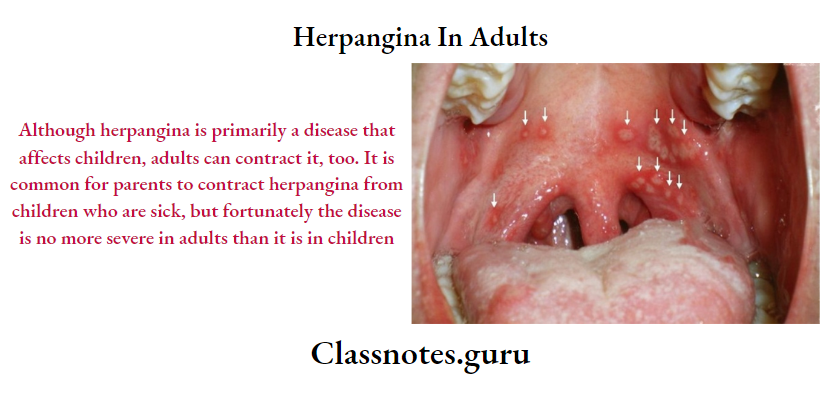
Herpangina In Adults

Infectious Mononucleosis Complications:

  • Airway obstruction.
  • Splenic rupture
  • Neurological involvement
  • Hemolytic anemia

Infectious Mononucleosis Diagnosis:

  • Positive Paul Bunnel test
  • Increase in WBC count

Infectious Mononucleosis Management:

  • For Oral Lesions
    • Topical anesthetic agent
    • Hydrogen peroxide rinses
  • For Fever And Pain
    • Antipyretics and Analgesics are prescribed
  • To Control Infection
    • Ganciclovir
    • Alfa interferon
  • To Avoid Complication
    • Corticosteroids are indicated

Oral Medicine Viral Infections Short Answers

Question 1. Herpes labialis.
Answer:

Herpes Labialis

Herpes Labialis occurs in patients with no prior infection with HSV-1 (Herpes Simplex Virus-1)

Herpes Labialis Clinical Features:

  • Age:
    • It occurs in children and young adults
    • Incubation period – 5 – 7 days
    • Prodromal generalized symptoms
    • Fever, malaise
    • Headache
    • Nausea, vomiting
    • Painful mouth
    • Sore throat
    • Irritability
    • Excessive drooling of saliva
    • Lack of tactile sensation
    • Cervical lymphadenopathy
    • Later oral symptoms
    • Numerous vesicle formations over the keratinized mucosa
    • Vesicles are thin-walled
    • They contain clear fluid
    • They rupture leaving multiple, small, punctuate shallow painful ulcers of size 2-6 mm
    • Ulcers are surrounded by a red ring of inflammation
    • Ulcers may become secondarily infected
    • Healing starts in about 3 days and is completed within 7-14 days

Herpes Labialis Treatment:

  • To prevent secondary infection – Antibiotics are given
  • To control fever – Antipyretics are given

Question 2. Post Herpetic Neuralgia.

Answer:

Post Herpetic Neuralgia

Post Herpetic Neuralgia is a complication of zoster infection

Post Herpetic Neuralgia Etiopathogenesis:

  • Nerve injury
    • Zoster virus attacks the peripheral nerve and leads to atrophy of dorsal horn cells in the spinal cord
  • Infection
    • Persistent infection of trigeminal ganglion

Post Herpetic Neuralgia Clinical Features:

  • Age and Sex – Common in older women
  • Presentation
    • Skin rashes appear
    • They are painful
    • Pain continues for weeks and months, more than 6 months
    • There may be paresthesia, hyperesthesia, and allodynia
    • There is also the presence of a sensory deficit

Post Herpetic Neuralgia Management:

  • Prevention: Use of live attenuated varicella-zoster vaccine
  • Topical Therapy: Use of topical agents like lidocaine, capsaicin
  • Drug Therapy: Use of Amitriptyline, Carbamazepine to minimize pain
  • Surgery: Carried out at the level of peripheral nerve or dorsal root
  • Steroid Therapy: Steroid injections are given to reduce the pain of the patient

Question 3. Herpetic Whitlow.
Answer:

Herpetic Whitlow

  • Herpetic Whitlow is caused by Herpes Simplex Virus
  • Herpetic Whitlow is the infection of a finger by the virus through the break in the skin
  • A dentist may experience it through contact with lesions of the mouth or saliva of patients who are asymptomatic carriers of HSV
  • The lesions are usually preceded by prodromal symptoms of burning or tingling sensation

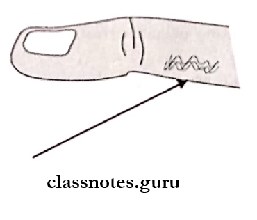
Oral Medicine Viral Infections Herpetic whitlow

Oral Medicine Viral Infections Viva Voce

  1. Herpes simplex virus causes oral ulcerations in immunocompromised patients
  2. Herpes virus shows prodromal symptoms preceding local lesions
  3. Acyclovir controls herpes infections by inhibiting DNA replication in HSV-infected cells
  4. Skin eruptions found in rheumatic fever are known as erythema marginatum
  5. Varicella Zoster virus is the most common viral infection in older patients
  6. Lipschutz bodies are seen in primary herpetic stomatitis
  7. Strawberry tongue is seen in scarlet fever
  8. Koplik’s spots are a characteristic feature of measles
  9. Rubella is teratogenic virus
  10. Herpetic whitlow occurs in fingers
  11. In herpes simplex there is ballooning degeneration of inclusion bodies called Lipschutz bodies
  12. Herpes simplex is caused by herpes simplex virus type 1
  13. Herpangina is caused by the coxsackie virus
  14. Measles is caused by the rubella virus, paramyxovirus

Cellular Adaptations Question And Answers

Cellular Adaptations Important Notes

1. Terms

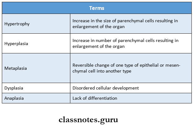
Cellular Adaptations terms

Cellular Adaptations Long Essays

Question 1. Hypertrophy
Answer:

Hypertrophy Definition: Hypertrophy is an increase in the size of parenchymal cells resulting in enlargement of the organ/tissue, without any change in the number of cells.

Hypertrophy Etiology:

Hypertrophy is caused by increased functional demand or by hormonal stimulation.

Hypertrophy Etiological types:

It may be classified into:

  • Physiologic hypertrophy: Enlarge the size of the uterus in pregnancy.
  • Pathologic hypertrophy: Hypertrophy of cardiac muscle: In cardiovascular disease.
    • Systemic hypertension
    • Aortic valve disease
    • Mitral insufficiency.
  • Hypertrophy of smooth muscle:
    • Cardiac achalasia (in the esophagus)
    • Pyloric stenosis (in the stomach)
    • Muscular arteries in hypertension.
  • Hypertrophy of skeletal muscle:
    • A hypertrophied muscle in athletes and manual laborers
  • Compensatory hypertrophy: This may occur in an organ when the contralateral organ is removed.
    • After nephrectomy on one side in young patients there is compensatory hypertrophy of the kidney as well as nephrons on the other side.

Morphologic features:

  • The affected organ is enlarged and heavy.
  • There is an enlargement of muscle fibers as well as of nuclei

Read And Learn More: Pathology Question And Answers

Question 2. Metaplasia
Answer:

Metaplasia Definition:

Metaplasia is defined as a reversible change of one type of epithelial or mesenchymal adult cells to another type of adult epithelial or mesenchymal cells, usually in response to abnormal stimuli.

Metaplasia Types:

Metaplasia is divided into the following two types.

  1. Epithelial
  2. Mesenchymal.

1. Epithelial Metaplasia:

  • Squamous metaplasia There is a transformation of various types of epithelium into squamous epithelium due to chronic irritation
  • For example:
    • In bronchus in chronic smokers.
    • In uterine endocervix in prolapse of the ureters and in old age.
    • In renal pelvis and urinary bladder in chronic infections.
  • Columnar metaplasia: There is the transformation of various epithelia into columnar epithelium.
  • For example:
    • Intestinal metaplasia in healed chronic gastric ulcer.
    • In Barrett’s esophagus, normal squamous epithelium changes to columnar.

2. Mesenchyme metaplasia: There is the transformation of an adult type of mesenchymal tissue to another.

  • Osseous metaplasia: Osseous metaplasia is the formation of bone in fibrous tissue, cartilage, and myxoid tissue.
  • For example:
    • In the arterial wall in old age (Monchkeberg’s medial calcific sclerosis]
    • In soft tissues in myositis ossifications.
    • In cartilage of larynx and bronchi in elderly people.
    • In fibrous stoma of the tumor.
  • Cartilaginous metaplasia: In the healing of fractures, cartilaginous metaplasia may occur where there is undue mobility.

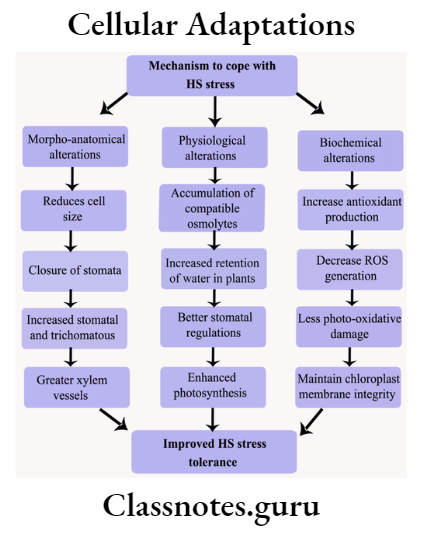
Cellular Adaptations

Question 3. Hyperplasia
Answer:

Hyperplasia Definition:

  • Hyperplasia is an increase in the number of parenchymal cells resulting in enlargement of the organ/tissue.
    • Hyperplasia occurs due to the increased recruitment of cells from the resting phase of the cell cycle to undergo mitosis when stimulated.
    • Hyperplasia persists as long as the stimulus in present.

Hyperplasia Etiology:

Hyperplasia may occur due to physiologic and path-logic causes.

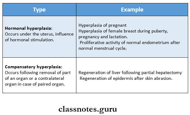
1. Physiologic hyperplasia: The two most common types are.

Cellular Adaptations Physiological Hyperplasia

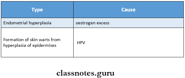
2. Pathologic hyperplasia: Due to excessive stimulation of hormones or growth factors.

Cellular Adaptations Pathologic Hyperplasia

Cellular Adaptations Short Question And Answers

Question 1. Atrophy
Answer:

Atrophy Definition:

Atrophy may be defined as the decrease in the number and size of parenchymal cells of an organ or its parts which was once normal.

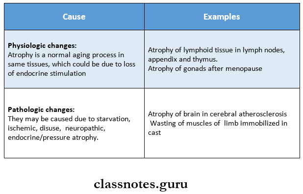
Atrophy Causes:

Cellular Adaptations Atrophy Causes

Question 2. Anaplasia
Answer:

Anaplasia Definition:

  • Anaplasia is a lack of differentiation and is a characteristic feature of most malignant tumors.
  • Depending upon the degree of differentiation, the extent of anaplasia is also variable i.e., poorly differentiated malignant tumors have a high degree of anaplasia.

Result of Anaplasia:

  • Loss of polarity
  • Pleomorphism
  • N: C ratio changes from 1:5 to 1:1
  • Anisonucleosis.
  • Hyperchromatism.
  • Prominent nucleolus
  • Tumor giant cells
  • Chromosomal abnormalities.

Pathology Hemodynamic Derangements Due To Obstructive Nature Short And Long Essay Question And Answers

Hemodynamic Derangements Due To Obstructive Nature Important Notes

1. Virchow’s triad includes 

  • Endothelial injury
  • Alteration in blood flow
  • Hypercoagulability of blood

2. Arterial thrombi

  • They are white, firm, mural
  • It produces ischemia

3. Venous thrombi

  • They are red, soft occlusive
  • Causes embolism
  • It may arise from thrombi in veins of the lower legs, pelvic veins, veins of upper limbs, cavernous sinus of the brain and right side of the heart
  • It leads to obstruction of pulmonary arterial circulation leading to pulmonary embolism

4. Infarcts
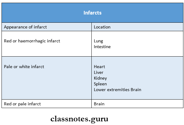
Hemodynamic Derangements Due To Obstructive Nature Infarcts

Hemodynamic Derangements Due To Obstructive Nature Long Essays

Question 1. Define thrombosis. Discuss etiopathogenesis of thrombus.
Answer:

Thrombosis:

  • Thrombosis is a process of the formation of solid mass in circulation from the constituents of flowing blood.
  • The mass itself is called a thrombus.

Thrombus Etiopathogenesis:

There are primary events that pre-dispose to thrombus formation. It is called as vir- chow’straid.

1. Endothelial injury:

The integrity of the blood vessel wall is important for maintaining normal blood flow.

  • Vascular injury exposes the subendothelial connective tissue (For example. Collagen, elastin, fibronectin, calamine, and glycosaminoglycans) which are thrombogenic and thus play an important role in initiating hemostasis as well as thrombosis.
  • Conditions and factors which cause endothelial injury and predispose to thrombogenesis are as follows:
    • Endocardial injury in myocardial infarction, myocarditis, cardiac surgery, prosthetic valves.
    • Ulcerated plaques in advanced atherosclerosis.
    • Haemodynamic stress in hypertension.
    • Arterial diseases
    • Diabetes mellitus
    • Endogenous chemical agents such as endotoxins, hypercholesterolemia
    • Exogenous chemical agents such as cigarette smoke.
  • Following endothelial cell injury, platelets come to play a central role sequence of events are:
    • Platelet Adhesion: Platelets in circulation recognize the site of endothelial injury and adhere to exposed sub endothelial collagen (Primary aggre¬gation).
    • Platelet release reaction: Activated platelets then undergo a release reaction by which platelet gran¬ules are released to the exterior.
    • Platelet aggregation: Two main types of platelet granules are.
      • Alpha granules containing fibrinogen, fibronectin, platelet factor 4, and cationic pro¬teins.
      • Dense bodies – ADP (Adenosine diphosphate), 5 – HT (serotonin), histamine, epinephrine, ionic calcium.

Read And Learn More: Pathology Question And Answers
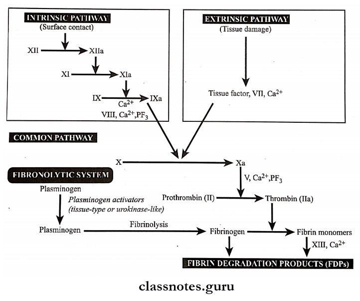
Hemodynamic Derangements Due To Obstructive Nature Intrinsic and extrinsic Pathway

Following the release of ADP, potent platelet aggregating agent, aggregation of additional platelets takes place (secondary aggregation).

  • This results in the formation of a temporary hemostatic plug.
  • The stable hemostatic plug is formed by fibrin, thrombin, and thromboxane A2.
  • The coagulation system is involved in both the hemostatic process and thrombus formation.
  • The coagulation mechanism is the conversion of the plasma fibrinogen into a solid mass of fibrin.
  • Pathways of the coagulation system and fibrinolytic system.

2. Alteration of blood flow:

  • Normally, there is the axial flow of blood in which a rapidly moving central stream – consists of leu¬cocytes and red cells. Slow-moving laminar stream – adjacent to central stream – consists of platelets peripheral stream – consists of most slow-moving cell-free plasma.
  • In thrombosis, turbulence and stasis occur. Where normal flow is disturbed when blood slow down, blood cells including platelets mar- migrate to the periphery and kind of pavement closed to the endothelium. (Margination and pave- meeting).
  • Stasis allows a higher release of oxygen from the blood and initiates venous thrombi even without evidence of endothelial injury.
  • Turbulence injures the endothelium resulting in the deposition of platelets and fibrin – facilitating the formation of arterial and cardiac thrombi.

3. Hypercoagulability of blood:

  • Hypercoagulability of blood is loosely defined as any alteration of coagulation pathways that pre¬disposes to thrombosis.
  • It is brought about by following changes in the blood composition.
  • Increase in coagulation factors like fibrinogen, prothrombin, factor 7 a, 7 a, and Xa.
  • Increase in platelet count and their adhesiveness
  • Decreased levels of coagulation inhibitors like anti-thrombin 3, and fibrin split products.

Question 2. Mention types and discuss the fate of the thrombus.
Answer:

Types of Thrombi:

  • Thrombi can develop anywhere in the cardiovascular system i.e., in cardiac chambers, on valves, or in arteries, veins/capillaries.
  • The size and shape of the thrombus depend on the site of origin and the cause.

Various types of thrombi are as follows:

1. Mural thrombi:

  • Thrombi occurring in the heart chambers or in the aortic lumen are known as mural thrombi.
  • Abnormal myocardial contraction or endomyocar¬dial injury promotes cardiac mural thrombi.

2. Arterial thrombi:

  • They are white or pale in color.
  • Microscopically, distinct lines of Zahn composed of platelets, and fibrin with enlarged red and white blood cells are seen.
  • Arterial thrombi produce ischemia and infarction.

3. Venous thrombi:

  • Venous thrombi are red and occlusive
  • Microscopically, distinct lines of Zahn with more abundant red blood cells are seen.
  • Red (venous) thrombi have more abundant red cells, leucocytes, and platelets entrapped in fibrin meshwork. Thus, red thrombi closely resemble blood clots in vitro.
  • Venous thrombi cause embolism.

4. Post mortum thrombi:

They are gelatinous with a dark red dependent portion where red cells have settled by gravity and a yellow chicken fat supernatant and they are usually not attached to the underlying wall.

5. Vegetation:

  • Thrombi occurring on the heart valves are called vegetation.
  • Bacterial or fungal blood-borne infections can cause valve damage, leading to large thrombotic masses.
  • Sterile vegetation can also develop on non-infected valves in a hypercoagulable state, so-called non-bacterial thrombotic endocarditis.

The fate of Thrombus:

1. Resolution:

  • Thrombus activates the fibrinolytic system with the consequent release of plasmin which may dissolve the thrombus completely resulting in resolution.
  • Usually, lysis is complete in small venous thrombi while large thrombi may not be dis¬solved.

2. Organisation:

  • If the thrombus is not removed, it starts getting organized phagocytic cells (neutro¬phils and macrophages) appear and begin to phagocytose fibrin and cell debris. Capillaries grow into the thrombus from the site of the attachment and fibroblasts start invading the thrombus.
  • Thus, fibrovascular granulation tissue is formed which subsequently becomes dense and less vascular and is covered over by endothelial cells.
    • This way thrombus is excluded from the vascular lumen and becomes part of the vessel wall.
    • The new vascular channels in it may be able to re-establish the blood flow, called recanaliza¬tion.
    • The fibrosis thrombus may undergo hyalinization and calcification example phlebitis in the pelvic reins.

3. Propagation:

Thrombus may enlarge in size due to more and more deposition from the constitu¬ents of flowing blood. In this way, it may ultimately cause obstruction of some important ves¬sel.

4. Thrombo embolism:

Thrombi in the early stage and infected thrombi are quite friable and may get detached from the vessel wall. These are released in the bloodstream as emboli which produce ill effects at the site of their lodgement.

Question 3. Define embolism. Give an account of various types of embolism.
Answer:

Embolism: Embolism is the partial/complete obstruc¬tion of some part of the cardiovascular system by any mass carried in the circulation, the transported intravas¬cular mass detached from the site of origin is called an embolus.

Types of Emboli:

1. Depending upon the matter in the emboli.

  • Solid
    • Example: Detached thrombi (thrombotic emboli), tissue fragments, parasites, and foreign bodies.
  • Liquid
    • Examples: Fat globules, amniotic fluid, bone marrow.
  • Gaseous
    • Example: Air, other gases

2. Depending upon whether infected or not.

  • Bland – when sterile
  • Septic – when infected

3. Depending upon the source of emboli.

  • Cardiac emboli
    • Example: Bmboli originating from atrium and atrial appendages, infarct in the left ventricle.
  • Arterial emboli
    • Example: In systemic arteries in the brain, spleen, kidney, and intestine.
  • Venous emboli
    • Example: In systemic arteries in the brain, spleen, kidney, and intestine.
  • Lymphatic emboli can also occur.

4. Depending upon the flow of blood.

  • Paradoxical embolus: An embolus which is carried from the venous side of circulation to the arterial side/vice versa is called paradoxical/crossed em¬bolus.
  • Retrograde embolus: An embolus which travels against the flow of blood is called retrograde em-bolus.

Question 4. Define infarction. Describe the pathology of the infarct.
Answer:

Infraction:

Definition: Infarction is the process of tissue necrosis resulting from some form of circulatory insufficiency, the localized area of necrosis of developed is called an “infarct.

Infarct Etiology:

  • Interrupted arterial blood supply is called ischemic necrosis.
  • Less commonly venous obstruct (stagnant hypoxia).
  • Sudden complete and continuous occlusion by thrombosis/embolism produces infarcts.
  • Also by mono occlusive circulatory insufficiency.
  • Example: Incomplete atherosclerotic narrowing of coro¬nary arteries may produce myocardial due to acute coronary insufficiency.

Types of infarcts:

1. Acc to colour:

  • Pale/Anaemic: due to arterial occlusion And are seen in compact organs example., In the kidneys, heart, and spleen.
  • Read/Haemorrhagic: Seen in soft loose tissue And are caused either by pulmonary arterial obstruction (example, in the lungs) or by arterial/venous occlusion (example In the intestines)

2. Acc. to age:

  • Recent/Fresh
  • Old/healed.

3. Acc. to presence/absence of infection: Bland, when free of bacteria contamination septic, when infected.

Infarct Pathogenesis:

  • Localized hyperemia occurs immediately after ischemia. Within a few hours, the affected part becomes swol¬len because of edema And hemorrhage.
  • Early changes are cloudy swelling And degeneration. Pro-gressive autolysis of necrotic tissue And hemolysis of red cells follows.
  • An acute inflammatory reaction And hyperemia appear in the. surrounding tissue. Blood pigments are deposited in the infarct.
  • There is progressive ingrowth of granulation tissue. Finally, the infarct is replaced by a fibrous scar.

Gross Appearance:

  • All infarcts tend to be usually wedge-shaped, the apex pointing towards the occluded vessel And the wide base on the surface of the organ.
  • Infarct due to arterial occlusion is pale while those due to venous obstructions are hemorrhagic.
  • The recent infarcts are generally slightly elevated over the surface while the old infarcts are shrunken And de-pressed under the surface of the organ.

Histologic Characteristics of Infraction:

  • The dominant histologic characteristic of infarction is ischemic coagulative necrosis of the affected area.
  • It generally contains the same amount of hemorrhage.
  • At the periphery, there is inflammatory reactions pre-dominated by neutrophils initially later even macro¬phages and fibroblasts appear.
  • Most infarcts are ultimately replaced by fibrous tissue, which at times may show calcification. The brain is an exception. In CNS, liquefactive necrosis takes place.

Question 5. Describe the morphology and microscopic structure of cardiac infarction.
Answer:

Cardiac infarction Morphology:

  • Infarcts of solid organs are usually wedged shaped
  • Apex points towards the occluded artery and base on the surface of the organ
  • Infarcts due to arterial occlusion appear pale and due to venous obstruction are hemorrhagic
  • Red cells are lysed
  • Cerebral infarcts
    • Poorly defined
    • Central softening
  • New infarcts appear slightly elevated over the surface
  • Old infarcts are shrunken and depressed under the surface

Cardiac infarction Microscopic Structure:

  • It shows coagulative necrosis of the affected area of tissue or organ
  • Shows some amount of hemorrhage
  • At the periphery inflammatory reaction is seen
  • Cerebral infarcts show liquefaction necrosis
  • Gliosis occurs later
  • Initially
    • Neutrophils are predominately present
    • Other cells present are macrophages and fibro¬blasts
  • Later
    • The necrotic area is replaced by a fibrous scar
    • May show dystrophic calcification

Hemodynamic Derangements Due To Obstructive Nature Short Essays

Question 1. Fat emboli
Answer:

  • Obstruction of arterioles and capillaries by fat globules constitutes fat embolism.
  • If the obstruction in the circulation is by fragments of adipose tissue, it is called a fat-tissue embolism.

Fat emboli Etiology:

1. Traumatic causes:

  • Trauma to bones- a most common cause.
    • Example: In fractures of long bones leading to the passage of fatty marrow in circulation, after orthopedic surgi¬cal procedures, etc.
  • Trauma to soft tissue
    • Example: Laceration of adipose tissue and in puerperium due to injury to pelvic fatty tissue.

2. Non-traumatic causes:

  • Extensive burns
  • Diabetes mellitus
  • Fatty liver
  • Pancreatitis
  • Sickle cell anemia
  • Decompression sickness
  • Inflammation of bones and soft tissues
  • Extrinsic fat/oils are introduced into the body.

Fat emboli Pathogenesis:

There are few mechanisms to explain the pathogenesis.

  1. Mechanical theory: Mobilisation of fluid fat following trauma to bones or soft tissues Releases fat glob¬ules which may enter venous circulation.
  2. Emulsion instability theory: In non-traumatic cases, fat emboli are formed by aggregation of plasma lipids due to disturbance in the natural emulsification of fat
  3. Intravascular coagulation theory: In stress, the release of some factors activates intravascular coagulation (DIC) and aggregation of fat emboli.
  4. Toxic injury theory: Blood vessels of the lungs are chemically injured resulting in increased vascular permeability and consequent pulmonary edema.

Fat emboli Consequences:

  1. Pulmonary fat embolism: Widespread obstruction can result in sudden death.
  2. Systemic fat embolism: Petechial skin rash, petechial hemorrhages, decreased glomerular filtration, renal insufficiency, etc.

Question 2. Caissons disease
Answer:

  • Decompression sickness is a specialized form of gas embolism and is known as Caisson’s disease
  • It occurs when individuals are exposed to sudden changes in atmospheric pressure
  • As seen in the deep sea, in underwater construction workers, and in individuals in unpressurized aircraft

Fat emboli Pathogenesis:

Produced when the individual decompresses suddenly either from high atmospheric pressure to normal level or from normal to low level

Fat emboli Features:

  • Formation of minute gas bubbles in
    • Skeletal muscles
    • Supporting tissues in and about the joints
  • This creates bends
  • In the lungs, edema, hemorrhage, and focal atelectasis or emphysema may appear
  • It may lead to sudden respiratory distress called chokes

Fat emboli Treatment:

Place the individual in a compression chamber which permits slow decompression of the individual

Question 3. Air emboli
Answer:

  • Air embolism occurs when air is introduced into venous or arterial circulation.
  • Gas bubbles within the circulation can obstruct vascular flow.

Venous air embolism: Air may be sucked into systemic veins during.

  • Operations on head and neck and trauma
  • Obstetrical operations and trauma
  • Intravenous infusion of blood and fluids
  • Angiography.

Arterial air embolism: Entry of air into pulmonary veins or its tributaries may occur in the following conditions:

  • Cardiothoracic surgery and trauma
  • Paradoxicalair embolism
  • Arteriography.

Hemodynamic Derangements Due To Obstructive Nature Short Question And Answers

Question 1. Atrial embolism
Answer:
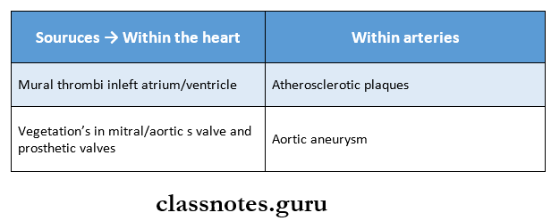
Hemodynamic Derangements Due To Obstructive Nature Atrial embolism


Arterial emboli may lead to infarction, myocardial infarc¬tion, gangrene, and sudden death.

Question 2. Pulmonary embolism
Answer:

Definition: Pulmonary embolism is the most common and fatal form of venous thromboembolism in which there is occlusion of a pulmonary arterial tree by thrombotic emboli.

Pulmonary embolism Etiology:

Thrombi originating from large veins of lower legs are 95%, less commonly thrombi originate from varicosities superficial veins of legs, and pelvic veins.

Pulmonary embolism Pathogenesis:

Detachment of thrombi produces thrombo-embolus that flows through venous drainage into large veins draining into right side of the heart

Pulmonary embolism Consequences:

  • Acute corpulmonale
  • Pulmonary hypertension
  • Pulmonary infarction
  • Chroniccorpulmonale
  • Pulmonary hemorrhage
  • Sudden death.

Question 3. Thromboembolism
Answer:

A detached thrombus or part of a thrombus forms thromboembolism
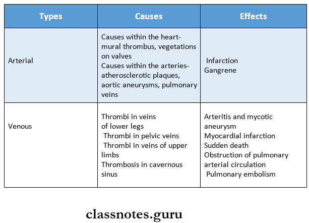
Hemodynamic Derangements Due To Obstructive Nature Thromboembolism

Question 4. Causes of ischemia
Answer:

Ischemia is defined as a deficiency of blood supply to a part of a tissue.

Etiology: Various causes of ischemia are as follows.

1. Causes in the heart: Inadequate cardiac output may result from.

  • Heart block
  • Ventricular arrest
  • Fibrillation.

This may lead to ischaemic injury to the brain.

2. Causes in the arteries: These are

  • Luminal occlusion such as due to
    • Thrombosis
    • Embolism
  • Causes in the arterial wall such as
    • Vaso spasm
    • Hypothermia
    • Arteriole sclerosis
    • Poly arteritis nodosa.
  • Outside pressure on the artery such as ligature tour¬niquet torsion tight bandages.

3. Causes in the veins: Blockage of venous drainage may lead to engorgement and obstruction to arterial supply resulting in ischemia.

Examples:

  • Luminal occlusion as in.
    • Thrombosis of mesenteric veins
    • Cavernous sinus thrombosis.
  • Causes in vessel wall- varicose veins of legs.
  • Outside pressure on the vein.
    • Strangulated hernia
    • Intussusceptions’.
  • Causes in the microcirculation.
    • Luminal occlusion.
      • By red cells example In sickle cell anemia
      • By white cells example In chronic myeloid leu¬kaemia
      • By fibrin, for example, defibration syndrome o by fat embolism.
    • Causes in microvasculature walls such as.
      • Vasculitis example Arthus reaction and
      • Frostbite injures wall of small blood ves¬sels.
    • Outside pressure on microvasculature as in bed sores.
Pathology Hemodynamic Derangements Due To Deranged Volume Short And Long Essay Question And Answers

Hemodynamic Derangements Due To Deranged Volume Important Notes

1. Hyperaemia

  • Increased volume of blood from arterial and arteriolar dilatation is called hyperemia
  • It is seen in
    • Inflammation
    • Blushing
    • Menopausal flush
    • Muscular exercise
    • High-grade fever
  • The affected organ is pink or red

2. Caissons disease

  • It is a form of gas embolism
  • It is produced when an individual decompresses suddenly either from high atmospheric pressure to normal level or from normal pressure to low atmospheric pressure

3. Lines of Zahn

  • It consists of alternate layers of aggregated platelets within fibrin meshwork and red cells
  • Lines of Zahn are distinct in arterial thrombi

4. In hypovolemic shock, kidneys conserve body fluids by

  • Reducing GFR
  • Releasing aldosterone and ADH – aldosterone increases renal retention of sodium and water

Hemodynamic Derangements Due To Deranged Volume Long Essays

Question 1. Define shock. Discuss classification, pathogenesis, and pathology of shock.
(or)
Discuss Etiology, pathogenesis, and pathology of shock.
Answer:

Shock: Shock is a life-threatening clinical syndrome of cardiovascular collapse characterized by:

  • An acute reduction of effective circulating blood volume [Hypotension) and
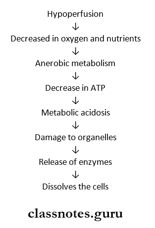
  • Inadequate perfusion of cells and tissues [Hypoperfusion).

Types Of shock:

Classification and etiology of shock:

1. Hypovolaemic Shock

  • Acute hemorrhage
  • Dehydration from vomiting, diarrhoea
  • Burns
  • Excessive use of diuretics
  • Acute pancreatitis

2. Cardiogenic Shock

  • Deficient emptying example
    • Myocardial infarction
    • Cardiomyopathies
    • Rupture of the heart, ventricle or papillary muscle
    • Cardiac arrhythmias
  • Deficient filling example
    • Cardiac tamponade from haemopericardium
  • Obstruction to the outflow example
    • Pulmonary embolism
    • Ball valve thrombus
    • Tension pneumothorax
    • Dissecting aortic aneurysm

Read And Learn More: Pathology Question And Answers

3. Septic Shock

  • Gram-negative septicemia (endotoxic shock) example Infection with E. coli, Proteus, Klebsiella, Pseudomonas and Bacteroides
  • Gram-positive septicemia (exotoxic shock) example Infection with streptococci, pneumococci

4. Other Types

  • Traumatic shock
    • Severe injuries
    • Surgery with marked blood loss
    • Obstetrical trauma
  • Neurogenic shock
    • High cervical spinal cord injury
    • Accidental high spinal anesthesia
    • Severe head injury
  • Hypoadrenal Shock
    • Administration of high doses of glucocorticoids
    • Secondary adrenal insufficiency (example in tubercu¬losis, metastatic disease, bilateral adrenal hemorrhage, idiopathic adrenal atrophy)

Pathology of Shock:

  • Shock is characterized by multi-system failure.
  • The morphogenic changes in shock are due to hypoxia resulting in degeneration and necrosis in various or¬gans.
  • Predominant morphologic complications in shock are as follows:

1. Hypoxic Encephalopathy:

  • An altered state of consciousness may be produced by cerebral ischemia in compensated shock.
  • In prolonged shock and cardiac arrest, the brain suffers from severe ischaemic damage with loss of cortical functions, coma, and a vegetative state.
  • Dead and dying nerve cells are replaced by gliosis.

2. Heart in shock:

  • The heart is more vulnerable to the effects of hypoxia than any other organ.
  • 2 important morphogenic changes in the heart are Hemorrhages and necrosis – There may be small/large ischaemic areas of infarcts, particularly located in the subepicardial and subendocardial regions.
  • Zonal lesions- these are opaque transverse con-traction bands in the myocytes near the intercalated disc.

3. Shock lung:

  • Due to dual blood supply lungs are not affected by hypovolaemic shock but are affected by septic shock. The lungs become heavy and wet.
  • Microscopically, changes of adult respiratory distress syndrome (ARDS) are seen. Briefly, the changes include congestion, interstitial and alveolar edema, lymphocyte infiltration, fibrin, and platelet thrombi in the microvasculature.

4. Shock kidney:

  • Irreversible renal injury is one of the most important complications of shock.
  • Acute tubular necrosis occurs in which there are tubular lesions.
  • The end result is generally anuria and death.

5. Adrenal changes in shock:

  • Adrenals show stress syndrome which includes the release of aldosterone, glucocorticoid, and catecholamines like adrenaline.
  • In severe shock, adrenal high can occur.

6. Haemorrhagic Gastroenteropahty:

The hypoperfusion of the alimentary tract in conditions such as shock and cardiac failure may result in mucosal and mural infarction called hemorrhagic gastro- teropathy.

7. Liver in shock:

Due to hypoxia vasodepressor ma¬terial is released which causes vasodilation, focal necrosis, fatty liver, and impaired liver functions grossly, faint nutmeg appearance is seen.

8. Other organs: Other organs such as lymph nodes, spleen, and pancreas may also show foci of necrosis in shock.

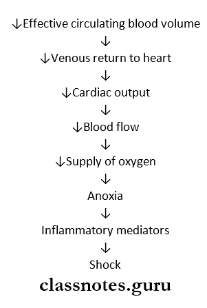
Pathogenesis: There are 2 basic features in the pathogenesis of shock.

1. Reduced effective circulating volume: It may result in either.

  • By actual loss of blood volume as in hypovolemic shock or.
  • By decreased cardiac output without actual loss of blood as in cardiogenic and septic shock.

2. Tissue Anoxia:

  • Following the reduction in effective cir¬culating blood volume, there is decreased venous return and hence Tsedcardic output.
  • This leads to reduced 02 supply, hence tissue anoxia which sets in cellular injury.

3. Release of inflammatory mediators: In response to cellular injury, inmate immunity of the body gets ac¬tivated and there is the release of inflammatory media¬tors but eventually these agents themselves become the cause of cell injury.

Pathogenesis

Hemodynamic Derangements Due To Deranged Volume Release of immatory mediators

Question 3. What are the factors leading to irreversible shock?
Answer:

Factors Leading To Irreversible Shock:

1. Progressive Vasodilatation

  • Anoxia damages the capillary and venular wall
  • Arterioles do not respond to vasoconstrictors
  • This results in Vasodilatation and peripheral pool¬ing of blood

2. Increased vascular permeability

  • Tissue damage occurs due to anoxia
  • This releases inflammatory mediators
  • Results in increased vascular permeability

3. Myocardial depressant factor

  • Release of Myocardial depressant factor causes
  • Fall in blood pressure
  • Persistent reduced blood flow to myocardium a Coronal insufficiency
  • Myocardial ischemia

4. Worsening pulmonary hypoperfusion Causes respiratory distress

5. Anoxic damage to the heart, kidney, and brain

  • Due to anaerobic glycolysis, tissue anoxia causes
    • Severe metabolic acidosis
    • Release of inflammatory mediators
    • Ischaemic cell death

6. Hypercoagulability

  • Tissue damage activates the coagulation pathway which causes
    • Release of clot-promoting factor, thromboplastin
    • Release of platelet aggregator
    • ADP
    • Slowing of bloodstream
    • Vascular thrombosis

Hemodynamic Derangements Due To Deranged Volume Short Essays

Question 1. Irreversible shock
Answer:

When the shock is so severe and no recovery takes place in spite of compensatory mechanism, therapy, and control of the etiologic agent which caused the shock, it is called decompensated/irreversible shock.

Irreversible shock Pathogenesis:

  • Progressive vasodilation
  • ↑Vascular permeability
  • Myocardial depressant factor (MDF]
  • Pulmonary hypoperfusion
  • Anoxic damage
  • Hyper coagulability

Irreversible shock Complications:

  • Brain – hypoxic encephalopathy
  • Heart – focal myocardial necrosis
  • Lungs – ARDS
  • Kidney – ATN
  • GI – Haemorrhagic gastro enteropathy
  • Liver – necrosis
  • Blood-DIC.
  • Adrenals – necrosis

Question 3. Chronic venous congestion of the liver.
Answer:

Liver Etiology:

  • It is seen in
  • Right heart failure
  • Occlusion of inferior vena cava and hepatic vein

Liver Macroscopic Appearance:

  • Enlarged liver
  • The tensed capsule is present

Liver Cut Surface:

  • Shows a nutmeg liver appearance
  • It shows the red and yellow mottled appearance
  • The red part corresponds to the congested center of lob¬ules
  • The yellow part corresponds to the fatty peripheral zone

Microscopic Appearance:

  • Centrilobular zone
    • Shows severe hypoxia
    • Distended central veins and adjacent sinusoids
    • Degeneration of hepatocytes
    • Hemorrhagic necrosis
    • Fibrosis and regeneration of hepatocytes in long-standing cases
  • Peripheral zone
    • Less effected
    • Fatty change in hepatocytes is seen

Question 4. Septic shock.
Answer:

Septic shock Etiology:

  • Severe bacterial infection – more commonly Gram-negative
  • Septicemia

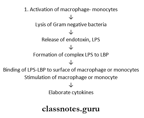
Septic shock Pathogenesis:

Hemodynamic Derangements Due To Deranged Volume Activation Of Macrophage Monocytes

Septic shock Result:

  • Produces more neutrophils which liberate free radical causing vascular injury
  • Promotes NO synthase for vasodilatation and hypoten¬sion

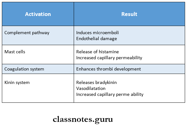
2. Activation of other inflammatory response

Hemodynamic Derangements Due To Deranged Volume Activation of other inflammatory response

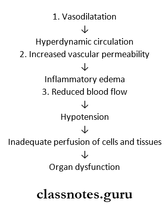
Activation of other inflammatory response Results

Hemodynamic Derangements Due To Deranged Volume Activation Of Other Inflammatory Response Result

Hemodynamic Derangements Due To Deranged Volume Short Question And Answers

Question 1. Stages of shock
Answer:

Deterioration of the circulation in shock is a progressive phenomenon and can be divided into the following 3 stages.
1. Initial nonprogressive stage: During this stage reflex compensatory mechanisms are activated and perfusion of vital organs is maintained.

2. Progressive stage: This stage is characterized by tissue hypoperfusion and the onset of worsening circula¬tory and metabolic imbalances.
3. Irreversible stage: It sets in after the body has in¬curred cellular and tissue injury so severe that even if the hemodynamic defects are corrected, survival is not possible
Question 2.Anaphylactic shock
Answer:

Anaphylactic shock Definition:

  • Anaphylaxis is a state of rapidly developing immune response to an antigen to which the individual is previously sensitized.
  • Anaphylactic shock develops due to vasodilation and peripheral pooling of blood.

Anaphylactic shock Pathogenesis:

  • IgE antibodies sanitize basophils of peripheral blood/most cells of tissue leading to the release of anaphy¬lactic mediators like histamine, serotonin, VIP, and chemotactic factors of anaphylaxis.
  • The effects produced are increased vascular permeability, smooth muscle contraction, and early vasoconstriction followed by vasodilation and shock.
  • Increased vascular permeability and vasodilation lead to peripheral pooling and blood. This results in a reduction of effective circulating volume and shock.
  • Example: Systemic anaphylaxis due to administration of antisera drugs like penicillin.

Clinical features of systemic anaphylaxis include itching, erythema, contraction of respiratory bronchioles, pulmonary edema, shock, and death.

Question 3. Mechanism of shock in burns
Answer:

  • Reduction in blood volume due to fluid loss in burns in¬duces hypovolemic shock.
  • The major effects of hypovolemic shock are due to de¬creased cardiac output and low intracardiac pressure.
  • Accordingly, clinical features are increased heart rate (Tachycardia), low blood pressure (hypotension), low urinary output, and alteration in mental state (agitated to confused to lethargic).

Question 4. End result in a shock
Answer:

  • Acute respiratory distress syndrome
  • Disseminated intravascular coagulation
  • Acute renal failure
  • Multiple organ dysfunction syndrome
  • Stupor
  • Coma
  • Death

Question 5. Nutmeg liver.
Answer:

  • It is seen in the cut surface of chronic congestion of the liver
  • It shows the red and yellow mottled appearance
  • The red part corresponds to the congested center of lobules
  • The yellow part corresponds to the fatty peripheral zone

Question 6. Neurogenic shock.
Answer:

Neurogenic shock Pathophysiology:

Hemodynamic Derangements Due To Deranged Volume Neurogenic Shock Pathophysiology

Neurogenic shock Damage to Organs:

  • Increase in myocardial contractibility
  • ARDS
  • Increase in blood pressure
  • Toxaemia
  • Bacteraemia
  • Renal failure
  • Upper GI bleeding

Neurogenic shock Management:

  • Administration of oxygen
  • Vasoactive drugs – Phenoxybenzamine
  • Inotropic agents – Dopamine
  • Corticosteroids – Prednisolonel5 mg/kg
  • Mechanical ventilation
  • Fluid replacement

Haemodynamic Derangements Question And Answers

Haemodynamic Derangements Of Body Fluids Important Notes

Haemodynamic Derangements

1. Oedema

  • It is an abnormal and excessive accumulation of fluid in the interstitial tissue spaces and serous cavities
  • Causes
    • Decreased plasma oncotic pressure
    • Increased capillary hydrostatic pressure
    • Increased capillary permeability
    • Lymphatic obstruction
    • Sodium and water retention

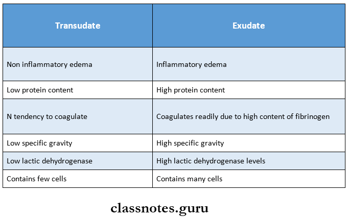
2. Transudate and exudate

Derangments Of Body fluids Transudate And Exudate

Derangements Of Body Fluids Long Essays

Question 1. Define oedema. Discuss pathogenesis and causes of oedema,
(or)
Discuss etiopathogenesis of oedema.
Answer:

Oedema: Oedema may be defined as abnormal and exces¬sive fluid accumulation in interstitial tissue spaces and serous cavities.

Types of Oedema:

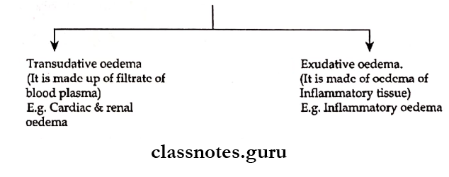
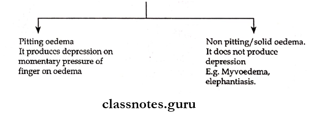
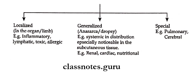
1. Three main types of oedema:

Derangments Of Body fluids Types Of Oedema

2. Subcutaneous oedema is of 2 types.

Derangments Of Body fluids Subcutaneous oedema


Read And Learn More: Pathology Questions and Answers

3. Depending on oedema fluid.

Derangments Of Body fluids Depending on oedema fluid

Etiopathogenesis:

Oedema is the result of an increase in the forces that tend to more fluids from the intravascular compartment to the interstitial space

The following six mechanisms may be operating singly/in combination to produce oedema.

1. Decreased plasma oncotic pressure:

  • Plasma oncotic pressure exerted by the total amount of plasma proteins tends to draw fluid into the vessels normally.
  • A fall in the total plasma protein level/hypoproteinemia less than 5g/dl lowers plasma oncotic pressure and thus it cannot effectively counteract the hydrostatic pressure of blood.
  • This results in Tsed outward movement of fluid from the capillary wall and anodised inward movement of fluid from the interstitial space causing oedema.
  • Example: Oedema of renal disease: Nephrotic syndrome, acute glomerulonephritis.
  • Ascites of liver disease: cirrhosis.

2. Increased capillary hydrostatic pressure:

  • Capillary hydrostatic pressure is the force that normally tends to drive fluid through the capillary wall into interstitial space by counteracting the force of plasma oncotic pressure.
  • A rise in the hydrostatic pressure at the venular end to a level more than the plasma oncotic pressure results in minimal/no reabsorption of fluid at the venular end consequently leading to oedema.
  • Example: Oedema of cardiac diseases→ Congestive cardiac failure, constrictive pericarditis.
  • Passive congestion → Mechanical obstruction due to thrombosis of veins of lower limbs, varicosities, pressure by interns, and tumours.

3. Lymphatic obstruction:

Normally the interstitial fluid in the tissue space escapes through lymphatic and thus obstruction to the outflow of the lymphatics causes localized oedema known as lymphoedema.

4. Tissue factors:

They can cause oedema when the oncotic pressure of the interstitial fluid is elevated due to increased vascular permeability and inadequate removal of proteins by lymphatics.

5. Increased capillary permeability:

  • Intact capillary endothelium acts as a semipermeable membrane permitting free flow of water and crystalloids and allowing only a minimal passage of tissue proteins.
  • Sometimes, capillary endothelium may be injured, in such cases there is development of gaps increasing the capillary permeability to plasma proteins.
  • Thus, the oncotic pressure of plasma is reduced and that of interstitial fluid is elevated producing oedema.
  • Example: Generalized oedema in systemic infections, poisonings localized oedema. Inflammatory oedema – infections, allergic reactions. Angioneurotic oedema.

6. Sodium and water retention:

  • Excessive retention and decreased renal excretion of sodium and water occur in response to hypovoleamia.
  • Example: Oedema of cardiac disease – congestive cardiac failure.
  • Ascites of liver disease – cirrhosis.

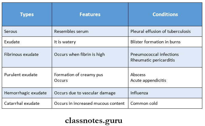
Question 2. What are the types of exudation? Describe the sequelae of pyogenic abscesses.
Answer:

Exudate:

Exudate is inflammatory oedema which is protein-rich with a specific gravity usually greater than 1.020

Exudation Types:

Derangments Of Body fluids Types Of Exudation

Sequence of Pyogenic Abscess:

  • Drainage
    • Abscess may be discharged to the surface due to increased pressure inside
  • Healing by fibrous scarring
  • Calcification
    • If the abscess is not drained, it gets organized by dense fibrous tissue

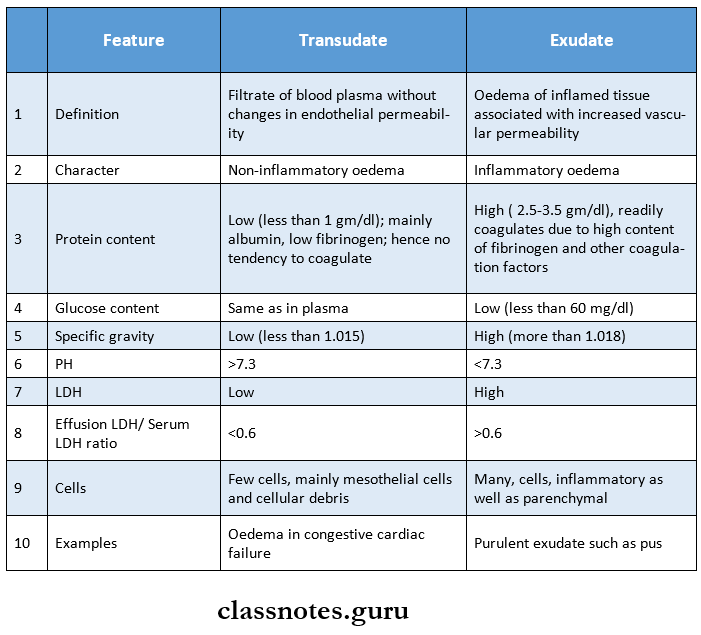
Question 3. Classify inflammatory exudate. Give example. What are the differences between exudate and transudate?
Answer:

Derangments Of Body fluids Differences between Exudate and Transudate

Derangements Of Body Fluids Short Essays

Question 1. Angioedema
Answer:

  • Angioedema is an autosomal dominant disorder which manifests as a form of local anaphylaxis
  • It Is characterised by laryngeal oedema, oedema of eyelids, lips, tongue and trunk
  • The response is mediated by humoral antibodies of IgE type
  • It involves the release of
    • Histamine
    • Serotonin
    • Leukotrienes
    • Prostaglandin
    • Platelet-activating factors
  • These act as anaphylactic mediators
  • They are responsible for changes associated with angioedema

Derangements Of Body Fluid Short Question And Answers

Question 1. Renal oedema
Answer:

Generalised oedema occurs in certain diseases of renal origin such as

1. Nephrotic syndrome:

In nephrotic syndrome, there is persistent and heavy proteinuria, there is hypo-al-buminemia causing decreased plasma oncotic pressure resulting in severe generalised oedema.

2. Glomerulonephritis:

Nephritic oedema is usually mild as compared to nephrotic oedema. Nephritic oedema is due to excessive reabsorption of sodium and water in the renal tubules via the resin angiotensin-aldosterone mechanism.

3. Acute tubular necrosis:

Acute tubular injury following shock/toxic chemicals results in gross oedema of the body.

Question 2. Pulmonary oedema
Answer:

  • Pulmonary oedema develops from left heart failure.
  • There is fluid accumulation not only in tissue spaces but also in the pulmonary arteriole.
  • It causes serious functional impairment

Etiopathogenesis: Can result from either elevation of pulmonary hydrostatic pressure/increased capillary permeability.

  • Elevation of pulmonary hydrostatic pressure.
    • An increase in hydrostatic pressure of pulmonary capillaries and the resulting imbalance between palm hydrostatic press and plasma oncotic pressure causes excess fluid to move into the interstitium.
    • Interstitial oedema develops later with a prolonged elevation of hydrostatic pressure, alveolar lining cells develop and alveolar oedema results.
    • This mechanism is seen in left heart failure, mitral stenosis and thyrotoxicosis.
  • Increased capillary permeability: Damage to alveolar-capillary membrane causes increased permeability so that excess fluid and plasma proteins leak out, initially into the interstitium and later into the alveoli.
  • Example: Fulminant pulmonary infections, radiation injury, inhalation of ototoxic substances.

Pathology Infections And Infestation Short And Long Essay Question And Answers

Infections And Infestation Short Essays

Question 1. Lab diagnosis of enteric fever
Answer:

Enteric fever Lab Diagnosis:

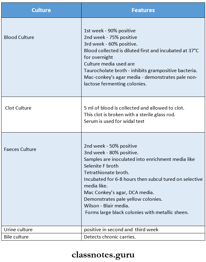
1. Isolation of bacilli.

  • For isolation of bacteria, specimens are obtained from blood, faeces, urine, aspirated duodenal fluid, bile, bone marrow or rose spot.
  • These specimens are then cultured.

Infections And Infestation Isolation of bacilli

2. Demonstration of Antibodies

1. Widal test.

It is an agglutination for the detection of agglutinins H and 0 in patients with enteric fever

Widal test Method:

  • Equal volumes f0.4 ml) serial dilutions of serum from 1:10 to 1: 640 and H and O antigens are mixed.
  • One control tube containing antigen and normal saline is used.
  • All these tubes are incubated in a water bath at 37°C.

Read And Learn More: Pathology Question And Answers

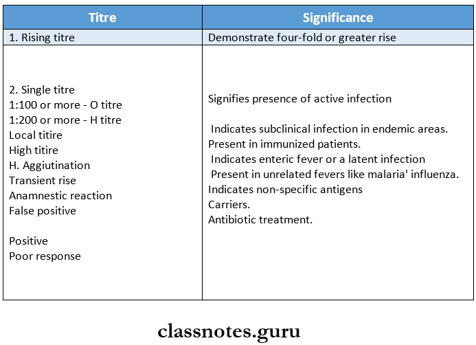
Widal test Results:

Infections And Infestation Widel test Results

Widal test Interpretation:

Infections And Infestation Widel test Interpretation

3. Demonstration of circulating antigen.

Done by counterimmunoelectrophoresis and ELISA.

Question 2. Urinary sediment
Answer:

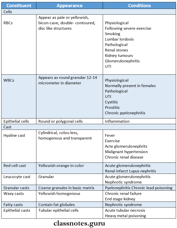
Urinary sediment contains the following constituents

Infections And Infestation Urinary sediment

  • It also contains
    • Crystals of
      • Calcium oxalate
      • Uric acid
      • Amorphous urate
      • Tyrosine
      • Calcium carbonate
    • Miscellaneous structures like
      • Spermatozoa
      • Parasite
      • Fungus
      • Tumour cells

Infections And Infestation Short Question And Answers

Question 1. Amoebiasis
Answer:

  • Caused by entamoeba histolytica
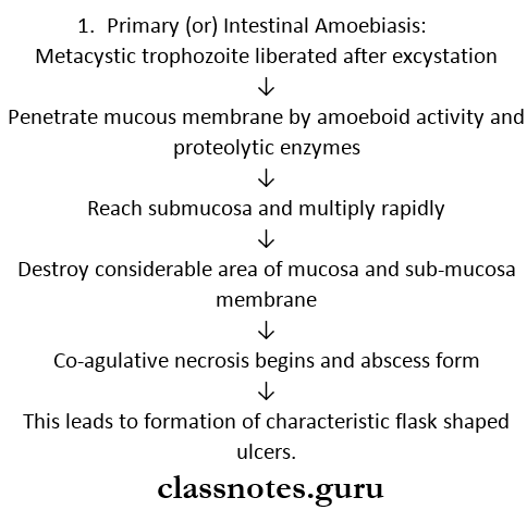
  • E. Histolytica can cause two types of pathological lesions as follows

Infections And Infestation Primary Amoebiasis

  • This condition is called as amoebic dysentery.
  • These ulcers may be generalised (or) may be localised to the ileo-caecal (or) sigmoidorectal region.
  • Occasionally ulcers may involve deeper tissues and may cause perforation (or) peritonitis.
  • Erosion of blood vessels may lead to haemorrhage.
  • The superficial lesions generally heal without scarring.
  • Deep ulcers form scars leading to strictures, partial obstruction and thickening of gut wall.

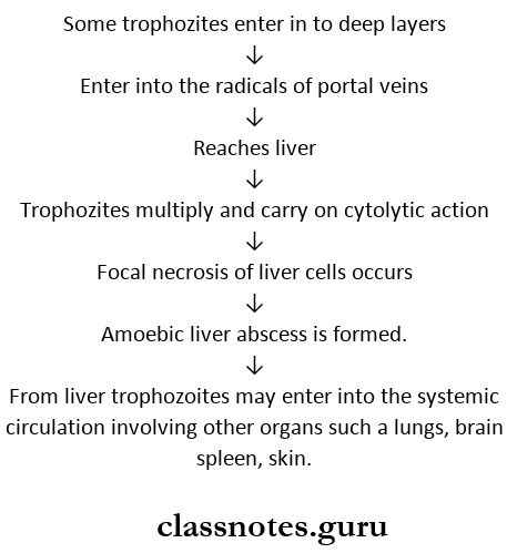
2. Extra-intestinal amoebiasis (or) secondary (or) metastatic lesions.

Amoebic liver abscess:

Infections And Infestation Amoebic liver abscess

Question 2. Candidiasis
Answer:

  • Candidiasis is caused by Candida albicans and occasionally by other Candida species
  • It is an opportunistic endogenous infection.

Candidiasis Predisposing Factors:

  • Diabetes
  • Immunodeficiency
  • Malignancy
  • Prolonged administration of antibiotics
  • Patients on immunosuppressive drugs and intravenous catheters

Candidiasis Treatment:

  • Removal of predisposing factors
  • For superficial infections- Topical application of polyene and imidazole is used
  • For systemic infections- Amphotericin B + 5- fluoroscopy- tosine is used

Question 3. Rhinosporidiosis
Answer:

Rhinosporidiosis is a chronic granulomatous disease.

Rhinosporidiosis Causative Organism:

Rhinosporidium seeberi.

Rhinosporidiosis Mode of Infection:

Frequent contact with stagnant water.

Rhinosporidiosis Features:

  • Friable polyps
  • Sites involved- nose, mouth and eye
  • Oral manifestations are Oropharyngeal lesions
  • They appear as soft red polypoid growth and spread to the pharynx and larynx.
  • These lesions contains mucoid discharge and are vascular.

Rhinosporidiosis Diagnosis:

  • H and E stained tissue sections shows a large number of endospores within the sporangia
  • These are embedded in a stroma of connective tissue, and capillaries

Question 4. Cysticercus cellulose
Answer:

  • Cysticercus cellulose is the larval stage of taenia solium
  • It develops in the muscles of the pig which is intermediate host
  • A mature cyst is an opalescent ellipsoidal body and the long axis of the cyst is parallel to the muscle fibre.
  • A dense milky white spot is present at the side where the scolex with its hooks and suckers remains invalidated.
  • The cyst develops further when ingested by man which is the definitive host
  • It may develop in any organ but are usually present in the subcutaneous tissues and muscles.

Various features of cysticercosis:

  • They causes palpable nodule in sub-cutaneous tissues and muscles
  • In brain leads to epileptic attacks.
  • Neurocysticercosis involving the nervous system is the most serious form

Question 5. Fungi infecting hair
Answer:

  • Dermatophytes are the group of fungi affecting the hair.
  • Favus is a chronic type of ringworm involving the hair follicles

Fungi infecting hair Features:

  • Alopecia
  • Scarring
  • Sparse hyphal growth
  • Formation of air spaces within the hair shaft

Fungi infecting Hair Types:

Infections And Infestation Fungi infecting hair Types

Question 6. Urinometer.
Answer:

  • It is an equipment for determining urine specific gravity
  • It is composed of
    • Float – It is air filled glass tube
    • Weight – It is a bulb filled with ball bearings
    • Stem – It has calibrated graduation and numbers marked off to indicate specific gravity measurements
  • Urinometer is placed in a tube of urine and where the meniscus of the urine reaches displays the specific gravity of the urine

Pathology Of Immune System Question And Answers

Diseases Of The Immune System Important Notes

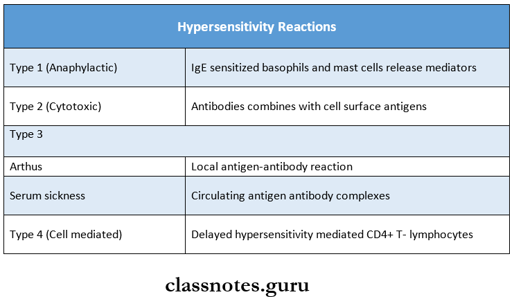
1. Hypersensitivity reactions

Diseases Of The Immune System Hypersensitivity Reactions

Diseases Of The Immune System Long Essays

Question 1. Define hypersensitive reaction. Explain type IV hypersensitive reaction.
Answer:

  • Hypersensitivity or allergy is defined as a state of exaggerated immune response to the antigen, which may lead to tissue damage, disease or even death following contact with specific antigens.
  • Lesions of hypersensitivity i.e., immunologic tissue injury are produced due to the interaction between antigen and product of immune response.
  • Depending upon the rapidity, duration, and type of immune response, hypersensitivity reactions are classified into.

1. Immediate type:

It includes 3 types.

1. Type 1 – Anaphylactic reaction.

  • Example: Systemic anaphylaxis/local anaphylaxis.

2. Type 2 – Cytotoxic reaction.

  • Example: Erythroblastosis fetal, leucopenia.

3. Type 3 – Immune complex-mediated reaction

  • Example: Arthritis and skin diseases.

2. Delayed type:

Type 4- Hypersensitivity reaction.

Type 4/delayed hypersensitivity reaction is tissue injury by cell-mediated immune response without the formation of antibodies but is instead a slow and prolonged response of specifically sensitized T- lymphocytes.

Etiology and pathogenesis: Type 4 reaction involves the role of most cells and basophils, macrophages and CDQ + T cells. The mechanism of type 4 reaction is:

  • The antigen is recognized by CDg + T cells (cytotoxic T cells) and is processed by antigen-presenting cells.
  • Antigen-presenting cells migrate to the lymph node where antigen is presented to helper T-cells (CD4 + T cells).
  • Helper T cells release cytokines that stimulate T cell proliferation and activate macrophages.
  • Activated T cells and macrophages release pro-inflammatory mediators and cause cell destruction.

Read And Learn More: Pathology Question And Answers

Examples:

  • Reactions against mycobacterial infection.
    • Examples: Tuberculin reaction, granulomatous reaction in tuberculosis, leprosy.
  • Reaction against virally infected cells
  • Reaction against malignant cells in the body.
  • Reaction against organ transplantation example transplant rejection, graft versus heart reaction.

Diseases Of The Immune System Short Essays

Question 1. Anaphylaxis
Answer:

  • Anaphylaxis or type I hypersensitivity is defined as a state of rapidly developing an immune response to an antigen (i.e., allergen) to which the individual is previously sensitized.
  • The reaction appears within 15-30 min of exposure to antigen.

Anaphylaxis Etiology: Type I reaction is mediated by humeral antibodies of IgE type or regain antibodies in response to antigen.

The definite cause is not known, but the following may be responsible.

  • Environmental pollutants
  • Genetic basis
  • Concomitant factors – Allergic response may be linked to the occurrence of certain viral infections of the upper respiratory tract.

Anaphylaxis Pathogenesis: Type 1 reaction includes the participation of B lymphocytes and plasma cells, mast cells and basophils, neutrophils, and eosinophil It mechanism is:

  • During first contact, with antigen, sensitization takes place.
  • During the second contact, IgE antibodies on the surface of mast cells – basophils are firmly bound to FQ receptors and set in cell damage and degranulation of mast cells occurs.
  • Released granules contain important chemicals and enzymes with pro-inflammatory properties – histamine, serotonin, vasoactive, intestinal peptide, prostaglandins, platelet-activating factor, etc. The effects of these agents are.
    • Increased vascular permeability
    • Smooth muscle contraction
    • Vasoconstriction followed by vasodilation
    • Shock
    • Increased gastric secretion
    • Increased nasal and lacrimal secretions
    • Eosinophilia and neutrophilia.

Anaphylaxis Examples:

  • Reactions against mycobacterial infection.
    • Examples: Tuberculin reaction, granulomatous reaction in tuberculosis, leprosy.
  • Reaction against virally infected cells
  • Reaction against malignant cells in the body.
  • Reaction against organ transplantation example, transplant rejection, graft versus host reaction.

Diseases Of The Immune System Short Question And Answers

Question 1. Atopy
Answer:

  • Atopy is a form of type I hypersensitivity reaction
  • The antigens commonly involved in it are pollens, house dust, and food
  • These induce IgE antibodies

Atopy Features:

  • Atopy shows marked familial distribution
  • Atopy sensitization is developed spontaneously following natural contact with opens
  • The reaction occurs at the site of entry of antigen
  • For example
    • Inhalation of pollens affects the lungs
    • Contact leads to local allergy

Atopy Manifestations:

  • Conjunctivitis
  • Rhinitis
  • Bronchospasm
  • GI symptoms
  • Dermatitis
  • Cutaneous eruptions

Question 2. B lymphocytes
Answer:

  • Lymphocytes are of 2 major types. B lymphocytes (10 – 15% of T lymphocytes (75 – 80%).
  • Lymphocytes undergo maturation and differentiation in the bone marrow and form B cells.
  • B cells are involved in humeral immunity by inciting antibody response.
  • On coming in contact with an antigen, B cells are activated to proliferate and transform into plasma cells.
  • Depending upon the maturation stage of B cells, specific CD molecules appear on the cell surface which can be identified by CD markers, common B cell markers include CD 19, 20, 21, 23.
  • These cells also possess B cells receptors for surface immunoglobulin (IgM and IgG) and FQ receptors for attaching to antibody molecules.

Question 3. Cell-mediated immunity
Answer:

  • It is also known as type 4 delayed hypersensitivity re-action.
  • It is defined as tissue injury by cell-mediated immune response without the formation of antibodies but is instead a slow and prolonged response of specifically – sensitized lymphocytes.
  • The reaction occurs about 24 hours after exposure to antigen and the effects is prolonged which may last up to 14 days.

Question 4. Routes of transmission of HIV
Answer:

Routes of transmission:

The virus is present in the body fluids like blood, lymph, and genital fluids and gets transmitted when a healthy individual’s body fluids come in contact with an affected individual.

  • Sexual transmission: This is the commonest form of transmission.
  • Transmission via blood and blood products: Next commonest form.
    • Intravenous drug abusers: By sharing needles, syringes, etc.
    • Hemophiliacs: Who received large amounts of factor 8 concentrates.
    • Recipients of HIV: Infected blood and blood products.
  • Perinatal transmission: It is also called vertical transmission, which occurs from the infected mother to the newborn during pregnancy or through breast milk.
  • Occupational transmission occurs in workers engaged