Complete Denture Impressions

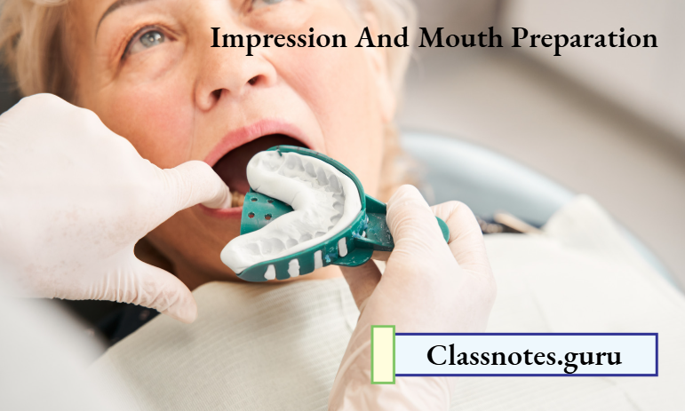
Impression And Mouth Preparation Short Answers

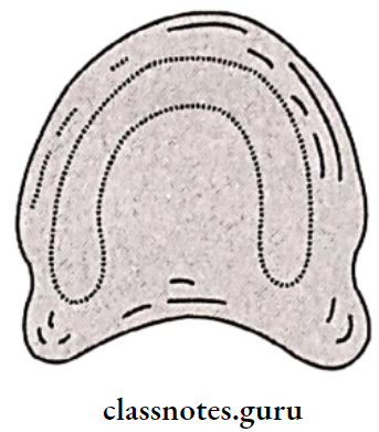
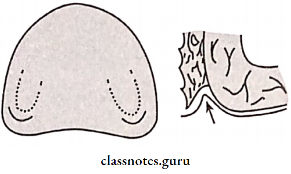
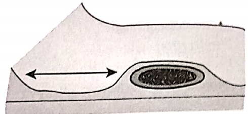
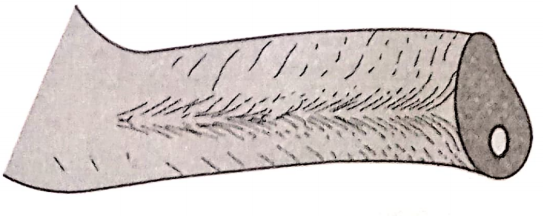
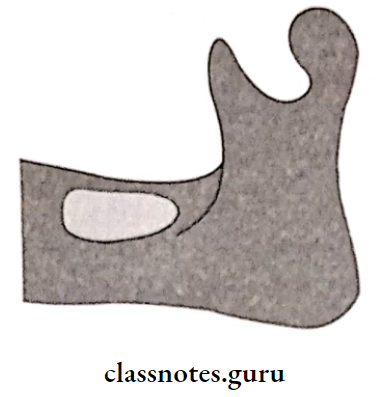
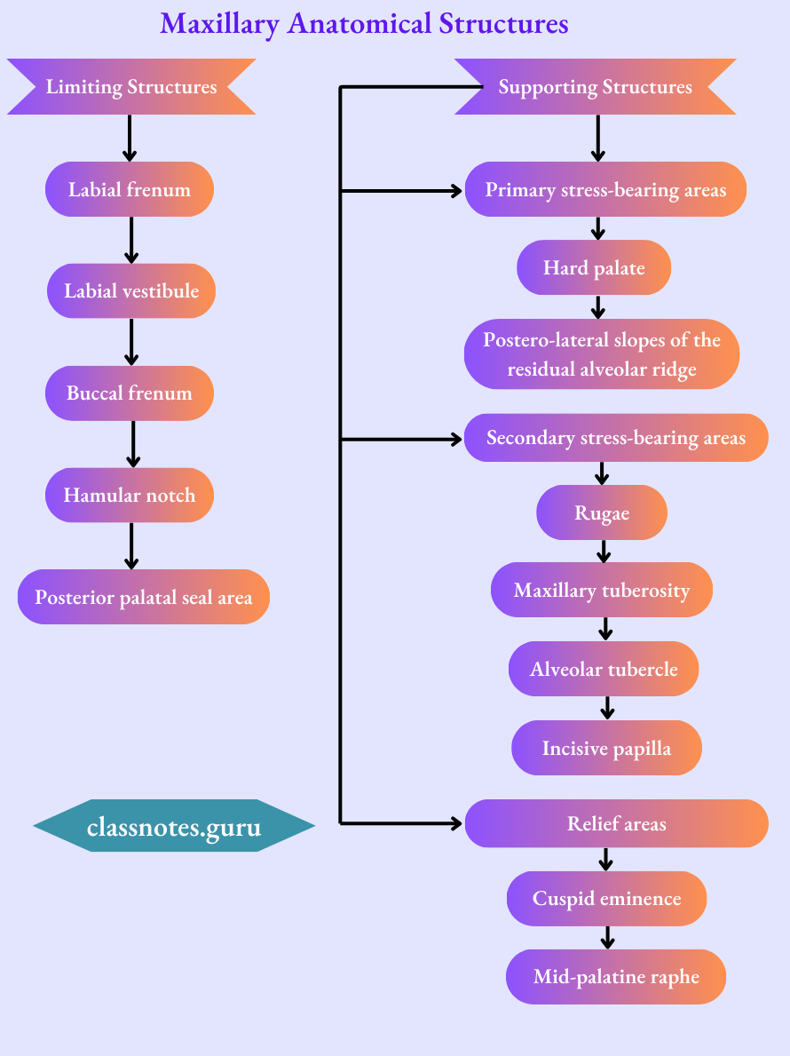
Question 1. Buccal shelf area
Answer:

Area Of Buccal Shelf :

Between the buccal frenum and the anterior border of the masseter

Boundaries of Buccal Shelf :

  • Medial crest of the ridge
  • Distal Retromolar pad
  • Lateral external oblique ridge

Significance Of Buccal Shelf :

  • Primary stress-bearing area of the mandible
  • The width of it increases as resorption continues

Read And Learn More: Prosthodontics Question And Answers

Impression And Mouth Preparation Buccal Shelf Area

Impression and Mouth Preparation in Complete Dentures

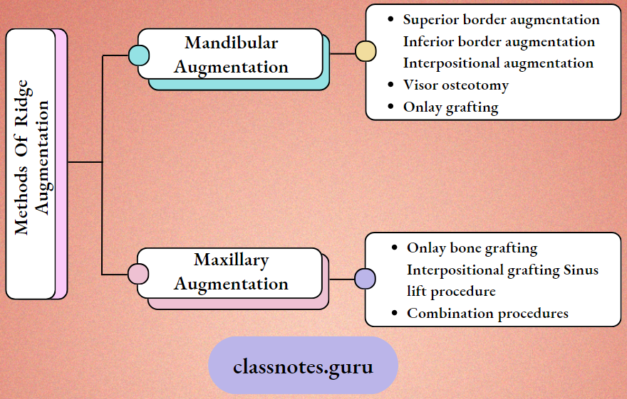
Question 2. Ridge Augmentation
Answer:

Ridge Augmentation Definition:

Any procedure designed to enlarge or increase the size, extent, or quality of a deformed residual ridge is referred to be ridge augmentation procedures

Impression And Mouth Preparation Methods Of Ridge Arguments

Question 3. “Cohesion” in complete dentures.
Answer:

  • Cohesion is the physical attraction of like molecules to each other
  • Acts within the film of saliva
  • Normal quality and adequate quantity of saliva are cohesive
  • When the quantity of saliva is excessive and the quality is watery, cohesion is decreased
  • When the quantity of saliva is decreased and its viscosity is increased, cohesion is decreased due to an increase in the thickness of saliva

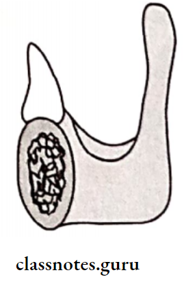
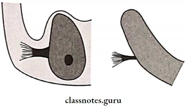
Question 4. Mylohyoid ridge  importance
Answer:

  • The mylohyoid Ridge is a rough bony crest extending from the third molar to the second bicuspid region
  • The mylohyoid muscle is attached to the mylohyoid ridge
  • In the anterior region, the mylohyoid ridge with attached mylohyoid muscle lies close to the inferior border of the mandible
  • Posteriorly, it is superior in position and the lingual flange of the denture may extend below the mylohyoid ridge if it drops vertically or slopes at 45 degrees towards the tongue

Short Questions on Mouth Preparation and Impressions

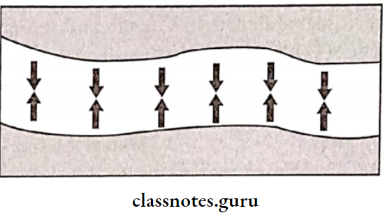
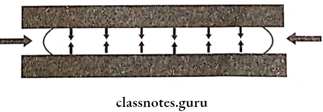
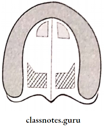
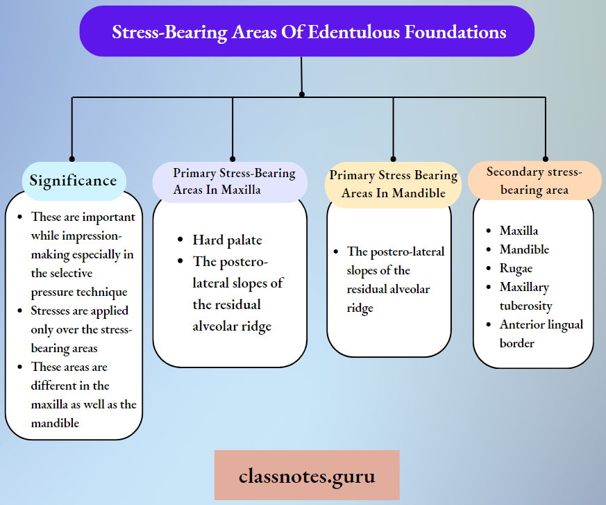
Question 5. Stress-bearing areas of edentulous foundations
Answer:

These are those areas where stresses are directed at are right angle

Impression And Mouth Preparation Stress Bearing Areas Of Identification Foundations

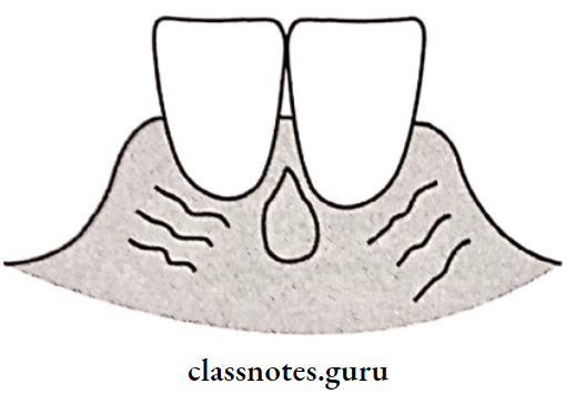
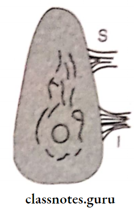
Question 6. Retromolar papilla
Answer:

  • Retromolar Papilla is described as a pear-shaped papilla. It is a small elevation
  • Retromolar Papilla is a residual scar formed after the extraction of the third molar
  • Retromolar Papilla  lies along the line of the ridge
  • The denture should terminate at the distal end of the pear-shaped papilla
  • Being in this area improves retention

Question 7. Objectives of complete denture impressions
Answer:

  • Retention: It is the resistance to displacement away from the tissue surface. It is a mucosa-borne phenomenon.
  • Support: It is the resistance to the occlusal forces in the vertical direction. It is a bone-borne phenomenon.
  • Stability: It is resistant to lateral shifting.
  • Preservation of remaining structures.

BDS Prosthodontics Viva on Impressions

Question 8. Diagnostic casts.
Answer:

Diagnostic Casts Requirements:

  • Diagnostic Casts Should Be:
    • Free of voids
    • Smooth surface
    • Provide good denture support
    • Walls should be tapering outward
    • Smooth tongue space
    • Free of moisture
    • The occlusal table is parallel to the floor.

Diagnostic Casts Uses:

  • Measure the undercuts. Determine the path of insertion
  • Treatment planning
  • Perform mock surgery, evaluate the arch
  • Assess retention & stability
  • Determine secondary retentive forms

Question 9. Saliva’s influence on denture retention & stability.
Answer:

  • Thick and ropy saliva, loss of retention
  • Thin and watery saliva compromised retention
  • Watery serous saliva is more retentive
  • In xerostomia there is no adhesion
  • Ptyalism leads to gagging
  • Xerostomia: Soreness and irritation

Question 10. Retention in the  maxilla
Answer:

Retention factors present in the maxilla are

  1. Large denture-bearing area
  2. Thick and ropy saliva
  3. Interfacial surface tension

Impression Techniques in Complete Dentures

Question 11. Maxillary anatomical structures
Answer:

Impression And Mouth Preparation Maxillary Anatomical Structures

Question 13. Final impression methods in complete denture
Answer:

  1. Dry gauze is placed on the floor of the mouth to remove the saliva
  2. It is removed just before making an impression
  3. Impression material is manipulated and loaded onto the tray
  4. The tray is rotated in the horizontal plane and inserted into the mouth using the anterior handle
  5. The tray is seated completely by applying alternating pressure over the posterior handles
  6. The patient is asked to touch his upper lip with his tongue
  7. Passive movements are performed
  8. After the material gets set and examined for any defects

Question 14. Realeff effect
Answer:

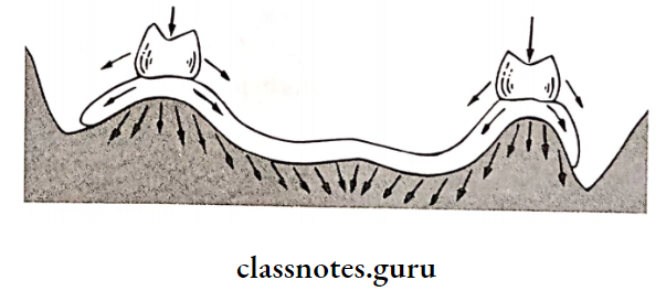
Complete dentures rest on the basal seat area, which is primarily the  oral mucosa and the residual alveolar ridge

  • The oral mucosa is displaceable and compressibl.e Hanau described this factor as “resiliency and like effect”- Realeff effect
  • It plays an important role in all steps of complete denture fabricate,on as in
  • Primary impression
  • Border moulding
  • Final impression
  • Jaw relation
  • Try in
  • Remount
  • Follow up
  • Mechanical or pathological reasons cause fibrous changes in the residual ridge, making it resilient
  • It is more commonly seen in Mandibular residual ridges
  • Maxillary anterior ridges

Factors Affecting the Real Effect

  • Consistency of mucosa
  • Excess bone loss during extraction
  • A person’s general health
  • Elderly tissues
  • Smaller forces produce distinct compression
  • Parafunctional habits
  • Single complete denture

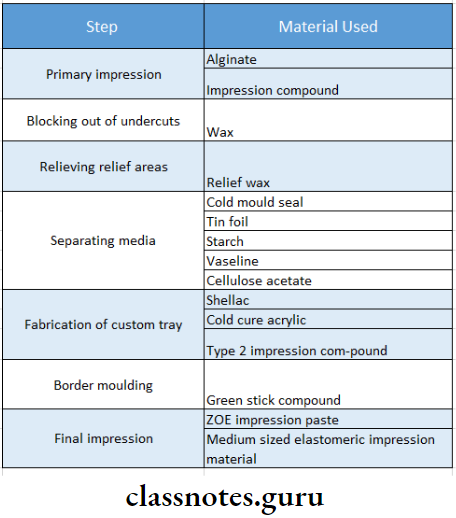
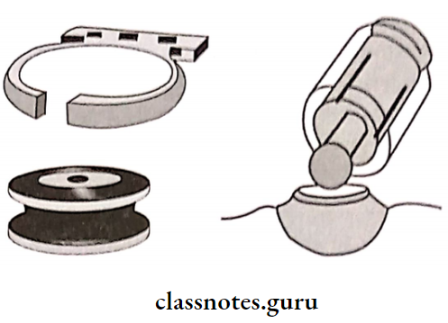
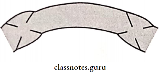
Question 15. Materials used for the master impression.
Answer:

Master Impression Requirements:

  • Low viscosity
  • Form a thin layer of impression.
  • Should be uniform

Master Impression Materials Used:

  • Zinc oxide eugenol paste impression
  • Medium-bodied elastomeric impression materials

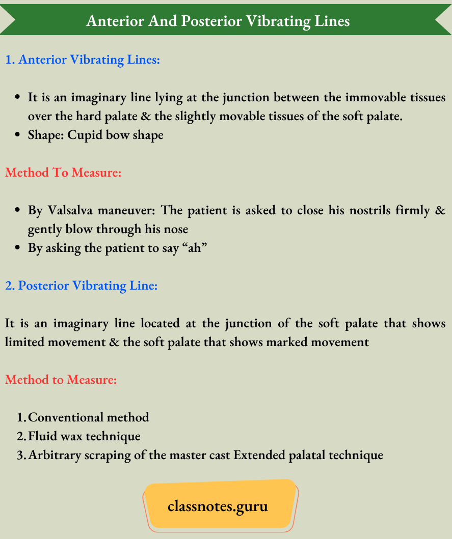
Question 16. Anterior and posterior vibrating lines.
Answer:

Impression And Mouth Preparation Anterior And Posterior Vibrating Lines

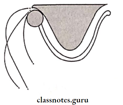
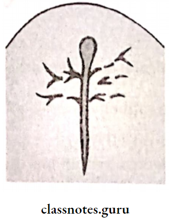
Question 17. Modiolus.
Answer:

The modiolus is a point where 8 muscles meet at the angle of the mouth

Modiolus Are:

  • Depressor anguli oris
  • Levator anguli oris
  • Risorius
  • Orbicularis oris
  • Buccinator
  • Zygomatic major
  • Quadralus labii superioris
  • Quadralus labii inferioris

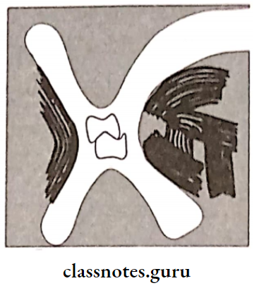
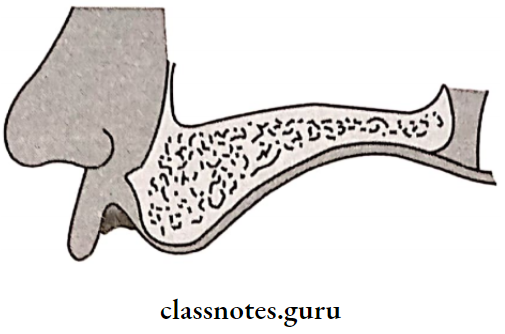
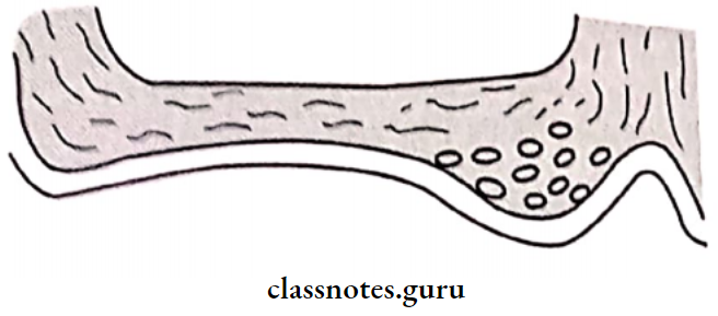
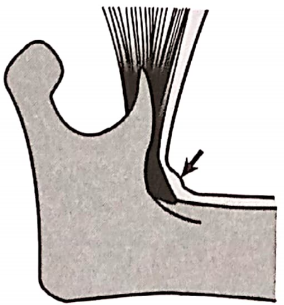
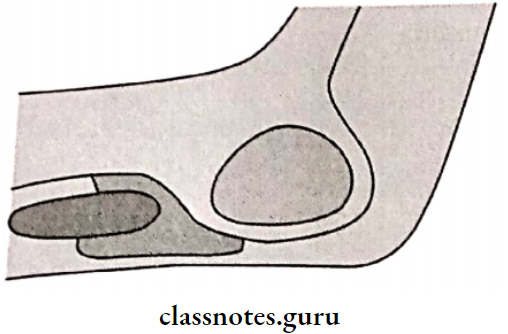
Question 18. Hamular notch.
Answer:

  • The Hamular Notch is the depression situated between the maxillary tuberosity and the hamulus of the medial pterygoid plate. It is a soft area of loose connective tissue
  • The tissues in this area can be safely displaced to achieve the posterior palatal seal
  • The distolateral border of the denture base rests in the hamular notch

Hamular Notch Significance:

  • The denture border should extend to the hamular notch
  • If the border is located anteriorly near the maxillary tuberosity, the denture will not have retentive properties, as the denture will lie on non-resilient tissues in such cases

Complete Denture Pre-Prosthetic Steps

Question 19. Retromylohyoid fossa.
Answer:

  • The Retromylohyoid Fossa belongs to the posterior part of the alveolar lingual sulcus
  • Retromy lohyoid Fossa lies posterior to the mylohyoid muscle

Retromylohyoid Fossa Boundaries:

  • Anteriorly retro mylohyoid curtain
  • Posterolaterally superior constrictor of the pharynx
  • Posteromedially palatoglossus and the lateral surface of the tongue
  • Inferiorly sub mandibular gland

Impression And Mouth Preparation Retromylohyoid Fossa

Question 20. Syneresis and imbibition.
Answer:

  • The process of water sorption by hydrocolloids is known as imbibition
  • Syneresis is a process where the gel may lose water by exudation of fluid
  • Syneresis & imbibition can result in dimensional changes & therefore inaccurate casts
  • To avoid this, hydrocolloids should be poured immediately

Question 21. Advantages of the perforated stock tray.
Answer:

  • Perforated Stock Tray is used for impression materials like alginate
  • Holes present in it are advantageous
  • Perforated Stock Tray helps in retaining the material while impression-making
  • Perforated Stock Tray retains the material by mechanical interlocking


Impression And Mouth Preparation

Impression And Mouth Preparation Long Essays

Question 1. Define impression. Discuss in detail the most widely accepted technique of making an impression in complete dentures.
Answer:

Impression:

A complete denture impression is a negative registration of the entire denture-bearing, stabilizing & border seal areas present in the edentulous mouth.

Impression Techniques:

  1. Mucostatic
  2. Mucocompressive
  3. Selective pressure technique
    • It is the most widely accepted impression technique
    • In this technique, forces are confined to stress-bearing areas, whereas non-stress-bearing areas are relieved

Read And Learn More: Prosthodontics Questions And Answers

Impression Technique:

  • The primary impression is made with overextended borders. The primary cast is prepared from it
  • Undercuts are blocked with wax on the cast. Relief wax is applied over relief areas on the cast
  • A spacer is adapted throughout the extent of the special tray
  • Separating media like cold mold seal or tin foil is applied overcast
  • A special tray with a handle is fabricated
  • Impression should be 2 mm short of the sulcus
  • Materials used for it are: Shellac, cold-cure acrylic, Type II impression compound
  • The wax spacer is then scraped off
  • Border molding is done using greenstick compound
  • Relief wax is removed
  • Escape holes are made over the tray
  • Secondary impression material is loaded over the tray
  • Impression is then seated in the patient’s mouth, and the secondary impression is made.

Impression and Mouth Preparation in Complete Dentures


Impression And Mouth Preparation

Impression Materials Used:

Impression And Mouth Preparation Materials Used And Step

Question 2. Define impression.
Or
Discuss various impression procedures in the complete denture.
Or
Add a note on the selection of impression materials.

Answer:

Impression:

A complete denture impression is a negative registration of the entire denture-bearing, stabilizing & border seal areas present in the edentulous mouth.

Complete Dentures Impression Procedures

Impression Selection Of Materials:

  • Impression should be fluid enough to adapt to oral tissues
  • Impression should be viscous enough to be contained in the tray that is seated in the mouth
  • While in the mouth, they should set into a rubbery or rigid solid in a reasonable amount of time
  • The set impression should not tear or distort when removed
  • The impression made should be dimensionally stable
  • Stability should be maintained after the removal of a cast so that a second or third cast can be made
  • The material should be biocompatible
  • Impression should be cost-effective
  • Should be nontoxic
  • Impression should be acceptable to patients with a pleasant odor and color
  • Should have adequate shelf life, Easy to use with minimum equipment, should have adequate strength

Complete Denture Pre-Prosthetic Preparation

Question 3. Define retention, stability, and support in the complete denture. Discuss various factors affecting retention.
Or 

Define complete denture retention. Enumerate various factors of retention.
Or
Write in detail about retention in complete dentures.

Answer:

  • Retention: That quality inherent in the prosthesis that resists the force of gravity, adhesiveness of foods, and the forces associated with the opening of the jaws
  • Stability: The quality of the denture to be firm, steady, and constant, to resist displacement by functional stresses & not to be subject to change of position when forces are applied
  • Support: Resistance to vertical forces of mastication, occlusal forces, and other forces applied in a direction toward the denture-bearing area

Factors Affecting Retention:

1. Anatomical Factors:

  • Size Of Denture Bearing Area:
    • The retention increases with an increase in the size of the denture-bearing area
    • Maxillary dentures are more retentive than mandibular as it has 24 cm2 compared to that 14 cm2

Impression And Mouth Preparation Maxillary Denture And Mandibular Denture

  • Quality Of Denture Bearing Area:
    • Displaceability of the tissues influences the retention of dentures
    • Displacement of tissues during impression-making
    • Result in tissue rebounding
    • This leads to a loss of retention of dentures

2. Physiological factors:

  • Saliva:
    • Thick & ropy saliva-loss of retention
    • Thin & watery saliva- compromised retention
    • Ptyalism- gagging
    • Xerostomia-Soreness & irritation

3. Physical factors:

  • Adhesion:
    • The physical attraction of unlike molecules to one another
    • The amount of adhesion present is proportional to the denture base area
    • In xerostomia there is no adhesion

Impression And Mouth Preparation Schematic Representation Of Adhesion

  • Cohesion:
    • The physical attraction of like molecules for each other
    • Act within the film of saliva
    • Watery serous saliva- more retentive

Impression And Mouth Preparation Schematic Representation Of Cohesion

  • Interfacial Surface Tension:
    • The tension or resistance to separation possessed by the film of liquid between two well-adapted surfaces
    • Present within the film of thin saliva
    • Useful in the retention of the maxillary denture
    • Depends on the presence of air at the margins of liquid & solid contact

Impression And Mouth Preparation Interfacial Surface Tension

Impression And Mouth Preparation Surface Tension Present In The Maxillary Denture

    • To attain maximum interfacial tension
    • Saliva should be thin
    • Perfect adaptation
    • Covering a large denture area
    • The presence of good cohesive and adhesive forces
  • Capillarity Attraction:
    • That quality or state, because of surface tension, causes elevation or depression of the surface of a liquid that is in contact with a solid
    • The presence of close adaptation between the denture and the mucosa increases surface contact
    • Results in an increase in retention
    • Capillarity Attraction Factors:
      1. Close adaptation
      2. Greater surface area
      3. A thin film of saliva

Impression Techniques in Prosthodontics

  • Atmospheric Pressure And Peripheral Seal:
    • Atmospheric Pressure And Peripheral Seal prevent air entry between the denture and soft tissue
    • To obtain it denture borders should rest on soft and resilient tissues
    • On application of forces, a vacuum is created that aids in retention
    • This is a natural suction of the denture
    • Atmospheric Pressure and Peripheral Seal are directly proportional to the denture base area

Impression And Mouth Preparation Atmosphoric Pressure

4. Mechanical Factors:

  • Undercuts
  • Retentive springs
  • Magnetic forces
  •  Denture adhesives
  • Suction chambers and suction discs

Impression And Mouth Preparation Other Mechanical Attachments

5. Muscular Factors:

  • The balance should be between the forces acting from the buccal musculature and the tongue

Impression And Mouth Preparation Posterior Teeth Arrangement

Question 4. Define complete denture impression. Discuss impression theories and techniques in treating complete denture patients.
Answer:

Complete Denture Impression:

A complete denture impression is a negative registration of the entire denture-bearing, stabilizing, and border seal areas present in the edentulous mouth.

Complete Denture Impression Theories

Techniques Of Impression:

Mouth Preparation Before Complete Dentures

1. Custom Tray Impression:

  • Design a custom tray with a spacer
  • It should be 2 mm short of the sulcus
  • Border molding is done
  • It results in recording the sulcus in dynamic function
  • Spacer is scraped
  • The impression is recorded by ZOE
  • It results in recording the stress-bearing areas under pressure, and the non-stress-bearing areas are relieved

Impression And Mouth Preparation The Wax Spacer Near The Periphery Of The Tray

Impression And Mouth Preparation The wax Spacer Near The Periphery Of The Tray.

Impression And Mouth Preparation Border Moulding The Cheek
Impression And Mouth Preparation A Finished Zinc Oxide Eugenol Impression

2. Tray Compound:

  • The impression made using the tray impression compound
  • The impression is refined and trimmed
  • The metal wire is attached to the tray compound to act as a handle
  • Treat it as a custom tray
  • Border moulding done
  • Wash the impression made on it

Question 5. Define impression. Discuss biological considerations for a maxillary impression.
Answer:

Complete Denture Impression:

A complete denture impression is a negative registration of the entire denture-bearing, stabilizing, and border seal areas present in the edentulous mouth.

Biological Considerations Of Maxillary Impression :

  • The anatomy of the edentulous ridge in the maxilla is important in designing a complete denture
  • Some parts of the ridge are capable of withstanding forces compared to others
  • Thus, it is considered before impression-making

Impression Limiting Structures:

  • Labial Frenum:
    • Labial Frenum is the limiting structure of the Maxilla and Mandible
    • Labial Frenum is a fibrous band
    • Covered by a mucous membrane
      • Extension: Labial aspect of the residual ridge to the lip
      • Maxillary: Passive due to the absence of muscle fibers

Recorded In Impression:

Recorded in impression as a V-shaped notch

  • Requirement: Notch should be narrow and deep

Impression And Mouth Preparation Labial Frenum

  • Labial Vestibule:
    • That portion of the oral cavity which is bounded on one side by the teeth, gingival, and alveolar ridge and on the other side by the lips and cheeks
    • Presence of orbicularis oris
    • Labial Vestibule has an indirect displacing effect on the denture
  •  Buccal Frenum:
    • Buccal Frenum separates the labial and buccal vestibule
    • Muscle attachments present: Levator anguli oris, orbicularis oris, and buccinator
    • Buccal Frenum needs greater clearance on the buccal flange of the denture

Impression And Mouth Preparation Buccal Frenum

  • Buccal Vestibule:
    • Buccal Vestibule extends from the buccal frenum to the hamular notch
    • Its size varies with: the contraction of muscle, the position of mandible and amount of bone loss in the maxilla
  • Hamular Notch:
    • Depression present between maxillary tuberosity and hamulus of medial pterygoid plate
    • Can be easily displaced to achieve posterior pala tal seal

Impression And Mouth Preparation Hamular Notch

Impression Supporting Structures:

1. Primary Stress Bearing Areas:

  •  Hard Palate:
    • Trabeculae pattern of it perpendicular to the direction of forces acting on it
    • Thus it acts as a primary stress-bearing area

Impression And Mouth Preparation Hard Palate

  • Postero-Lateral Slopes Of Residual Ridge:
    • Ridge rapidly resorbs following extraction of teeth
    • Resilient submucosa over provides support to the denture

2. Secondary Stress Bearing Areas:

  1. Rugae:
    • Rugae is a secondary support area in Maxilla
    • Rugae is covered by a thin mucosa

Impression And Mouth Preparation Rugae

    • Rugae Location:
      1. In the anterior region of the palatal mucosa
      2. At the angle of the occlusal plane of the residual ridge
    • Rugae Significance: Important in speech
    • Precautions During Fabrication:
      1. Should not distort this area while impression-making
      2. Metal dentures should reproduce this area to make it comfortable
  1. Maxillary Tuberosity:
    1. Bulbous extension of residual alveolar ridge in 2nd 3rd molar region
    2. It is least likely to resorb

Impression And Mouth Preparation Maxillary Tuberosity

Secondary Stress Relief Areas:

  • Incisive papilla
  • Relief Areas Location:
    • Midline behind central incisors
  • Reason For Relieving:
    • The reason For Relieving is the exit point of nasopalatine nerves and vessels
    • If it is not relieved it compresses vessels and nerves
  • Reason For Relieving Results:
    • Necrosis of the area
    • Paraesthesia of the anterior palate

Impression And Mouth Preparation Incisive Papilla

1. Cuspid Eminence:

  • Bony elevation on residual alveolar ridge
  • Location: between canine and 1st premolar

Impression And Mouth Preparation Cuspid Eminence

2. Mid-Palatine Raphe:

  • Median suture area
  • As it is covered by thin mucosa, it should be relieved

Impression And Mouth Preparation Mid Palatine Raphe

3. Fovea Palatine:

  • Coalescence of ducts of mucous glands
  • Determines the position of the posterior border of the denture
  • Relieved because of the presence of ducts

Impression And Mouth Preparation Fovea Palatina

Question 6. Describe mandibular anatomical structures.
Answer:

Limiting Structures:

  • Labial Frenum:
    • Active due to the presence of muscle incisive and orbicularis oris
    • It is a fibrous band
    • Covered by mucous membrane
    • Extention: labial aspect of the residual ridge to lip
    • Recorded In Impression: Recorded in impression as a V-shaped notch
    • Requirement: Notch should be narrow and deep
      Impression And Mouth Preparation Labial Frenum
  • Labial Vestibule:
    • That portion of the oral cavity which is bounded on one side by the teeth, gingiva, and alveolar ridge and on the other side by the lips and cheeks
    • Influences retention of dentures
  • Buccal Frenum:
    • Contains fibers of the buccinator
    • Prevent displacement of denture

Impression And Mouth Preparation Buccal Frenum.

Buccal Vestibule:

    • Extends from buccal frenum to retromolar region
    • Bound by alveolar ridge and buccinator
    • Influenced by masseter
    • Notch is produced in denture flange called masseteric notch
  • Lingual Frenum:
    • Effects stability of denture high frenal attachment is called tongue tie

Alveololingual Sulcus:

  • Has 3 Regions:
    1. Anterior Region
      • From lingual frenum to pre-mylohyoid fossa
    2. Middle Region
      • From the pre-mylohyoid fossa to the distal part of the mylohyoid ridge
    3. Posterior Region
      • In the region of retro mylohyoid fossa
      • Determines lateral throat form

Impression And Mouth Preparation Anterior Portion Of Alveololingual

Impression And Mouth Preparation Middel Portion Of The Alveololingual

Impression And Mouth Preparation Posterior Portion Of Alveololingual

  • Retromolar Pad:
    • Forms posterior seal of denture
    • Location: Distal to the third molar
    • Consists of: Loose connective tissues, mucosal glands
    • Retromolar Pad Boundaries:
      1. Posteriorly: temporalis
      2. Lateral: buccinator
      3. Medial: Pterygomandibular raphe and superior constrictor

Impression And Mouth Preparation Pear Shaped Pad

  • Pterygomandibular Raphe:
    • Extend: Hamular process to the mylohyoid ridge
    • Muscles Attached:
      • Postero- medially- superior constrictor
      • Antero-laterally-buccinator

Impression And Mouth Preparation Pterygomandibular Raphe

Alveololingual Sulcus Supporting Structures:

1. Buccal Shelf Area:

  • Between the buccal frenum and anterior border of the masseter
  • Buccal Shelf Area Boundaries:
    • Medial: crest of the ridge
    • Distal: retromolar pad
    • Lateral: external oblique ridge
  • Buccal Shelf Area Significance: Width increases with resorption of the ridge
  • Serves as primary stress-bearing area

Impression And Mouth Preparation Buccal Shelf Area

2. Residual Alveolar Ridge:

  • Flat with concave denture-bearing surface
  • On resorption inclines outward

Impression And Mouth Preparation Residual Alveolar Ridge

Relief Areas Mylohyoid Ridge:

  • Lies close to the inferior border of the mandible
  • Covered with mucosa so should be relieved

Impression And Mouth Preparation Mylophyoid Ridge

3. Mental Foramen:

  • Location: Between 1st & 2nd premolar region
  • Relieved as it may lead to paraesthesia

Impression And Mouth Preparation Mental Foraman

4. Genial Tubercle:

  • Location: anterior on the lingual side of the body of the mandible
  • Increases due to resorption

Impression And Mouth Preparation Genial Tubercles

5. Torus Mandibularis:

  • Location: on lingual side near premolar region (b)
  • Covered by a thin mucosa
  • Relieved or surgically removed

Impression And Mouth Preparation Tori On The Mandibular Premolar Area

Question 7. Discuss principles and objectives of making impressions for completely edentulous patients.
Or

What are the objectives of impression-making
Answer:

Principles Of Impression Making:

  1. Presence of healthy oral tissues
  2. Inclusion of all supporting and limiting tissues
  3. Borders within anatomical and physiological limitations
  4. Border molding – physiological type
  5. Space between material and tray
  6. Not damaging the tissues
  7. Application of selective pressure technique
  8. Use of guiding mechanism
  9. Use of dimensionally stable materials
  10. Similarity to the form of dentures

Objectives Of Impression Making:

1. Retention:

“That quality inherent in the prosthesis which resists the force of gravity, adhesiveness of foods, and the forces associated with the opening of the jaws”.

  • Retention Factors:
    1. Anatomical Factors:
      • Size of denture-bearing area
      • Quality of denture-bearing area
    2.  Physiological Factors:
      • Saliva
    3.  Physical Factors:
      • Adhesion
      • Cohesion
      • Interfacial surface tension
      • Capillary attraction
      • Atmospheric pressure
    4. Mechanical Factors:
      • Undercuts
      • Retentive springs
      • Magnetic forces
      • Denture adhesives
      • Suction chambers and suction discs
    5. Muscular Factors:
      • The balance should be between the forces acting from the buccal musculature and tongue

2. Stability:

  • The quality of the denture to be firm, steady, and constant, to resist displacement by functional stresses and not to be subject to change of position when forces are applied
  • Stability withstands horizontal forces
    • Stability Factors:
      1. The vertical height of the residual ridge
      2. Quality of soft tissue
      3. Quality of impression
      4. Occlusal rims
      5. Teeth arrangement
      6. Shape of denture

3. Support:

  • Resistance to vertical forces of mastication, occlusal forces and other forces applied in a direction toward the denture-bearing area
  • The denture base should cover as much denture-bearing area as possible
  • Results in the distribution of forces over a wider area
  • This leads to a reduction of force per unit area

Called Snow Shoe Effect:

Impression And Mouth Preparation Snow Shoe Effect Dencture

4. Aesthetics:

  • Thicker flange leads to fullness of the mouth

5. Preservation of Remaining Structures:

  • Muller stated that the preservation of what remains is more important rather than to replace what is lost.
  • Stress is provided over the stress-bearing area.
  • Relief is provided over non-stress-bearing areas.
  • Prevent damage to oral structures.
  • Avoid overextension of dentures.

Question 8. Define stability and discuss factors affecting it.
Answer:

Stability:

  • “The quality of denture to be firm, steady and constant, to resist displacement by functional stresses and not to be subject to change of position when forces are applied”.
  • Stability withstands horizontal forces

Stability Factors:

1. Vertical Height Of Residual Ridge:

Impression And Mouth Preparation Vertical Height Of Residual Ridge

  • A ridge with an adequate height of ridge provides sufficient support
  • Resorbed ridge causes loss of stability or reduced stability of the denture

Full Denture Clinical Steps – Impression and Prep

2. Quality Of Soft Tissue:

  • Adequate submucosa is required for good stability
  • Excessive submucosa results in poor stability

3. QualitOf Impression:

  • The Impression Should Be:
    • Accurate
    • Smooth surface
    • Devoid of voids
    • Not wrap on removal, Dimensionally stable

4. Occlusal Plane:

  • Should be parallel to the ridge
  • Should divide interarch space equally

Impression And Mouth Preparation The Plane Of Occulation

5. Teeth Arrangement:

  • Teeth are arranged in a neutral zone
  • That is balance is achieved between tongue and buccal musculature

6. Shape Of Denture:

  • The polished surface should resemble oral structures
  • Should not interfere with the functioning of oral structures

Complete Dentures Short Question and Answers

Complete Dentures Short Answers

Question 1. Parts of a complete denture
Answer:

Parts Of Complete Denture

Question 2. Steps in the fabrication of a complete denture
Answer:

  • Diagnosis and treatment planning
  • Making diagnostic casts
  • Mouth preparation
  • Primary impression
  • Primary cast
  • Fabrication of custom trays
  • Secondary impression
  • Master cast
  • Fabrication of occlusal rims
  • Recording jaw relations
  • Articulation
  • Arrangement of artificial teeth
  • Try in
  • Processing of denture
  • Insertion
  • Post-insertion review and maintenance

Read And Learn More: Prosthodontics Question And Answers

Question 3. Effects of aging.
Answer:

  • Young people have lighter teeth due to translucent enamel
  • Old people have dark & opaque teeth due to secondary dentin formationShiny teeth in old people
  • Teeth with a brownish tinge in older people. Certain diseases are limited to certain ages
  • Thus, age can be used to rule out certain systemic conditions
  • Age also determines the prognosis.

Complete Dentures: Short Questions and Answers

Question 4. Metal dentures.
Answer:

Complete Denture Metal Dentures

Question 5. Gagging
Answer:

The gag reflex is a normal, healthy defense mechanism to prevent foreign bodies from entering the trachea

Causes of Gagging:

  • Over-extended denture borders
  • Unstable occlusal conditions
  • Systemic conditions
  • Alcoholism, smoking

Dental Prosthodontics MCQs

Features of Gagging:

  • Causes the displacement of the denture
  • Triggered by tactile stimulation of the soft palate, the posterior part of the tongue, and the fauces

Site of Gagging:

  • Posterior part of the maxillary denture
  • Distolingual part of mandibular denture

Introduction To Complete Dentures Paltal Over Extension Of A Maxillary Denture And Distolingual Over Extension Of A Mandibular

Gagging Treatment:

  • Correction of overextended dentures
  • Correction of occlusal vertical dimensions and occlusal prematurities

Question 6. Angular cheilitis.
Answer:

Angular Cheilitis Etiology:

  • Angular Cheilitis occurs at the angle of the mouth among persons having deep commissural folds secondary to the overclosure of the mouth
  • Angular Cheilitis can occur among persons with lip-licking habits, denture-wearing, or deficiency of riboflavin, vitamin B12, and folic acid

Complete Dentures Angular Cheilitis

Question 7. Kelly’s combination syndrome.
Answer:

Kelly’s Combination Syndrome was identified by Kelly in 1972 in patients wearing a maxillary complete denture opposing a mandibular distal extension prosthesis

Complete Denture FAQs

Kelly’s Combination Syndrome Pathogenesis:

  • It occurs in a sequential manner
  • The patient tends to concentrate the occlusal load on the remaining natural teeth
  • Results in more force acting on the anterior portion of the maxillary denture
  • Increased resorption of the anterior part of the maxilla
  • The labial flange will displace and irritate the labial vestibule
  • Posteriorly, there will be fibrous overgrowth in the maxillary tuberosity
  • The shift of the occlusal plane posteriorly downwards processes resorption in the mandibular distal extension denture-bearing area
  • Mandible shifts anteriorly during occlusion
  • There is
    • Decrease in vertical dimension at occlusion
    • Decrease in retention and stability of a denture
    • Disocclusion of lower anterior
    • Reduced periodontal support of anterior teeth

Question 8. Infection control
Answer:

Infection Control Methods:

  • Immunization:
    • Vaccination for Hepatitis B for all dental personnel
  • Personal Barrier:
    • Hand washing before gloving and after degloving
    • Gloves protect against contamination
    • Changed between patients and for the same patient, is torn or worn out
      • Face Mask:
        1. Protects from inhalation of aerosols
        2. Prevents spatter from the patient’s mouth
        3. Prevents droplets from the contaminated solution
        4. Protective clothing
        5. Gown with long sleeves, high neck & long knee length
      • Eyewear:
        1. Prevention of bacterial/viral contact
  • Surgical Asepsis:
    • Preparation of the surgical site
    • Draping the patient
    • Isolation
  • Surface Asepsis:
    • Sterilization of instruments
    • Use of disposable instruments
  • Disposal Of Waste: Methods
    • Incineration
    • Burial in a landfill
    • |Sterilization

Question 9. Hypermobile ridge tissue.
Answer:

Hypermobile Ridge Tissue Cause:

  • Due to excessive residual ridge resorption, Management:
  • Small tissues that do not interfere with tissue functioning:
    • Left Untreated: Impression is made using the mucositis technique
    • Large Tissues: removed surgically

Denture Objective Questions

Question 10. Polished surface.
Answer:

  • The Polished Surface is defined as that portion of the surface of a denture that extends in an occlusal direction from the border of the denture and includes the palatal surfaces
  • The Polished Surface is the part of the denture base that is usually polished, and it includes the buccal and lingual surfaces of the teeth
  • Polished Surface refers to the external surfaces of the lingual, buccal, and labial flanges and the external palatal surface of the denture
  • The Polished Surface should be well polished and smooth to avoid the collection of food debris
  • The Polished Surface should be harmonious with the oral structures
  • They should not interfere with the action of oral mus- culture

Question 11. Denture irritation hyperplasia.
Answer:

The hyperplastic reaction of mucosa over the borders of the denture

Complete Dentures Denture Irritation Hyperplasia

Question 12. Xerostomia.
Answer:

Xerostomia refers to a subjective sensation of a dry mouth, but is not always associated with salivary hypofunction

Xerostomia Etiology:

  1. Developmental:
    1. Salivary gland aplasia
  2. Water/metabolic Loss:
    • Impaired fluid intake
    • Hemorrhage
    • Vomiting/diarrhea
  3. Iatrogenic:
    • Medications:
      • Antihistamines: Diphenhydramine
      • Decongestants: Pseudoephedrine
      • Antidepressants: Amitriptyline
      • Antipsychotic: Haloperidol
      • Antihypertensive: Methyldopa, CCB
      • Anticholinergic: Atropine
  4. Radiation therapy of the head and neck:
    1. Both stimulated and unstimulated salivary flow decreases with increasing radiotherapy.
    2. Systemic Diseases:
      • Sjogren’s syndrome
      • Diabetes mellitus
      • Diabetes insipidus
      • HIV infections
      • Psychological disorders.
      • Graft-versus-host disease
  5. Local Factors:
    • Decreased mastication.
    • Smoking
    • Mouth breathing

Question 13. Functions of saliva.
Answer: 

Introduction To Complete Dentures Function Of Saliva

Question 14. Combination syndrome.
Answer:

Combination Syndrome was identified by Kelly in 1972 in patients wearing a maxillary complete denture opposing a mandibular distal extension prosthesis

BDS Prosthodontics Viva Questions

Combination Syndrome Pathogenesis:

  • Combination Syndrome occurs in a sequential manner
  • The patient tends to concentrate the occlusal load on remaining natural teeth resulting in more force acting on the anterior portion of the maxillary denture
  • Increased resorption of the anterior part of the maxilla
  • The labial flange will displace and irritate the labial vestibule. Posteriorly, there will be fibrous overgrowth in the maxillary tuberosity
  • The shift of the occlusal plane posteriorly downwards processes resorption in the mandibular distal extension denture-bearing area
  • Mandible shifts anteriorly during occlusion
  • Decrease in vertical dimension at occlusion
  • Decrease in retention and stability of the denture
  • Disocclusion of lower anterior
  • Reduced periodontal support of anterior teeth.

Diseases Of The Nervous System Long Essays

Diseases Of The Nervous System Long Essays

Question 1. Describe the etiology, clinical features, and localization of infra-nuclear facial nerve palsy.
(or)
Describe the etiology, clinical features, and management of facial palsy

Answer:

Facial Palsy

  • Idiopathic paralysis of the facial nerve of sudden onset

Etiology: 5 Hypothesis:

  • Rheumatic
  • Cold
  • Ischaemia
  • Immunological
  • Viral

Facial Palsy Clinical Features:

  • Pain in post auricular region
  • Sudden onset
  • Unilateral loss of function
  • Loss of facial expression
  • Absence of wrinkling
  • Inability to close the eye “ Watering of an eye “
  • Inability to blow the cheek
  • Obliteration of nasolabial fold
  • Loss of taste sensation
  • Hyperacusis
  • Slurring of speech

Diseases of the nervous system long essays

Facial Palsy Management:

  • Physiotherapy
  • Facial exercises
  • Massaging
  • Electrical stimulation a Protection for the eye
  • Covering of eye with bandage
    • Medical management
      • Prednisolone 60-80 mg per day
      • 3 tablets for 1st 4 days
      • 2 tablets for 2nd 4 days
      • 1 tablet for 3rd 4 days
    • Surgical treatment
      • Nerve decompression
      • Nerve grafting

Question 2. Classify epilepsy. Describe clinical features, diagnosis, and management of grand mal epilepsy.

Answer:

Epilepsy:

  • Epilepsy is a group of disorders of cerebral functions characterized by chronic, recurrent, paroxysmal, nonsynchronous discharge of cerebral neurons

Grand Mal Epilepsy Classification:

  1. Partial or focal seizures
    • Simple partial seizures
      • Motor
      • Sensory
      • Visual
      • Versive
      • Psychomotor
    • Complex partial seizures
      • Temporal lobe
      • Frontal lobe
    • Secondary generalized partial seizures
  2. Primary generalized seizures
    • Tonic-clonic
    • Tonic
    • Absence
    • Akinetic
    • Myoclonic
  3. Unclassified seizures
    • Neonatal seizures
    • Infantile spasms

Grand Mal Epilepsy:

  • It is a common type of epilepsy

grand Mal Epilepsy Clinical Features:

  • Phases of grand mal epilepsy
  1. Prodromal phase
    • Symptoms are uneasiness or irritability
    • It lasts for hours or days before an attack
  2. Aura
    • Occurs when partial seizure becomes generalized
    • Symptoms Are:
      • Visual disturbances
      • Hallucinations
      • Nausea
      • Epigastric discomfort
      • Alteration in psychic functions
  3. The tonic and clonic phase
    • Symptoms Are
      • Tonic contraction of muscles
      • Flexion of arms
      • Extension of legs
      • Cry due to spasm of respiratory muscles
      • It lasts for 10-30 seconds
      • Clonic phase causes:
      • Violent jerking of face and limbs
      • Biting of the tongue
      • Incontinence of urine and feces
      • It lasts for 1-5 minutes
  4. Postictal phase
    • Symptoms are:
    • Deep unconsciousness with flaccid limbs
    • Loss of corneal reflex
    • Plantar extensor
    • It lasts for a few minutes to several hours

Grand Mal Epilepsy Diagnosis:

  • History of patient
  • Clinical symptoms
  • Blood test to assess metabolic disorders
  • Brain imaging

Grand Mal Epilepsy Management:

  1. Elimination of causative agent
  2. Protection of patient
    • Protected from a hot and sharp object
    • Use of padded mouth gag
    • Airway maintenance
    • 4 administration of Diazepam 5-10 mg
  3. Long-term drug therapy
    • Phenytoin sodium-200-400 mg daily
    • Carbamazepine- 600-1800 mg daily in divided dose
    • Sodium valproate- 0-2000 mg daily
    • Phenobarbitone-60-180 mg daily
    • Primidone-750-1500 mg daily in a divided dose

Long essay questions on neurological disorders

Question 3. Describe the etiology and clinical features of meningitis. How would you proceed to establish the diagnosis?
(or)
What are the causes of meningitis? Describe clinical features, complications, and treatment of pyogenic meningitis.
(or)
Discuss the etiology, clinical features, and investigations of pyogenic meningitis.

Answer:

Meningitis:

  • Meningitis is defined as inflammation of the pia-arachnoid and the fluid contained in the space

Pyogenic Meningitis:

  • Pyogenic Meningitis is bacterial meningitis

Etiology:

  • Gram negative bacilli
  • Group B streptococci
  • Listeria monocytogenes
  • H. influenzae
  • Neisseria meningitidis
  • Mycobacterium tuberculosis

Read And Learn More: General Medicine Question and Answers

pyogenic Meningitis Clinical Features:

  • Classical triad- fever, headache, and neck rigidity
  • Tachycardia, tachypnoea
  • Convulsions in children
  • Headache
  • Blurring of vision
  • Papilloedema
  • Ecchymosis
  • Associated lung, ear, and sinus infection

Pyogenic Meningitis Diagnosis

Diseases Of The Nervous System Pyogenic Meningitis Diagnosis

Pyogenic Meningitis Complications:

  • Neurological deficiencies- hemiplegia, aphasia, blindness, deafness
  • Mental deterioration
  • Brain abscess
  • Auditory impairment
  • Subdural empyema
  • Internal hydrocephalous

Pyogenic Meningitis Management:

  • Ceftriaxone provides adequate coverage against infection

Diseases Of The Nervous System Pyogenic Meningitis Management

Central nervous system diseases long essays

Question 4. Discuss clinical features, complications, and management of tubercular meningitis.

Answer:

Tubercular Meningitis:

  • Meningeal involvement by the mycobacterium tuberculosis causes tubercular meningitis

Tubercular Meningitis Clinical Features:

  • Insidious in onset
  • Headache
  • Vomiting
  • Low-grade fever
  • Confusion
  • Lassitude
  • Visual disturbances
  • Papilloedema
  • Neck rigidity
  • Cranial nerve palsies
  • Hydrocephalus

Tubercular Meningitis Complications:

  • Hydrocephalous
  • Focal deficits
  • Cranial nerve palsies

Tubercular Meningitis Management:

  1. General management
    • Maintenance of nutrition
    • Electrolyte balance
    • Care of bowel and bladder
  2. Drug therapy
    • Anti-tubercular drugs
      • Injection of streptomycin 1 g IM daily
      • Tab icons 600-900 mg/ day
      • Tab ethambutol
    • Steroids
      • 20-30 mg prednisolone daily for a few weeks

Question 5. Discuss etiopathogenesis, and clinical features of Parkinsonian disease. Outline the drugs used in its treatment.

Answer:

Parkinsonian Disease:

  • Parkinsonian Disease is a syndrome consisting of akinesia and brake- nesia, rigidity, and tremors

Etiopathogenesis:

  • There is a loss of pigmented cells in the substantianigra
  • Dopamine levels in the striatum get depleted

Parkinsonian  Clinical Features:

  • Both sexes are equally affected
  • Age- The fifth decade and later age group are affected
  • Muscle ache
  • Depression
  • Slow activity
  • Tremors
  • Rigidity
  • Hypokinesia

Parkinsonian  Treatment:

  1. Anticholinergics
    • Trihexyphenidyl
    • Benzhexol
    • Phenadrine
  2. Amantadine
  3. I-dopa- it is administered orally
  4. Dopamine receptor agonists
    • Bromocriptine
    • Lisuride
    • Pergolide
  5. Selegiline
    • Catechol-o-methyltransferase inhibitors

Long descriptive answers on nervous system diseases

Question 6. Discuss various factors you consider in evaluating a patient for general anesthesia

Answer:

Preoperative Evaluation For General Anaesthesia:

  • The patient’s history is asked
  • Physical evaluation of the patient
  • General examination of a patient
    • Weight and height
    • Pulse rate
    • Rhythm
    • Volume
    • Blood pressure is measured
    • Temperature is measured
    • Movements of eyeballs
    • Feel the carotid arteries
  • Systems examined
    • Cardiovascular
    • Respiratory
    • Nervous system
    • GIT
    • Genitourinary tract

Question 7. Mention causes of cerebral embolism. What are its manifestations? Describe the principles of management.

Answer:

Cerebral Embolism:

  • The emboli travel to the brain and cause cerebral embolism

general anesthesia Causes:

  • Atherosclerosis
  • High cholesterol level
  • High blood pressure

General Anesthesia Clinical Features:

  • Focal motor deficit
  • Changes in sensorium
  • Visual and sensory deficits
  • Respiratory arrest
  • Seizures
  • Severe headache

General Anesthesia Treatment:

  • Use of anticoagulants like heparin and warfarin
  • Use of thrombolytics like streptokinase to dissolve the clot

Neurology long essay questions with answers

Question 8. Describe the pathogenesis, differential diagnosis, and management of coma

Answer:

COMA

Pathogenesis:

Diseases Of The Nervous System Pathogenesis

Coma Differential Diagnosis:

  • Cerebral anaemia
  • Mechanical injury of the brain
  • Convulsive attacks
  • Cerebral vascular attacks
  • Poisons
  • Local infection of the brain and meninges

Coma Management:

  • Treatment of the underlying cause
  • Provide proper nutrition
  • Maintain patient’s physical health
  • Prevention of infection
  • Physiotherapy to prevent bone, joint, and muscle deformities

Question 9. Describe the clinical features of intracerebral hemorrhage

Answer:

Clinical Features Of Intracerebral Haemorrhage:

  • Hypertension
  • Fever
  • Cardiac arrhythmias
  • Nuchal rigidity
  • Subhyoid retinal haemorrhages
  • Altered level of consciousness
  • Focal neurological deficits
  • Seizures
  • Headache
  • Nausea and vomiting

Nervous system pathology long answer questions

Question 9. Enumerate the causes of headaches. Discuss clinical features and management and prevention of migraine

Answer:

Causes Of Headache:

  1. Migraine headache
  2. Tension-type of headache
  3. Cluster headache
  4. Miscellaneous headache
  5. Traumatic headache
  6. Headache due to vascular causes- hematoma
  7. Headache due to nonvascular causes- due to increased pressure
  8. Headache due to substance abuse- alcohol
  9. Headache due to systemic infection
  10. Headache due to metabolic disorders
  11. Headache due to referred pain- from the ear, etc
  12. Cranial neuralgia- trigeminal neuralgia
  13. Unclassified headache

Migraine:

  • Migraine is characterized by an episodic, hemicranial, or unilateral throbbing headache and is often associated with nausea, vomiting, and visual disturbances

Headache Clinical Features:

  • Starts after puberty
  • Common in females
  • Headache occurs at regular intervals
  • Each attack lasts for hours to days
  • Prodromal symptoms
  1. Photophobia
  2. Visual disturbances
  3. Dysphagia
  4. Tinnitus
  5. Hemiparesis
  6. Hemianaesthesia
  7. Severe and throbbing headache

Long essay topics in nervous system disorders

Headache – Management:

  • Removal of aggravating factors like alcohol, oral contraceptives, dietary factors
  • Aspirin-600-900 mg/day.
  • Paracetamol lg/day
  • Anti-emetics like metoclopramide
  • Ergotamine tartrate 0.5-1 mg sublingually orally or rectally
  • Serotonin agonist sumatriptan 50-100 mg orally 2-3 times a day

Headache Prevention:

  • Beta-blockers- propranolol- 80-120 mg/ day
  • Pizotifen-1.5-3 mg at night
  • Antidepressant- amitriptyline 50-100 mg at bedtime
  • Flunarizine- 10 mg daily
  • These all block 5-HT receptors

Diseases Of The Nervous System Short Essays

Diseases Of The Nervous System Short Essays

Question 1. Status epilepticus

Answer:

Status Epilepticus

  • Status Epilepticus is a condition in which a series of seizures occur in the patient without regaining consciousness in between successive attacks

Status Epilepticus Precipitating Factors:

  • Sudden withdrawal of drugs
  • Irregular use of anti-convulsants
  • following intracranial pathology

Status Epilepticus Management:

  • Loosen clothes around neck
  • Maintain airway
  • Administration of high concentration of oxygen
  • Diazepam 10-20 mg IV over 1-5 minutes
  • Monitor BP, ECG, and blood gases
  • Diazepam 10 mg IV repeat once after 15 minutes
  • Start infusion drip of phenytoin, 18 mg/ kg at the rate of 50 mg/min
  • If seizure are not controlled, start infusion drip of chloromethiazol 4 0.5-1.2 g/hour
  • If seizures are not still controlled start 4 drip of thiopentone sodium 20 mg/kg 4 at 50-100 mg/min

Diseases of the nervous system short essays

Read And Learn More: General Medicine Question and Answers

Question 2. Anti-epileptic drugs

Answer:

Anti-Epileptic Drugs Classification:

Diseases Of The Nervous System Anti Epileptic Drugs

Anti-Epileptic Drugs – Mechanism Of Action:

  • Blockade of sodium channels
  • Prolongation of their inactive state
  • Blockade of low threshold calcium current in the thalamic neurons
  • Enhancing GABA-mediated inhibition

Question 3. Trigeminal neuralgia

Answer:

Etiology:

  1. Pathological
    • Dental pathosis
    • Traction on divisions of trigeminal nerve
    • Ischaemia
    • Aneurysm of internal carotid artery
  2. Environmental
    • Allergic
    • Irritation to the ganglion
    • Secondary lesions

Trigeminal Neuralgia Clinical Features:

  • Age: Around 35 years
  • Sex: Common in female
  • Site: Right lower portion of the face, usually unilateral
  • Duration: Few seconds to few minutes
  • As time passes duration between the cycles decreases
  • Nature: stabbing or lancinating
  • Aggravating Factors: Activation of TRIGGER ZONES
  • These are Vermillion border of lip, around the eyes, ala of nose

Interference With Other Activities:

  • Patient avoids shaving, washing face, chewing, brushing, as these may aggrevate pain
  • These lead to poor lifestyle
  • Extreme Cases: leads to FROZEN OR MASK-LIKE FACE

Trigeminal Neuralgia Management:

1. Medical

  • Carbamazepine: initial dose: 100mg twice daily until relief is achieved
  • Dilantin: 300-400mg in single or divided doses
  • Combination Therapy: Dilantin + carbamazepine

2. Surgical

  • Injection of alcohol in gasserian ganglion
  • Nerve avulsion: Performed on lingual, buccal or mental nerve
  • Part of nerve is sectioned
  • Electrocoagulation of gasserian ganglion: diathermy is done
    • Rhizotomy: Trigeminal sensory root is sectioned
    • Newer Technique: TENS
  • Low-intensity current is used at high frequency is applied to the skin through electrodes attached by a conduction paste

Short essay on nervous system disorders

Question 4. Etiology and clinical manifestations of depression

Answer:

Etiology

  • Depression is a common psychiatric disorder

Diseases Of The Nervous System Etiology And Clinical Manifestations Of Depression

Etiology Clinical Manifestations:

  1. Emotional symptoms
    • Sadness
    • Misery
    • Hopelessness
    • Low self esteem
    • Loss of interest
    • Suicidal thoughts
  2. Biological symptoms
    • Fatigue
    • Apathy
    • Loss of libido
    • Loss of appetite
    • Lack of concentration
    • Sleep disturbances
  3. Symptoms of bipolar depression
    • Over enthusiasm
    • Overconfidence
    • Irritation
    • Aggression

Question 5. Petit mal epilepsy

Answer:

Petit Mal Epilepsy

  • This form of epilepsy is seen in children

Petit Mal Epilepsy Features:

  • Child stops working
  • Looks confused
  • Stares in space
  • May blink or roll up eyeballs
  • Fails to respond to verbal commands
  • Attack is brief

Petit Mal Epilepsy Diagnosis:

  • EEG changes shows spike and wave complexes at a frequency of 3 Hz per second

Question 6. Peripheral neuropathy

Answer:

Peripheral Neuropathy

  • Peripheral neuropathy is the disorder of peripheral nerves either sensory, motor, or mixed, symmetrical, and affecting distal parts of limbs

Diseases Of The Nervous System Peripheral Neuropathy

Neurological disorders short essay questions

Question 7. Causes of epilepsy

Answer:

Causes Of Epilepsy

Diseases Of The Nervous System Causes Of Epilepsy

Question 8. Hypertensive encephalopathy

Answer:

Hypertensive Encephalopathy

  • Hypertensive encephalopathy is characterized by a very high blood pressure and neurological disturbances including transient abnormalities in speech, vision, paresthesia, disorientation, fits, loss of consciousness, and papilloedema

Hypertensive Encephalopathy Treatment:

  • Intravenous sodium nitroprusside-0.3-1 micro- gratn/kg/ min
  • Parenteral labetelol- 2 mg/min
  • Hydralazine-5-10 mg every 30 min
  • Bed rest
  • Sedation
  • Diuretics

Nervous system diseases short note essays

Question 9. Discuss the differential diagnosis of headache

Answer:

The Differential Diagnosis Of Headache

  1. Migraine headache
  2. Tension type of headache
  3. Cluster headache
  4. Miscellaneous headache
  5. Traumatic headache
  6. Headache due to vascular causes- hematoma
  7. Headache due to non vascular causes- due to increased pressure
  8. Headache due to substance abuse- alcohol
  9. Headache due to systemic infection
  10. Headache due to metabolic disorders
  11. Headache due to referred pain- from ear, etc
  12. Cranial neuralgia- trigeminal neuralgia
  13. Unclassified headache

Diseases Of The Nervous System Important Notes

Diseases Of The Nervous System Important Notes

  1. Bell’s Palsy
    • Bell’s Palsy is idiopathic paralysis of facial nerve
    • Etiology
      • Rheumatic – Cold
      • Ischaemia
      • Immunological
      • Viral
    • Clinical features
      • Pain in post auricular region
      • Sudden onset
      • Unilateral loss of function
      • Loss of facial expression
      • Absence of wrinkling
      • Inability to close eye
      • Watering of eye
      • Inability to blow
      • Obliteration of nasolabial fold
      • Loss of taste sensation
      • Hyperacusis
      • Slurring of speech
  2. Grand Mal Epilepsy
    • Phases
      • Prodromal phase
      • Aura
      • Tonic and clonic phase
      • Postictal phase
  3. Nerve LesionDiseases Of The Nervous System Nerve Lesion
  4. lower Motor Neuron v/s Upper Motor NeuronDiseases Of The Nervous System Lower Neuron Or Upper Motor Neuron
  5. Types Of SeizuresDiseases Of The Nervous System Types Of Seizures
  6. Pyogenic Meningitis
    • Etiology
      • Cram negative bacteria
      • Croup B streptococci
      • Listeria monocytogenes
      • H. influenza
      • N. meningitides
      • M. tuberculosis
      • Drug of choice – ceftriaxone
  7. Features Of Horner’s Syndrome
    • Miosis
    • Ptosis
    • Anhydrosis
    • Enophthalmos
  8. Drug Of Choice In Different SeizuresDiseases Of The Nervous System Drug Of Choice In Different Seizure
  9. Disorders Of Basal Ganglia
    • Parkinson’s disease
    • Wilson’s degeneration
    • Chorea
    • Athetosis
  10. Parkinsonism
    • Parkinsonism is a syndrome consisting of akinesia and bradykinesia, rigidity, and tremors
    • Anticholinergics are used for it
  11. Migraine
    • Migraine is characterized by an episodic hemicranial or unilateral throbbing headache and is often associated with nausea, vomiting, and visual disturbances. It is the most common vascular headache
    • Causes pain of the face and jaws
    • Occurs due to vasoconstriction of intracranial vessels followed by vasodilation
  12. Status Epilepticus
    • Status Epilepticus is a condition in which a series of seizures occur in the patient without regaining consciousness in between successive attacks
    • Precipitating factors
      • Sudden withdrawal of drugs
      • Irregular use of anticonvulsants
      • Following major intracranial pathology
    • Treatment
      • Immediate treatment
        • Rest the patient on the bed
        • Loosen the clothes
        • Maintain airway
        • Administration of high concentration of 02
        • Administration of 20-25 ml dextrose
        • Transfer the patient to ICU
    • Late treatment
      • Diazepam 10 mg IV repeat once only after 15 min
      • Lorazepam 4 mg IV repeated after 10 min
        • If the patient does not respond then drip of phenytoin 15 mg/kg at the rate of 50 mg/min
        • If still not controlled then 4 drip of thiopentone sodium 20 mg/kg at 50-100 mg/min.

Diseases Of The Nervous System Nervous System

Diseases of the nervous system important notes

Read And Learn More: General Medicine Question and Answers

  1. Trigeminal Neuralgia
    • Occurs due to paralysis of the trigeminal nerve
    • Clinical features
      • Site: right lower portion of the face, usually unilateral
      • Duration: a few seconds to a few minutes
      • As time passes duration between the cycles decreases
      • Nature: stabbing or lancinating
      • Aggravating Factors: activation of TRIGGER ZONES
      • These are the Vermillion border of the lip, around the eyes, ala of nose
      • Interference with other activities:
      • The patient avoids shaving, washing face, chewing, and brushing, as these may aggravate pain
      • These lead to a poor lifestyle
      • Extreme cases: leads to “FROZEN OR MASK-LIKE FACE”
  2. Syncope
    • Syncope refers to generalized weakness of muscles, loss of postural tone, inability to maintain an erect posture, and loss of consciousness
    • Syncope Causes
      • Decreases cerebral perfusion
        • Inadequate vasoconstrictor mechanism
        • Hypovolemia
        • Reduction of venous return
        • Reduced cardiac output
        • Arrhythmias
        • Cerebrovascular disturbances
      • Noncirculatory causes
        • Hypoxia
        • Prolonged bed rest
        • Hypoglycaemia
        • Anaemia
        • Anxiety neurosis
        • Hyperventilation
    • Syncope Types
      • Vasovagal syncope
      • Postural hypotension with syncope
      • Micturition syncope
      • Cardiac syncope
      • Carotid sinus syncope
      • Cough syncope
      • Syncope of cerebrovascular disease
  3. Petit Mal Epilepsy
    • Typically seen in children
    • It is brief, and lasts only for seconds
    • They may left unnoticed by the people
    • Later it may develop into tonic-clonic seizures in adulthood

Neurology quick revision notes

Occurs Due To Developmental Abnormality Of Neuronal Control

  1. Babinski’ Sign
    • Extension of the great toe with fanning of other toes is called Babinski’s sign
  2. Headache
    • Classification
      • Migraine headache
      • Tension type of headache
      • Cluster headache
      • Miscellaneous headache
      • Traumatic headache
      • Headache due to vascular causes- hematoma
      • Headache due to nonvascular causes- due to increased pressure
      • Headache due to substance abuse- alcohol
      • Headache due to systemic infection
      • Headache due to metabolic disorders
      • Headache due to referred pain- from the ear, etc.
      • Cranial neuralgia- trigeminal neuralgia
      • Unclassified headache

Respiratory Diseases Long Essays

Diseases Of The Respiratory System Long Essays

Question 1. Describe the causes of bronchitis and chronic bronchitis and their clinical features, complications, and management.

Answer:

Bronchitis:

  • Bronchitis is an inflammation of the bronchi leading to increased bronchial secretions and airway obstruction

Bronchitis Causes:

  1. Smoke, atmospheric, and industrial pollutants
    • These are atmospheric pollutants
    • They damage the protective mucosal barrier of the nose
    • Results in hypertrophy of mucus-secreting glands
  2. Infection
    • It acts as a precipitating factor
  3. Physical factors
    • Sudden change in temperature and exposure to fog leads to acute attacks of bronchitis
  4. Genetic and familial
    • Alpha 1 antitrypsin deficiency leads to bronchitis

Bronchitis Management:

  1. Removal of etiological factors
    • Avoid smoking
    • Avoid exposure to pollutants
  2. Treatment of infection
    • Antibiotics used are:
      • Oxytetracycline or ampicillin 250-500 mg after every 6 hours for 5-7 days
      • Cotrimoxazole- 960 mg tablets for 5-7 days
  3. Bronchodilators
    • Oral theophylline- 150 mg BID is used
    • Inhaled beta-2 adrenoreceptor stimulants- salbutamol 200 mg every 6 hours is used
    • In severe cases, ipratropium bromide- 40-80 meg every 6 hours is used
  4. Mucolytic agents
    • These include bromhexine and carbocystein
  5. Corticosteroids
    • Prednisolone 30 mg/day for 2 weeks is used
  6. Oxygen therapy

Respiratory system disorders long answer

Chronic Bronchitis:

  • Chronic Bronchitis is defined as a cough with expectoration on most days at least three consecutive months in a year for more than two consecutive years

Bronchitis Clinical Features:

  • Productive cough
  • Dyspnoea
  • Wheezing
  • Occasionally chest pain, fever, and fatigue
  • Hemoptysis- the presence of blood streaks in sputum
  • Complications:
  • Type 1 and 2 respiratory failure
  • Pulmonary arterial hypertension and corpulmonale
  • Secondary infections
  • Secondary polycythemia

Question 2. Describe clinical features and management of bronchial asthma.
(or)
Discuss the etiology, clinical features, investigations, and management of bronchial asthma.

Answer:

Bronchial Asthma:

  • Bronchial Asthma is a disease of the airway in which there is chronic inflammation and increased responsiveness to a variety of stimuli leading to reversible airway obstruction.

Etiology:

  • Etiology is caused by various allergens, which are
  1. Most common
    • Pollens, house dust, feathers, animal dander, insect web, fungal spores, etc.
  2. Less common
    • Ingestion in predisposed individuals like fish, meat, milk, yeast, and wheat

Bronchial Asthma Clinical Features:

  • Symptoms may be episodic or persistent
  1. Episodic asthma
    • Sudden in onset
    • Triggered by allergens, exercise
    • Attacks may be spontaneous
    • Duration varies from a few hours to days and even two weeks
    • Characterized by wheezing, cough, and dyspnoea
    • Between attacks, the patients are usually asymptomatic
  2. Acute severe asthma
    • It is a severe life-threatening attack of asthma
    • The patient may have tachycardia, cyanosis, and active accessory respiratory muscles
  3. Chronic asthma
  • Triad of symptoms- chest tightness, wheeze or cough, and exertional dyspnoea occurs
  • Cough is productive
  • A prominent wheeze is audible
  • Breath sound is harsh vesicular with prolonged expiration

Pathophysiology of respiratory diseases essay

Read And Learn More: General Medicine Question and Answers

Bronchial Asthma Investigations:

Diseases Of The Respiratory System Bronchial Asthma Inverstigation

Bronchial Asthma Management:

  1. Management Of Acute Asthma
    • Avoidance of allergens
      • Avoid allergens that may aggravate asthmatic attack
  2. Hyposensitisation
  3. Prevention of mediators of bronchoconstriction
    • Sodium cromoglycate is administered by inhalation
    • Useful in some cases of non-atopic asthma
  4. Drug treatment
    • Use of salbutamol or terbutaline 100-200 meg is used
    • Use of beclomethasoneupto 800 meg twice a day is used along with it
    • A high dose of corticosteroids is used along with bronchodilators.

Respiratory Diseases Respiration Events

Respiratory diseases long questions with answers

Question 3. Describe the etiology, clinical features, and management of ARDS.

Answer:

Acute Respiratory Distress Syndrome- ARDS:

  • It describes the acute, diffuse pulmonary inflammatory response to either direct or indirect blood-borne insults.

Etiology:

  1. Direct-Inhalation
    • Aspiration of gastric contents
    • Toxic gases/ bum injury
    • Pneumonia
    • Blunt chest trauma
  2. Indirect-Blood Borne
    • Necrotic tissue
    • Multiple trauma
    • Pancreatitis
    • Severe bums
    • Drugs
    • Major blood transfusion reacion
    • Anaphylaxis
    • Fat embolism

Acute Respiratory Distress Syndrome Clinical Features:

  • Neutrophil sequestration in pulmonary capillaries
  • Increased capillary permeability
  • Protein-rich pulmonary edema
  • Alveolar collapse
  • Progressive pulmonary fibrosis
  • Multiple organ failure

Acute Respiratory Distress Syndrome Management:

  1. General
    • Mechanical ventilation through
      • Oro-tracheal intubation
      • Tracheostomy
        • Antibiotic therapy to treat infection
  2. Mechanical ventilation
    • For it, three parameters are used
      • Positive end-expiratory pressure- to maintain maximal recruitment of alveolar units
      • Mean airway pressure- to promote recruitment and predictor of hemodynamic effects
      • Plateau pressure- predictor of alveolar overdistention
  3. Fluid management
    • By diuresis or fluid restriction
  4. Corticosteroids
    • Methylprednisolone 2 mg/kg daily is used for 3-5 days
    • The dose is then tapered to 0.5-1 mg daily

Question 4. What is pneumothorax? What are the causes of pneumothorax? How do you manage tension pneumothorax?

Answer:

Pneumothorax:

  • The presence of air in the pleural cavity is known as pneumothorax

Pneumothorax Causes:

  • Rupture of subpleural blebs at the lung apices
  • Rupture of emphysematous bullae
  • Rupture of a subpleural tuberculous focus
  • Rupture of lung abscess
  • Pulmonary infarction
  • Bronchial asthma
  • Acute respiratory distress syndrome
  • Sarcoidosis

Pneumothorax Management:

  • Introduction of a wide-bore plastic cannula, one end is attached to a long rubber tubing, and the other is placed underwater in a bottle
  • Another method is the introduction of an intercostal catheter connected to a water seal drainage system

Respiratory diseases classification long form

Question 5. Describe pneumonia, Bacterial Pneumonia, complications, clinical features, and treatment of community-acquired pneumococcal pneumonia.

Answer:

Pneumonia:

  • Pneumonia is defined as an inflammation of the parenchyma of the lung.

Pneumococcal Pneumonia

  • Pneumococcal Pneumonia is characterized by the homogeneous consolidation of one or more lobes or segments

Bacterfial Pneumonia:

  • Etiology:
    • Streptococcal pneumonia Staphylococcal Aureus
    • Mycoplasma
    • Streptococcus pyrogens

Bacterial Pneumonia Complications:

  • Para pneumonic pleural effusion or Empyema
  • Lpbar collapse
  • Pneumothorax
  • Lung abscess
  • Hepatitis, meningitis
  • Pericarditis
  • Myocarditis
  • Septicaemia

Bacterial Pneumonia Clinical Features:

  • It is sudden in onset
  • Fever with chills and rigors occurs
  • Vomiting
  • Convulsions
  • Loss of appetite
  • Headache
  • Breathlessness
  • Chest pain
  • Central cyanosis
  • Haemoptysis
  • Weakness
  • Tachycardia, tachypnoea
  • Productive cough
  • Rust-colored sputum
  • Rapid and shallow breathing

Bacterial Pneumonia Management:

  1. Oxygen therapy
    • Delivered in high concentration through masks
  2. Analgesics
    • Pethidine-50-100 mg or morphine 10-15 mg is given intramuscularly
  3. Antibiotics
    • Oral amoxicillin 500 mg 8 hourly or
    • Erythromycin 500 mg 6 hourly or
    • Oral cephalosporin 250 mg 8 hourly
    • Cotrimoxazole 960 mg 2 times daily orally
  4. Physiotherapy
    • The patient is encouraged to cough and take a deep breath

Respiratory tract infections long answer questions

Question 6. Describe etiopathology, clinical features, complications, diagnosis, and treatment of lobar pneumonia

Answer:

Lobar Pneumonia:

  • Lobar pneumonia is a radiological and pathological condition referring to the homogenous consolidation of one or more lung lobes associated with pleural inflammation

Etiopathology:

  • Etiopathology is caused by viruses, bacteria, fungi and parasites

Lobar Pneumonia Clinical Features:

  • Sudden or insidious onset
  • Fever with chills and rigors
  • Headache
  • Productive cough
  • Breathlessness
  • Chest pain
  • Nausea, vomiting, diarrhea
  • Myalgia, arthralgia

Lobar Pneumonia Complications:

  • Para pneumonic pleural effusion
  • Emphysema
  • Lung abscess
  • Acute Respiratory
  • Distress Syndrome
  • Pneumothorax
  • Hepatitis
  • Multiorgan failure
  • Formation of ectopic abscess

Lobar Pneumonia Diagnosis:

Diseases Of The Respiratory System Lobar Pneumonia

Lobar Pneumonia Treatment:

  1. Oxygen therapy
    • High-concentration oxygen is delivered through masks
  2. Intravenous fluid administration
  3. Antibiotic therapy
    • Duration of treatment-10-14 days
    • Drugs used are:
    • Ceftriaxone 1-2 g/day plus macrolide or fluoroquinolone

Pneumonia – etiology, clinical features and management

Question 7. Define and classify pneumonias. Describe the investigations, complications, and treatment of community-acquired pneumonia.

Answer:

Definition: Pneumonia is defined as an inflammation of the parenchyma of the lung.

Community-Acquired Pneumonia Classification:

  • Pneumonia is classified into four types
  1. Lobar pneumonia
    • Generally, the entire lobe of the lung is involved
  2. Bronchopneumonia
    • There is neutrophilic exudate in the bronchi and bronchiole with the peripheral spread of infection to alveoli
  3. Interstitial pneumonia
    • There is predominant involvement of the in-interstitium, alveolar wall, and connective tissue around the broncho-vascular tree
  4. Military pneumonia
    • Occurs due to the homogenous spread of pathogens to the lungs

Community-Acquired Pneumonia:

  • It is acquired by inhalation of infected oropharyngeal secretions
  • Caused specifically by highly virulent organisms

Community-Acquired Pneumonias Classification Investigations:

Diseases Of The Respiratory System Pneumonias Investigation

Community-Acquired Pneumonia Classification Complication:

  • Persistent fever
  • Emphysema
  • Proximal bronchial obstruction
  • Recurrent aspiration

Pulmonary tuberculosis long essay

Question 8. Describe the clinical features of pulmonary tuberculosis, and write short-term chemotherapy.

Answer:

Pulmonary Tuberculosis:

  • Tuberculosis is a chronic infective disorder produced by mycobacterium tuberculosis characterised by the formation of granulomas and the development of cell-mediated hypersensitivity
  • The involvement of the lungs by tuberculosis is called pulmonary tuberculosis

Pulmonary Tuberculosis Clinical Features:

  • Loss of weight
  • Loss of appetite
  • Fever with night sweats
  • Tiredness, malaise
  • Amenorrhoea
  • Cough with purulent sputum
  • Haemoptysis
  • Chest pain
  • Breathlessness
  • Finger clubbing
  • Distended neck and chest veins
  • Pleural effusion

Chemotherapy Of Tuberculosis

Diseases Of The Respiratory System Chemotherapy Of Tuberculosis

Question 9. Describe the diagnosis, complications, and management of post-primary tuberculosis

Answer:

Post-Primary Tuberculosis:

Post-Primary Tuberculosis Complications:

  • Haemoptysis
  • Pneumothorax
  • Secondary infection of the cavity
  • Pleural effusion
  • Emphysema
  • Pulmonary fibrosis
  • Scar carcinoma
  • Respiratory failure
  • Amyloidosis
  • Anaemia

Post-Primary Tuberculosis Investigations:

Diseases Of The Respiratory System Post Primary Tuberculosis Inverstigation

Lung cancer classification and treatment long answer

Question 10. Describe the etiology, clinical features, and treatment of bronchial obstruction.

Answer:

Bronchial Obstruction:

  • Bronchial Obstruction is called bronchiectasis
  • Bronchial Obstruction is defined as an abnormal and irreversible dilatation of bronchi

Etiology:

  1. Infective causes
    • Bacterial- H. influenzae, staphylococcus aureus, E. coli, Tuberculosis, mycoplasma
    • Viral- measles, adenovirus, influenza virus
    • Fungal
  2. Obstructive causes
    • Endobronchial benign neoplasm
    • Foreign body aspiration
    • Chronic bronchitis
    • Enlarged lymph nodes
  3. Noninfective causes
    • Allergic
    • Cystic fibrosis

Bronchial Obstruction Clinical Features:

  • Chronic cough with massive expectations
  • Haemoptysis
  • Recurrent pulmonary infection
  • Dyspnoea
  • Fever, weight loss, anemia, and weakness
  • Oedema
  • Sepsis

Bronchial Obstruction Treatment:

  1. Physiotherapy
    • To keep bronchi dilated
  2. Antibiotic therapy
    • Ampicillin- 500 mg 8 hourly or
    • Amoxycillin 500 mg 8 hourly or
    • Oral cefaclor 250 mg 8 hourly
  3. Bronchodilators and nuicolytics
    • Bronchodilators used are theophylline or terbutaline
    • Mucolytics used arc bromhexine, carboys- teenie
  4. Surgery- indications
    • Those who do not respond to medical therapy
    • Those who have repeated massive hemoptysis
    • Young patients
    • Unilateral bronchiectasis

Respiratory disorders long essays for nursing students

Question 11. Describe the pathogenesis, clinical features, and diagnosis of lung abscess.
(or)
Write etiopathogenesis, clinical features, and treatment of lung abscesses.

Answer:

Bronchial Obstruction Lung Abscess:

  • Bronchial Obstruction Lung Abscess is a collection of purulent material ilocalizedised necrotic area of lung parenchyma

Etiopathogenesis:

  1. Infection without obstruction
    • Aspiration of nasopharyngeal contents
    • Involvement of various organisms like staphylococcus, Kleibsella, gram-negative and anaerobic organisms
    • Formation of abscess
    • Metastatic spread of infection
  2. Obstruction with or without infection
    • Bronchus obstruction due to tumor, foreign body, lymph node
    • Bronchial collapse
    • Abscess formation

Bronchial Obstruction Clinical Features:

  • High-grade fever with chills and rigors
  • Pleuritic chest pain
  • Dry cough
  • The presence of copious purulent discharge
  • Haemoptysis
  • Weight loss, anorexia
  • Emphysema

Bronchial Obstruction Investigations:

Diseases Of The Respiratory System Lung Abscess Inverstigation

Bronchial Obstruction Management:

  1. Postural drainage and chest physiotherapy
  2. Antibiotic therapy
    • Oral amoxicillin 50 mg 8 hourly for 5-7 days
    • Cotrimoxazole 960 mg twice daily
    • Oral metronidazole 400 mg 8 hourly
  3. Surgery
    • Resection of part of the lung is done
    • Indications:
      • Massive hemoptysis
      • Localized malignancy
      • Associated symptomatic bronchiectasis
      • Persistent abscess cavity

Obstructive vs restrictive lung disease long essay

Question 12. Describe the etiology, clinical features, investigations, and management of pulmonary eosinophilia.

Answer:

Pulmonary Eosinophilia:

  • Pulmonary Eosinophilia is a lesion in the lungs associated with blood eosinophilia

Etiology:

  • Parasitic infections- hookworm disease, ascariasis, strongyloidiasis
  • Allergic conditions- Hay fever, asthma, drugs like aspirin, aspergillosis
  • Skin disorders- eczema, dermatitis herpetiform
  • Tumors- lymphoma
  • Collagen vascular disorders- rheumatoid arthritis, polyarteritisnodosa
  • Hypereosinophilic syndromes- Loeffler’s syndrome, idiopathic
  • Miscellaneous- Sarcoidosis, Addison’s disease

Pulmonary Eosinophilia Clinical Features:

  • Dyspnoea or orthopnoea
  • Wheezing
  • Cough with mucoid expectoration
  • Haemoptysis
  • Types
    • Cryptogenic eosinophilic pneumonia
    • Idiopathic eosinophilic syndromes- includes
      • Loeffler’s syndrome
      • Hypereosinophilic syndrome
      • Churg Strauss syndrome
    • Tropical pulmonary eosinophilia

Pulmonary Eosinophilia Diagnosis:

  • Blood examination- High blood eosinophils count
  • Sputum shows eosinophilia
  • X-ray of the chest shows diffuse miliary mottling of the lungs

Pulmonary Eosinophilia Treatment:

  • Diethylcarbamazine- 2 mg/kg three times a day is used
  • Allergic reactions are controlled by the use of antichrist-mimics and steroids