Vomer
Vomer Terminology
‘Vomer’ is a Latin word. The term is used for the thin plate of bone between the nostrils.
Vomer Location
Vomer forms the posteroinferior part of the septum of the nose.
Vomer Features And Attachments
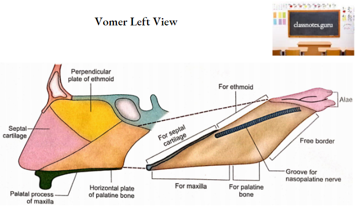
Vomer has got two surfaces (right and left) and four borders (superior, inferior, anterior, and posterior).
Vomer Surfaces
- It has small grooves for vessels.
- A large groove runs downwards and forwards. This is meant for nasopalatine nerve and vessels.
Vomer Borders
- Superior Border
- It is thick.
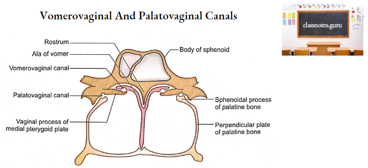
- Two lateral projections (alae) enclose a deep furrow that fits over the rostrum of the sphenoid.
- The margin of ala intervenes between the body of the sphenoid and the vaginal process of the medial pterygoid plate. Under the surface of the ala forms a vomer-ovaginal canal with the vaginal process.
- Inferior Border
- It articulates with the nasal crest formed by the maxillae and palatine bones.
- Anterior Border
- It is the longest border.
- Its upper half articulates with the posterior border of the perpendicular plate of the ethmoid bone.
- Its lower half is attached to septal cartilage.
- Posterior Border
- It is free.
- It is situated between two posterior nasal apertures (choanae).
Vomer Ossification
- Vomer develops by ossification of the membrane covering the median septal part of the cartilaginous nasal capsule.
- One center of ossification appears on each side of the cartilage at about the 8th week of intrauterine life. giving rise to two bony plates separated by a cartilage.
- Two bony plates fuse with each other in the lower part at about the 12th week of intrauterine life.
- The cartilaginous plate is gradually absorbed allowing the fusion of two bony plates which proceed upwards from below. Fusion is completed at puberty.
Vomer Applied Anatomy
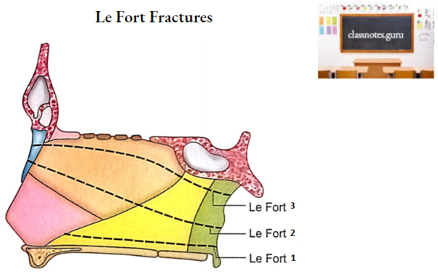
- The vomer is paper thin and does not resist much force responsible for the fracture.
- The vomer is involved in all three types of Le Fort fractures of the mid-facial skeleton.
- Vomer receives adequate blood supply from periosteal arteries and therefore all the fragments of fractured bone retain a periosteal blood supply.
- A transverse fracture of the vomer due to a direct blow on the nose can lead to deviation of the nasal septum (DNS).
- The vomer is clothed in mucosa over large areas of its surfaces, and therefore, its fracture opens into the nasal cavity with a potential risk of infection.
- Vomer may be deviated from the median plane as a result of birth injury or a congenital malformation.
- In case of severe deviation, the nasal septum comes into contact with the lateral wall of the nasal cavity. Surgical repair (submucosal resection-SMR) is usually necessary to correct the deviation.