Skull General Features Introduction
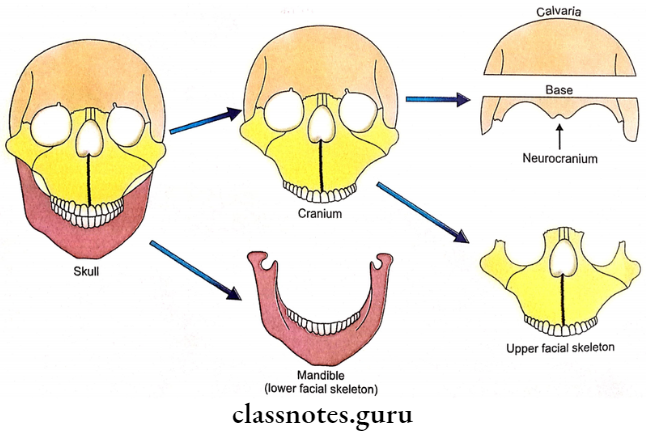
- The skull is the skeleton of the head.
- Cranium means skull minus the mandible.
- Neurocranium is the upper part of the skull that encloses the brain.
- The calvaria is the upper part of the cranium. It is also called the skull cap.
- The facial skeleton (viscerocranium) is the skull minus the calvaria.
- The facial skeleton is further divided into an upper facial skeleton and a lower facial skeleton (mandible).
Neurocranium
The bones which constitute the neurocranium can be classified as:
1. Paired Bones
- These include:
- Parietal bones.
- Temporal bones.
2. Unpaired Bones
These include:
- Frontal bone.
- Occipital bone.
- Sphenoid.
- Ethmoid.
Facial Skeleton (Viscerocranium Or Splanchnocranium)
It is composed of the following bones:
1. Paired Bones
These include:
- Maxillae.
- Zygomatic bones.
- Nasal bones.
- Lacrimal bones.
- Palatine bones.
- Inferior conchae.
2. Unpaired Bones
These include:
- Mandible.
- Vomer.
Anatomical Position Of Skull
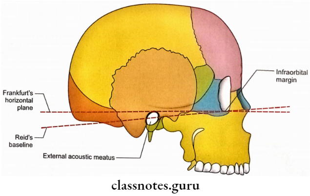
The skull can be kept in a normal anatomical position by Reid’s baseline or Frankfurt’s horizontal plane.
1. Reid’s Baseline
It is a horizontal line formed by the joining of the infraorbital margin with the center of the external acoustic meatus.
2. Frankfurt’s Horizontal Plane
It is marked by the horizontal line joining the infraorbital margin with the upper margin of the external acoustic meatus.
Note:
- Remember that Reid starts with ‘R’ which also stands for ‘Round’ opening of the external acoustic meatus, thus Reid’s baseline passes through the rounded external acoustic meatus.
- On the other hand ‘Frankfurt’ starts with ‘F’ which also stands for ‘Fly’ and anything that has to fly has to be above, therefore, Frankfurt’s horizontal plane passes above the external acoustic meatus.
Skull Applied Anatomy
Clinically the entire front of the skull is considered to be a facial skeleton. It is further divided into the following three parts:
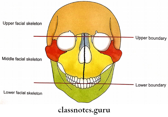
1. Upper Facial Skeleton
- It forms the skeleton of the forehead.
- It is comprised of frontal bone.
2. Lower Facial Skeleton
- It forms the skeleton of the lower jaw.
- It is comprised of a mandible.
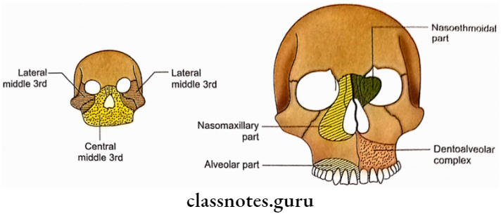
3. Middle Facial Skeleton
It is the complex middle 3rd of the facial skeleton. The region is of great clinical importance because the multiple bones constituting it are frequently involved in fractures.
1. Boundaries
- Upper: Transverse line passing through frontozygomatic, frontomaxillary, and frontonasal sutures.
- Lower: Incisal edge and occlusal plane.
- Posterior: Spheno-ethmoidal junction.
2. Bones Involved In The Fractures Of The Middle 3rd Of The Facial Skeleton Are As Follows:
- Maxillae.
- Palatine bones.
- Zygomatic bones.
- Zygomatic processes of temporal bones.
- Nasal bones.
- Lacrimal bones.
- Vomer.
- Ethmoid.
- Pterygoid processes of sphenoid.
3. Subdivisions
The middle 3rd facial skeleton can be divided into:
- Lateral middle 3rd also called zygomatico- maxillary part.
- Central middle 3rd, which can be further subdivided into:
- Alveolar part.
- Dentoalveolar complex.
- Nasomaxillary part.
- Nasoethmoidal part.