Ribs General Considerations
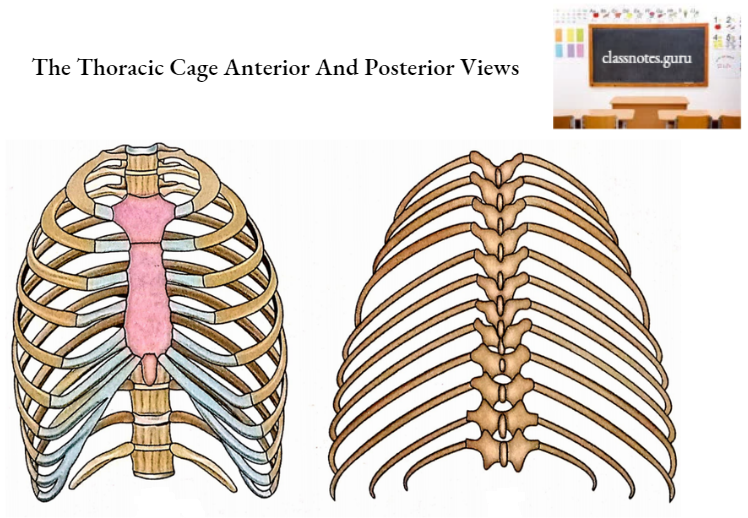
- Ribs are bilateral bony arches forming the greater part of the thoracic wall.
- Normally there are 12 pairs of ribs which are numbered from above downwards.
- The length of the ribs increases from the 1st to the 7th rib and then decreases from the 7th to the 12th rib. Therefore, the 7th rib is the longest rib.
- The ribs are arranged obliquely, i.e. the anterior end is at a lower level than the posterior end. The obliquity is maximum in the 9th rib.
- The 8th rib is the most laterally projected rib.
- The width of the rib gradually reduces from above downwards.
- Intercostal spaces (gaps between adjacent ribs) are deeper in front than behind and deeper in the upper part than the lower part.
First Ribs
Distinguishing Features
- It is the shortest.
- It is the broadest.
- It is most curved.
- It has no twisting.
- Angle coincides with tubercle.
- Head has got only a single facet.
- The costal groove is absent.
- The neck is rounded and elongated.
- It is flattened from above downwards and therefore has inner and outer borders and superior and inferior surfaces.
Side Determination
- Keep the larger end anteriorly and the smaller end posteriorly.
- Keep the surface of the shaft having two grooves separated by a ridge, superiorly.
- Keep the concave border towards the inner side and the convex border towards the outer side.
Note: Keep the rib on a flat surface considering its position in your own body.
The rib belongs to the side on which both ends touch the surface simultaneously. If the rib is placed on the wrong side then only the anterior end will be touching the table top.
Anatomical Position
- The posterior end is nearer the midline than the anterior end.
- The posterior end is 3.5 cm higher than the anterior end.
- The upper surface faces upwards as well as forwards.
Features And Attachments
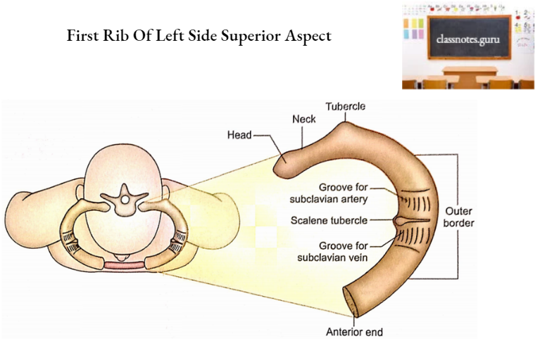
Just like a typical rib, the first rib is comprised of two ends (anterior and posterior) and a shaft.
- Anterior End
- It is the larger end.
- It meets with 1st costal cartilage.
- Posterior End
- It consists of the head, neck, and tubercle.
- Head
- It is small and rounded.
- It has a single rounded facet for articulation with the body of 1st thoracic vertebra to form a costovertebral joint.
- The capsular ligament of 1st costovertebral joint is attached to the margins of the facet.
- The radiate ligament is attached to the anterior margin of the head.
- Neck
- It is rounded.
- It is directed upwards, backward, and laterally.
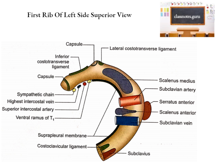
- The inferior costotransverse ligament is attached to its posterior surface.
- The following structures form the anterior relations of the neck from medial to lateral:
- Sympathetic chain.
- First posterior intercostal vein.
- Superior intercostal artery.
- First thoracic root (T) of brachial plexus.
- Note: Remember SVAN for the relations of the anterior aspect of the neck from medial to lateral in which the S-sympathetic chain, V-Vein,- Artery, and N-Nerve.
- Tubercle
- It is large and prominent.
- It articulates with the transverse process of 1st thoracic vertebra.
- The lateral costotransverse ligament is attached laterally to the tubercle.
- Head
- It consists of the head, neck, and tubercle.
- Shaft
- It consists of two borders (outer and inner) and two surfaces (upper and lower).
- Outer border
- It is convex.
- It is thick posteriorly and thin anteriorly.
- 1st digitation of the serratus anterior arises from its middle.
- It is related to the scalenus posterior muscle in its posterior part while clavipectoral fascia and pectoralis major muscle in its anterior part.
- Inner border
- It is concave.
- The scalene tubercle is situated near its middle.
- Sibson’s fascia (suprapleural membrane) is attached to it.
- Upper surface
- It is rough and irregular.
- It presents two shallow grooves separated by a ridge.
- The ridge continues medially with the scalene tubercle along the inner border.
- The scalenus anterior is inserted on the ridge and scalene tubercle.
- The subclavian vein lies in the groove anterior to the ridge.
- The subclavian artery along with the lower trunk of the brachial plexus occupies the posterior groove.
- Note: Remember ‘VAN’ is the sequence of structures occupying the grooves on the superior surface from anterior to posterior, i.e. Vein, Artery, and Nerve.
- The area anterior to the groove for the subclavian vein provides attachments to the subclavius muscle (anteriorly) and costoclavicular ligament (posteriorly). These attachments are located near the anterior end because they also extend over the costal cartilage.
- Scalenus medius is inserted on the rough area posterior to the groove for the subclavian artery.
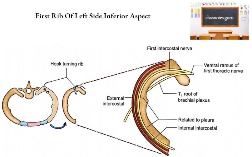
- Lower surface
- It is smooth.
- It is related to costal pleura.
- Intercostal muscles are attached to this surface near its outer border.
- 1st intercostal nerve and vessels are related to this surface mainly in its posterior part.
- Outer border
- It consists of two borders (outer and inner) and two surfaces (upper and lower).