Parietal Bones Terminology
The word parietal is derived from the Latin word ‘paries’ which means ‘wall’, because two parietal bones form a large part of the walls of the calvaria.
Parietal Bones Side Determination
- Keep the bone by the side of your own cranial vault in such a way that the outer surface is convex and the inner surface is concave.
- Inferior (squamosal) border is concave.
- The anteroinferior angle is prominent and has a vascular and narrow groove on its inner aspect.
- The posteroinferior angle has a shallow and wide groove for the sigmoid sinus on its inner aspect.
Parietal Bones Features And Attachments
1. Parietal Bones Surfaces
It has two surfaces, external and internal.
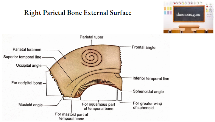
- External Surface
- It is relatively smooth.
- The most prominent part of this surface is called parietal tuberosity or eminence.
- There are two curved lines running anteroposteriorly. These are called superior and inferior temporal lines.
- The superior temporal line gives attachment to temporal fascia while the area below the inferior temporal line gives attachment to the temporalis muscle.
- The area above the superior temporal line is covered by galea aponeurotica.
- A foramen may be present near the posterior part of the sagittal border. This is called parietal foramen. It transmits the emissary vein.
- Internal Surface
- It is concave and exhibits elevations and depressions for cerebral sulci and gyri respectively.
- Near the sagittal border, there is a longitudinal half groove (to be completed with that of the opposite side) for the superior sagittal sinus. The margins of the groove provide attachment to falx cerebri
- Grooves for the branches of middle meningeal vessels are present at the anteroinferior angle and at the middle of the lower border of the bone.
- Adjacent to the groove for the superior sagittal sinus there are deep irregular pits (granular foveolae) produced by arachnoid granulations.
- The bone is grooved near the posteroinferior angle by the sigmoid sinus.
2. Parietal Bones Borders
It has four borders, superior, inferior, anterior, and posterior.
- Superior Border
- This is also called the sagittal border.
- It articulates with a similar border of opposite sides to form a sagittal suture.
- Inferior Border
- This is also called the squamosal border.
- It articulates with the following three bones from anterior to posterior:
- Greater wing of the sphenoid.
- Squamous part of temporal.
- Mastoid portion of temporal.
- Anterior Border
- This is also called the frontal border.
- It articulates with the frontal bone to form a coronal suture.
- Posterior Border
- This is also called the occipital border.
- It articulates with the squamous part of the occipital bone to form a lambdoid suture.
3. Parietal Bones Angles
The parietal bone has four angles (frontal, sphenoidal, occipital, and mastoid).
- Frontal Angle
- This is also called the anterosuperior angle.
- It corresponds to bregma, i.e. the junction of coronal and sagittal sutures.
- Sphenoidal Angle
- This is also called an anteroinferior angle.
- It corresponds to pterion, i.e. a small area enclosing four bones (frontal, temporal, parietal, and greater wing of sphenoid).
- Occipital Angle
- This is also called the posterosuperior angle.
- It corresponds to lambda, i.e. junction of sagittal and lambdoid sutures.
- Mastoid Angle
- This is also called posteroinferior angle.
- It corresponds to asterion, i.e. small area enclosing three bones, parietal, temporal, and occipital.
Parietal Bones Ossification
- Parietal bones ossify in the membrane.
- Each ossifies from two centers which appear at parietal tuberosity at about the 7th week of intrauterine life.
- The centers soon fuse with each other and then the ossification spreads radially.
- Angles are the parts last to be ossified explaining the existence of a fontanelle at each angle before the ossification is completed.
Parietal Bones Age Changes
- At birth
- Temporal lines are present at quite a lower level.
- Adult
- Higher and permanent positions of temporal lines are reached only after the eruption of permanent molar teeth.
Parietal Bones Applied Anatomy
- Occasionally the parietal bone is divided into upper and lower parts by an anteroposterior suture. The condition may be confused with fracture radiologically.
- The latter can be ruled out easily because the anomalous parietal suture is usually bilateral.
- Parietal bones are loosely attached to the adjacent bones at sutures during the intrauterine period allowing moulding (change in shape of calvaria) at the time of childbirth.
- Calvaria returns to normal shape within a few days after birth.
- Parietal bones undergo remodeling to allow enlargement of calvaria during childhood. This is only possible because of their loose attachments to the adjacent bones.
- Granular foveolae are more numerous and marked in aged parietal bones. This fact is of great medical importance.
- The regenerating capacity of the parietal bone is negligible due to the lack of a cambium layer in the periosteum.
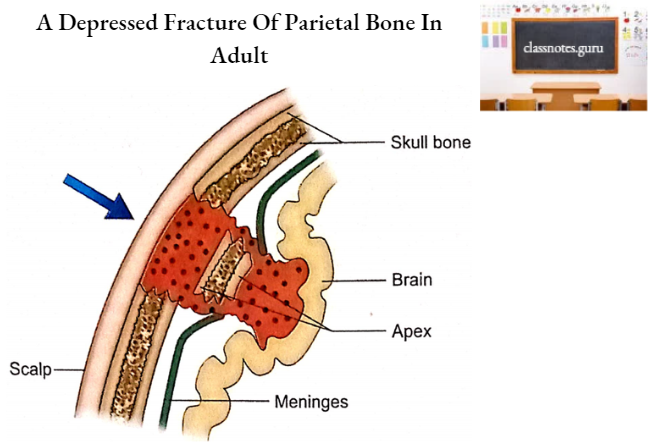
- In neonates, the parietal bone is pliable and soft, and, therefore, a depressed fracture (pond fracture) is like a dimple.
- In adults such fractures are produced by direct blows and always show an irregular line of fracture at the periphery of the depressed area. The depression of the inner table forms the lowest limit of the depressed area also
- Almost invariably all fractures the known as apex.
- A crack in the inner table of the parietal bone may damage a large diplomatic vein and produce a small epidural hematoma. parietal bone in children is associated with rupture of the dura mater.
- In adults, the parietal bone shows a fissured or linear fracture if the force is transmitted to this bone from frontal or occipital blows.