
Nasal Cavity
The nasal cavity is the beginning of the respiratory system. It is divided into right and left halves by a midline partition called nasal septum.
To study the cavity, a sagittal section of the skull is considered in which one and a half of the skull shows a lateral wall of the nasal cavity and the second half shows a nasal septum.
Nasal Cavity Features
Each half of the nasal cavity consists of a roof, floor, medial wall, and lateral wall.
1. Roof
It has anterior and posterior slopings and a middle horizontal part.
- Anterior Sloping
- It is formed by the nasal bone and nasal spine of the frontal bone.
- Posterior Sloping
- It is formed by the following bones from anterior to posterior:
- The anterior surface of the body of the sphenoid.
- Ala of the vomer.
- Sphenoidal process of palatine bone.
- It possesses an opening of the sphenoidal sinus.
- It is formed by the following bones from anterior to posterior:
- Middle Horizontal Part
- It is formed by the cribriform plate of the ethmoid bone.
- The number of foramina in it provides passages for filaments of the olfactory nerve.
- One of these perforations in its anterior part transmits the anterior ethmoidal nerve and vessels.
2. Floor
- It is formed by the superior surface of the bony palate, i.e. palatine process of the maxilla and horizontal plate of the palatine bone.
- Anteriorly near the septum, a small infundibular opening leads into an incisive canal.
- The nasopalatine nerve and greater palatine vessels traverse the incisive canal.
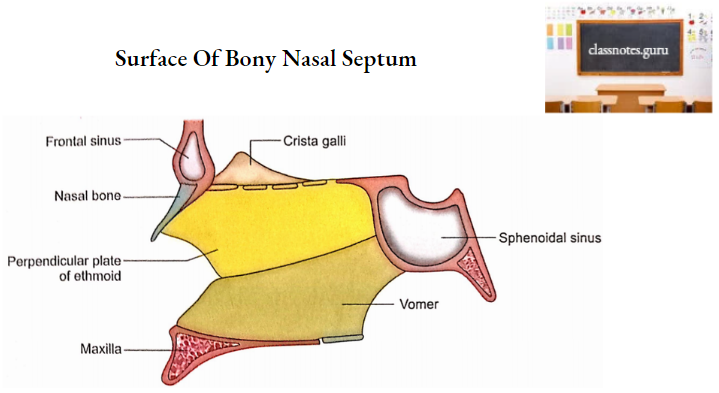
3. Medial wall
- It is formed by bony septum in a dried skull.
- The posteroinferior part of the bony septum is contributed by vomer.
- Its anterosuperior part is formed by a perpendicular plate of ethmoid.
- The nasal crest (below), sphenoidal crest, and rostrum (above and behind) provide minor contributions to the bony septum.
- A groove on each side of the vomer descends downwards and forwards towards the incisive canal. It lodges the nasopalatine nerve.
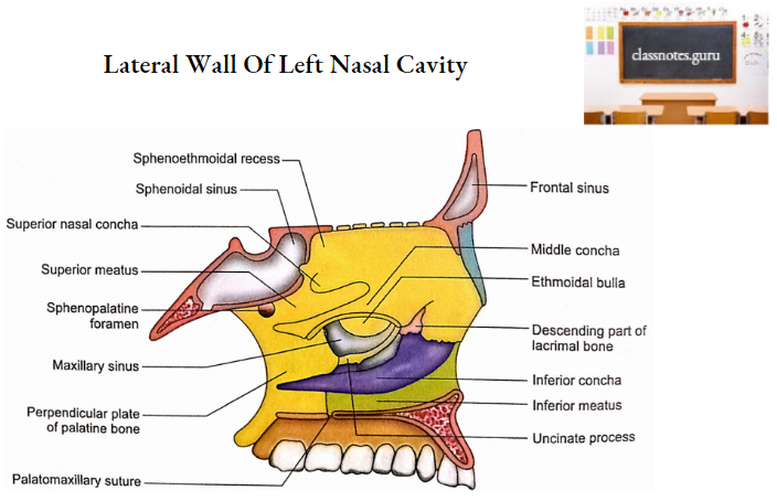
4. Lateral wall
- This is very irregular.
- This is marked by three bracket-like projections that run anteroposteriorly and lie one above the other. These projections are named from above downwards, superior, middle, and inferior conchae.
- Each concha forms the medial wall and roof of the corresponding meatus. Therefore, there are three meatus, superior, middle, and inferior.
- The part of the lateral wall above the superior concha is called sphenoethmoidal
recess. - The main bony contributions in the lateral wall are as follows:
- Below and in the front nasal surface of the maxilla.
- Behind-Perpendicular plate of palatine bone.
- Above-Ethmoidal labyrinth (with superior and middle conchae).
- Below-Inferior concha (an independent bone).
- Inferior meatus
- It lies below and lateral to the inferior concha.
- It receives an opening of the nasolacrimal duct in its anterior part.
- Middle meatus
- It is situated between the middle and inferior conchae.
- A rounded elevation in its upper part is called ethmoidal bulla produced by middle ethmoidal air cells.
- On the surface or just above the bulla is the opening of the middle ethmoidal sinus.
- Behind the bulla is the opening of the maxillary sinus.
- Anteroinferior to bulla is a curved, thin bony projection called the uncinate process of ethmoid.
- The uncertain process passes backward and encloses a curved gap between it and the bulla. This gap is called hiatus semilunaris.
- Upper end of hiatus semilunaris continues with ethmoidal infundibulum.
- The infundibulum of the ethmoid receives the opening of the anterior ethmoidal sinus and itself continues up as the frontonasal duct reaches the frontal sinus.
- Superior meatus
- It is situated between the superior and middle conchae.
- The opening of the posterior ethmoidal sinus is located in the superior meatus.
- Sphenoethmoidal recess
- It is situated between the roof and the superior concha.
- It receives the opening of the sphenoidal air sinus.
- Sphenopalatine foramen
- It is located just behind the superior concha.
- It transmits the sphenopalatine artery and nasal branches of the pterygopalatine ganglion.
Nasal Cavity Applied Anatomy
1. Fracture of the base of the skull involving the anterior cranial fossa might lead to communication between the nasal cavity and subarachnoid space resulting in CSF leak into the nose. This condition is called CSF rhinorrhoea.
2. An impact on the nasal bridge will lead to fracture and displacement of the nasal septum.
3. Fracture of the cribriform plate will result in anosmia (loss of smell sensation) due to damage to olfactory nerve filaments.
4. Fracture of the cribriform plate may cause infection to enter the cranial cavity from the nasal cavity resulting in meningitis.
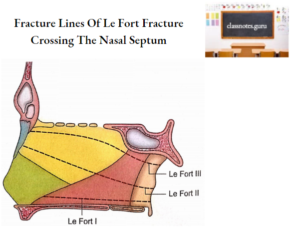
5. Le Fort fractures of the mid-facial skeleton always involve the nasal septum.
6. All the bones of the nasal cavity are clothed in the mucosa, therefore, their fractures open to the nasal cavity with the potential risk of infection.
7. The bones of the nasal cavity receive adequate blood supply from periosteal arteries and, therefore, all the fragments of fractured bone retain a periosteal blood supply.
8. Bony septum is papery thin and does not resist much to forces responsible for fracture.
9. The floor of the nasal cavity may be fractured in an uncommon central split of the palate. It is actually paramedian in nature because median sutures (intermaxillary and interpalatine) are relatively strong.
10. Nasal ethmoidostomy (an artificial opening to drain the ethmoidal air cell) is a common practice in several surgical procedures involving ethmoidal labyrinth.
11. Lacrimal bone is very fragile therefore an extra precaution should be taken to avoid trauma in cases of surgery of the lacrimal system.
12. In the management of congenital lacrimal defect, in fracture of the inferior concha is sometimes needed.
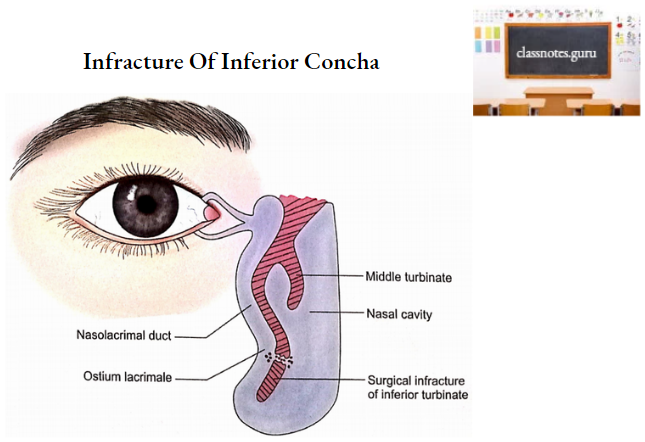
13. In cases of obstruction of the lacrimal sac or nasolacrimal duct, dacryocystorhinostomy is performed. In this operation, an artificial passage is made by breaking the lacrimal bone.
14. Congenital atresia of the choanae is an uncommon condition in which there is occlusion of the posterior naris by a bony or membranous diaphragm.
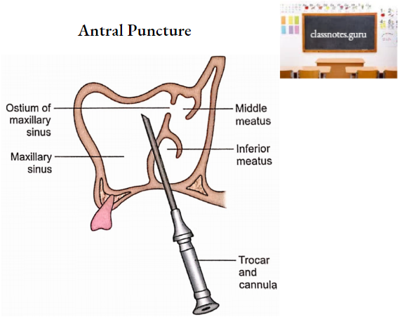
15. Antral puncture is a common procedure performed by ENT surgeons to wash out infected fluid in the maxillary sinus (antral lavage). The antrum is punctured through the inferior meatus by a trocar and cannula.
16. Deviation of nasal septum (DNS) is a common condition that may be developmental in origin or may arise from trauma.
17. A perforated septum may be due to septal surgery or syphilis or tuberculosis.
18. Sometimes a permanent opening is made in the inferior meatus to encourage the drainage of the pus in the maxillary sinus. This operation is called intranasal antrostomy.