
Oral Medicine Investigations Important Notes
1. Oral Medicine Investigations Biopsy:
- It is the removal of part of tissue for the purpose of histological examination And Analysis
Oral Medicine Investigations Types:
- Punch Biopsy.
- Incisional Biopsy.
- Excisional Biopsy.
- Needle Biopsy.
2. Oral Medicine Investigations Tests And Their Uses:
- Schilling test
- It is done to detect vitamin B12 deficiency as well as to distinguish and detect a lack of intrinsic factors and malabsorption
- The test is performed in three stages
- Stage 1 – without intrinsic factor
- Stage 2 – with intrinsic factor
- Stage 3 – test for malabsorption of vitamin B12
- Paget’s test
- It is used to examine the swelling
- Finger pressure is applied over the swelling
- Lugol’s iodine test
- It is used as an aid to the diagnosis of malignant lesions
- It contains iodine, potassium iodide, and distilled water
- Normal cells- stained brown black
- Inflammatory tissue- stained dark brown
- Schimmertest
- It is a diagnostic test for Sjogren’s syndrome
- A strip of filter paper is placed in between the eye And eyelid to determine the degree of tears measured in mm
- If it is < 5 mm in 5 min, it is positive
- Tzanck test
- Tzanck smear shows acantholysis of cells
- Patch test
- It is used to evaluate drug allergy
- The suspected allergen is placed on normal non-hairy skin i.e. on the upper portion of the back
- Paul Bunnel test
- It is a diagnostic test for infectious mononucleosis
- The normal titer is 1:8
- But the diseased person’s titer becomes 1:4096
3. Oral Medicine Investigations Bence Jones proteins:
- It is an unusual protein that coagulates when urine is heated to 40-60 degrees C and disappears when urine is boiled
- It reappears when urine is cooled
- It is also seen in patients with diseases such as
- Leukemia
- Polycythemia vera
- Multiple myeloma
- Solitary myeloma
Oral Medicine Investigations Short Essays
Question 1. Endocarditis prophylaxis regimen for dental procedures.
Answer:
1. Standard Prophylaxis:
- Amoxycillin
- Dose= Adult -2 gm
- Child- 50 mg 1 hour before surgery
2. Patient Unable To Take Orally:
- Ampicillin
- Dose: Adult – 2 gm IM/4
- Child- 50 mg 1 hour before surgery
Read And Learn More: Oral Medicine Question and Answers
3. Patient Allergic To Penicillin:
- Clindamycin
- Dose: Adult – 600 mg
- Child- 300 mg 1 hour before surgery
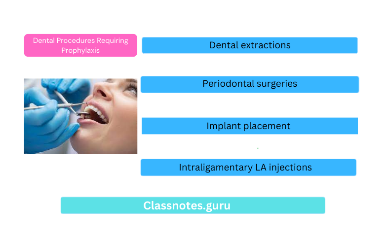
Dental Procedures Requiring Prophylaxis:
- Dental extractions
- Periodontal surgeries
- Implant placement
- Endodontic procedures beyond the apex
- Subgingival placement of fibers
- Intraligamentary LA injections
That Donot Require:
- Nonintraligamentary injections
- Intracanal endo treatment
- Placement of rubber dam
- Suture removal
- Placement of the removable prosthesis
- Making impressions
- Fluoride treatments
- Shedding of primary teeth
Question 2. Biopsy.
Answer:
Biopsy
Biopsy is the removal of part of tissue for the purpose of histological examination And Analysis
Biopsy Types:
1. Punch Biopsy: The sample is obtained with the help of a punch
Biopsy Indications:
- Mucosal lesions
- Inaccessible areas
2. Incisional Biopsy:
- Indication: large lesions
- Tumors: Edge biopsy is taken where the tumor cells can be compared with the normal cells
3. Excisional Biopsy:
- Indication: Small lesions
- The entire lesion is excised in a single sitting and sent for histological examination
4. Needle Biopsy:
- FNAC
- Indication: Cystic cavity:
- A 23-26 gauge needle is used to aspirate the contents of the lesion
Question 3. Exfoliative cytology.
Answer:
Exfoliative Cytology
Introduced by Papanicolaou And Traunt
Exfoliative Cytology Technique:
Scrap the surface of the lesion
↓
Collect it with the help of a wooden spatula
↓
Prepare a smear
↓
Stain it
↓
Observe under microscope Results:
↓
Class 1: Normal
↓
Class 2: atypical
↓
Class 3: Intermediate
↓
Class 4: Suggestive of cancer
↓
Class 5: Positive of cancer
Question 4. Aspiration biopsy.
Answer:
Aspiration Biopsy
- Needle Biopsy: technique
- FNAC:
- 23-26 gauge needle is inserted into the tissues
- Aspirate the needle
- Cystic fluid is collected in it
- Examine the fluid
- 23-26 gauge needle is inserted into the tissues
- Indication: cystic cavity:
- OKC:
Question 5. Schilling test.
Answer:
Schilling Test
Schilling Test is done to detect vitamin B12 deficiency as well as to distinguish and detect a lack of intrinsic factors and malabsorption The test is performed in three stages
Schilling Test Stage 1:
- Without intrinsic factor (IF)
- An oral dose of 0.5-1 pg of radioactively labeled vitamin B12 is administered orally
- After 2 hours a large dose(4 mg) of unlabelled vitamin B12 is given parenterally
- In normal individuals, more than 7% of 1 pg of an oral dose is excreted in a 24-hour urinary sample
- Patients with intrinsic factor deficiency excrete a lower quantity of it
Schilling Test Stage 2 (WITH IF):
- If the 24-hour urinary excretion of vitamin B12 is low, the test is repeated using the same procedure but with the addition of a high oral dose of IF is administered
- If the 24-hour urinary output is now normal the low value in the first test was due to IF deficiency
- Patients with pernicious anemia have abnormal tests even after treatment with vitamins due to IF defi¬ciency
Schilling Test Stage 3:
- Test for malabsorption of vitamin Bt:
- The same patient absorbed vitamin HI2 in water as was stipulated in the original test
- In conditions causing malabsorption, the test is repeated after a course of treatment with antibiotics or anti-inflammatory drugs
Question 6. Types and indications of biopsy.
(or)
Question 6. Biopsy
Answer:
Biopsy
Biopsy is the removal of part of tissue for the purpose of histological examination and analysis
Biopsy Types:
1. Punch Biopsy:
The sample is obtained with the help of a punch
- Indications:
- Mucosal lesions
- Inaccessible areas
2. Incisional Biopsy:
- Indication: large lesions:
- Tumors: Edge biopsy is taken where the tumor cells can be compared with the normal cells
3. Excisional Biopsy:
- Indication: small lesions:
- The entire lesion is excised in a single sitting and sent for histological examination
4. Needle Biopsy:
- FNAC:
- Indication: Cystic cavity:
- A 23-26 gauge needle is used to aspirate the contents of the lesion
Question 7. Paget’s test.
Answer:
Paget’s test
- It is used to examine the swelling
- Finger pressure is applied over the swelling
- It can be done for small swellings
- The center of the swelling becomes soft as it contains fluid
- While the periphery becomes hard
Question 8. Lugol’s Iodine test.
Answer:
Lugol’s Iodine Test
Lugol’s Iodine Test is used as an aid in the diagnosis of malignant lesions
Lugol’s Iodine Test Action:
- It will hinder glycogen present in the normal epithe¬lium
- It retains in normal squamous epithelial cells
- Thus it differentiates it from abnormal cells
Lugol’s Iodine Test Contents:
- Iodine 2 gm
- Potassium iodide 4 gm
- Distilled water- 100 cc
Lugol’s Iodine Test Effects:
- Normal cells- stained brown black
- Proliferating epithelium- inversely proportional to the degree of keratosis
- Inflammatory tissue- stained dark brown
Question 9. Investigation of oral cancer
Answer:
Investigation Of Oral Cancer – Clinical Methods:
1. Toulidlne Blue Staining:
Toulidlne Blue Staining is used as an aid in the diagnosis of oral cancer and potentially malignant lesions
Investigation Of Oral Cancer Method Of Use:
- Make the patient rinse the mouth with water twice for 20 seconds each
- Next, rinse with 1% acetic acid for 20 seconds
- Dry the area with the help of a gauze piece
- Apply 1% toluidine blue solution with a cotton swab
- Rinse again with acetic acid and water
- Observe the staining if present
Investigation Of Oral Cancer Advantages:
- Good sensitivity
- Very low false negative results
- It is effective in demonstrating dysplasia and early malignant lesions which is not clinically recognized and able
2. Lugol’s Iodine Test:
Lugol’s Iodine Test is used as an aid in the diagnosis of malignant lesions
Lugol’s Iodine Test Action:
- It will bind to glycogen present in the normal epithelium
- It retains in normal squamous epithelial cells
- Thus it differentiates it from abnormal cells
3. Acridine Binding Test
- In this method, the uptake of acriflavine by desquamated buccal cells is measured
- Since the DNA content of the dysplastic cells is higher, they will stain more intensely than normal cells
Photodiagnosis:
1. Autofluorescence Spectroscopy:
- Autofluorescence Spectroscopy is a non-invasive method
- Autofluorescence Spectroscopy is used for the detection of alteration in the struc¬tural and chemical composition of cells
2. Fluorescence Photography:
- Fluorescence Photography shows reduction and diminution of positive fluorescence associated with cancer regression and vice versa
Histopathological Methods:
1. Biopsy:
Biopsy is the removal of part of tissue for the purpose of histological examination and analysis
2. Exfoliative Cytology:
Exfoliative Cytology Technique:
Scrap the surface of the lesion
↓
Collect it with the help of a wooden spatula
↓
Prepare a smear
↓
Stain it
↓
Observe under microscope
Exfoliative Cytology Results:
- Class 1: Normal
- Class 2: Atypical
- Class 3: Intermediate
- Class 4: Suggestive of cancer
- Class 5: Positive of cancer
Exfoliative Cytology Molecular Methods:
1. Quantification Of Nuclear DNA Content:
- Quantitative analysis of DNA content reflects the total chromosomal content
2. Tumour Markers:
- Tumor markers may be produced by the host in response to cancerous substances
- They can be seen in blood circulation, body cavity fluids, cell membrane, and cell cytoplasm
Question 10. Coomb’s test.
Answer:
Coomb’s Test
Coomb’s Test was devised by Coombs, Mourant, and Race in 1945.
Coomb’s Test Method:
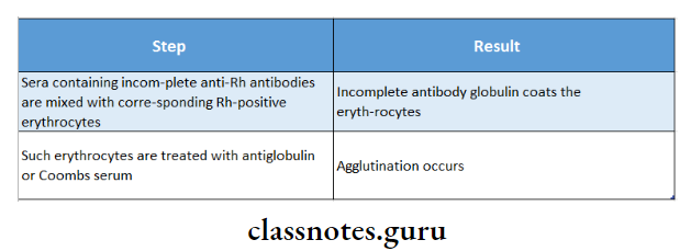
Coomb’s Test Types:
- Direct Coomb test
- Indirect Coomb test
Coomb’s Test Uses:
- Detects anti-Rh antibodies
- Demonstrates incomplete antibody
Question 11. Brush biopsy
Answer:
Brush Biopsy
This biopsy method utilizes an improved brush to obtain a complete transepithelial biopsy specimen with cellular representation from each of the three layers of the lesion: the basal, intermediate, and superficial layers.
- When used properly and rubbed against an area of suspect tissue aggressively [to the point of minor bleeding] the biopsy brush penetrates to the basement membrane, removing tissue from all three epithelial layers of the oral mucosa
- The oral brush biopsy does not require topical or local anesthetic and causes minimal bleeding and pain.
- The brush biopsy instrument has two cutting surfaces, the flat end of the brush and the circular border of the brush.
- Either surface may be used to obtain the specimen.
- Brush biopsies are utilized routinely in the detection of precancer and cancer in other organ systems.