
Exterior Of The Skull
Different views of the skull are considered from the description point of view. These are as follows:
- Norma vertical: This is the superior view.
- Norma occipitalis: This is a posterior view.
- Norma frontalis: This is the anterior view.
- Norma lateralis: This is a lateral (side) view.
- Norma basalis: This is the inferior view.
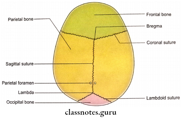
Norma Vertical
Norma Verticalis Definition
Observation of the skull from the superior aspect is called norma verticalis.
Norma Verticalis Shape
Norma’s vertical view of the skull appears ovoid in shape. It is relatively wider posteriorly.
Norma Verticalis Bones
The following bones contribute to the norma verticalis:
- Frontal bone (frontal squama): It lies anteriorly.
- Occipital bone (squamous part): It lies posteriorly.
- Parietal bones (paired): These lie on each side of the midline.
Junctions Of Bones (Sutures)
- Sutures are the immovable joints of the skull which are fibrous in nature.
- In norma verticalis following sutures can be seen:
- Coronal Suture: It is between the frontal and parietal bones.
- Sagittal Suture: It is between the two parietal bones.
- Lambdoid Suture: It is between the occipital and two parietal bones.
Norma Verticalis Features
1. Vertex
It is the highest point on the sagittal suture.
2. Vault
It is the arched roof of the skull.
3. Bregma
- It is situated at the intersection between coronal and sagittal sutures.
- Bregma is the site of a membranous gap in the foetal skull. This gap is known as anterior fontanelle.
- Anterior fontanelle closes by 18 months.
4. Lambda
- It is situated at the intersection of sagittal and lambdoid sutures.
- In the foetal skull, there is a membranous gap at the site of lambda. This gap is known as posterior fontanelle.
- Posterior fontanelle closes in 2-3 months.
5. Parietal Foramen
It pierces the parietal bone on each side of the midline about 3.5 cm in front of lambda. An emissary vein passes through it.
6. Obelion
It is the region on the sagittal suture between two parietal foramina.
7. Parietal Eminence
It is the area of maximum convexity of the parietal bone.
8. Temporal Lines
- There are two temporal lines on each side:
- Superior temporal line.
- Inferior temporal line.
- Both the temporal lines start as a single line from the zygomatic process of the frontal bone.
- The two lines arch backwards and upwards and cross the frontal bone, coronal suture and parietal bone.
- The superior temporal line fades out in the posterior part of the parietal bone.
- Epicranial aponeurosis and temporal fascia are attached to a superior temporal line.
- The inferior temporal line marks the upper limit of the origin of the temporalis muscle.
Norma Verticalis Applied anatomy
1. Fontanelles serve two important purposes:
- These allow moulding during birth.
- These allow the brain to grow.
2. The presence of anterior fontanelle is both clinically and therapeutically very significant.
- A bulge indicates increased intra-cranial tension (for example. in the case of a brain tumour).
- An abnormal depression indicates excessive loss of fluid (for example. in case of bleeding and dehydration).
- Diagnostic and therapeutic punctures could be carried out through anterior fontanelle.
- Ultrasonography of the brain in infants is performed through anterior fontanelle.
3. The osseous closure of the anterior fontanelle is an important milestone in the normal development of a child.
4. Soft and pliable bones of the neonate can withstand a considerable amount of compression and moulding, a fact clinically important during childbirth.
5. In neonates the flat bones of the vault are very soft and, therefore, a depressed fracture is like a dimple (pond fracture).
6. In adults there is some amount of elasticity in flat vault bones which often prevents fractures in cases of minor trauma. However, if the trauma force exceeds the minimal elasticity, fractures are bound to occur.
7. In adults a depressed fracture always shows an irregular line of fracture at the periphery of the depressed area.
8. Almost invariably all fractures of the vault of the skull in children are associated with the rupture of the dura mater.
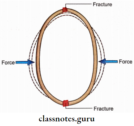
9. When the skull is compressed between two hard surfaces an axial shortening takes place along the line of the force and an axial lengthening takes place at a right angle. This results in the fracture of distant poles of the skull far from the actual site of the application of force.
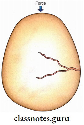
10. Fracture of the skull is usually due to a direct blow. A forceful hit on the forehead may cause a linear fracture of the vertex.
11. Cranium is clinically important because it reflects the size of the brain. Macrocephaly (enlargement of the head) can be due to hydrocephalus. Microcephaly may be hereditary or due to the maldevelopment of the brain.
12. Because of the lack of regenerating capacity of the flat bones of the vault, a gap in it should be filled with tantalum or titanium.
Norma Frontalis
Norma Frontalis Definition
When the skull is observed from the anterior aspect it is known as norma frontalis.
Norma Frontalis Shape
It is oval in shape being wider above than below.
Norma Frontalis Bones
Major bones contributing to the surface features of norma frontalis (excluding bones contributing to deeper orbits, nasal cavity and oral cavity) are as follows:
- Frontal bone (unpaired).
- Maxillae (paired).
- Nasal bones (paired).
- Zygomatic bones (paired).
- Mandible ethmoid (unpaired).
Junctions of Bones (Sutures)
- The junction between the zygomatic process of the frontal bone and the frontal process of the zygomatic bone is called the frontozygomatic suture. It is observed along the lateral margin of the orbital opening.
- A junction between the nasal part of the frontal bone and the frontal process of the maxilla (frontal-maxillary suture) is observed along the medial margin of the orbital opening in its upper part.
- The junction between the nasal part of the frontal bone and the nasal bones is called the frontonasal
sutures. - The junction between two nasal bones is a midline suture just above the anterior nasal aperture. This is called internasal suture.
- The junction between the maxilla and zygomatic bone (zygomaticomaxillary suture) is an oblique suture extending downwards and laterally from the lower border of each orbital opening.
- The junction between two maxillae is called an intermaxillary suture. It is a midline suture just below the anterior nasal aperture.
Note: Intermaxillary suture is also observed in the hard palate between the palatine processes of two maxillae.