
Norma Occipitalis
Norma Occipitalis Definition
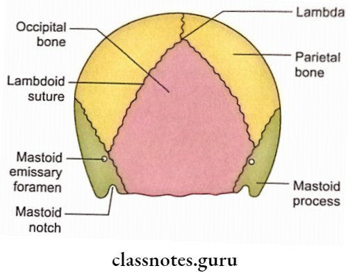
When the skull is observed from the posterior aspect, it is known as norma occipitalis.
Norma Occipitalis Shape
Norma occipitalis is convex upwards and flat below.
Norma Occipitalis Bones
The following bones contribute to the norma occipitalis.
- Parietal bones (paired).
- Squamous part of occipital bone (unpaired).
- Mastoid parts of temporal bones (paired).
Norma Occipitalis Sutures
1. Posterior Part Of Sagittal Suture
2. Lambdoid Suture
- It is between the two parietal bones and the occipital bone.
- The lower end of the lambdoid suture meets with the mastoid portion of the temporal bone at a point which forms the junction of occipitomastoid and parietomastoid sutures.
3. Occipitomastoid Suture
It is situated between the occipital bone and the mastoid part of the temporal bone.
4. Parietomastoid Suture
It is situated between the parietal bone and the mastoid part of the temporal bone.
Norma Occipitalis Features
1. External Occipital Protuberance
- It is a midline protuberance on the lower part of norma occipitalis.
- It marks the junction of the head and neck posteriorly.
- Inion is the most prominent point of external occipital protuberance.
- Trapezius originates from the upper part of the external occipital protuberance.
- Ligamentum nuchae is attached to the lower part of this protuberance.
2. Superior Nuchal Lines
- These are curved ridges passing laterally from the external occipital protuberance.
- These form the junction of head and neck posteriorly.
- The trapezius originates from the medial 1/3rd of the superior nuchal line.
- The Sternocleidomastoid is inserted on the lateral part of the superior nuchal line.
- Splenius capitis is also inserted on the lateral part of this line below the attachment of the sternomastoid.
3. Highest Nuchal Lines
- These are situated about a ‘cm’ above the superior nuchal lines.
- The epicranial aponeurosis is attached to their medial parts.
- The occipital belly of occipitofrontalis originates on each side from its lateral 2/3rd.
4. Mastoid Foramen
- It is located near the occipitomastoid suture.
- It opens internally into the sigmoid sulcus.
- The following structures transverse through it:
- Meningeal branch of occipital artery.
- Emissary vein.
5. Occipital Point
- It is situated in the midline a little above the inion.
- It is farthest from the glabella.
Norma Occipitalis Applied anatomy
Craniostenosis is a condition in which there is premature closure of the cranial sutures.
- When lambdoid and coronal sutures are involved, the skull grows vertically leading to the tower skull.
- The squamous part of the occipital bone is prone to both fissured and depressed fractures.
- A crack in the inner table of the squamous part of the occipital bone may damage the large diploic vein and produce a small epidural haematoma.
- Almost invariably fractures of the occipital squama in children are associated with rupture of the dura mater.
- A gap in the occipital squama is usually filled with tantalum or titanium due to a lack of regeneration in this part whose periosteum is devoid of a cambium layer.
- The inner table of cranial vault bones (including the squamous part of the occipital bone) is more brittle than the outer table, therefore, fractures are more extensive in the inner table.