Norma Lateralis
Norma Lateralis Definition
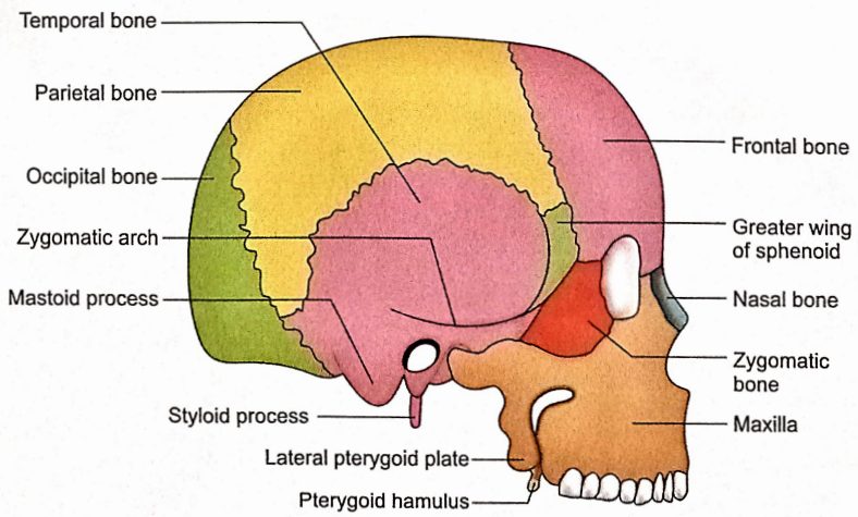
When the skull is observed from the side, it constitutes norma lateralis.
Norma Lateralis Bones
The following bones can be visualized in this view:
- Frontal
- Parietal
- Occipital
- Nasal
- Zygomatic
- Temporal
- Sphenoid
- Maxilla
Norma Lateralis Features And Attachments
1. Temporal Lines
There are two temporal lines, superior and inferior.
- Superior Temporal Line
- It commences at the frontal process of the zygomatic bone.
- It arches upwards and backwards across the parietal bone.
- It fades away on the temporal bone.
- Temporal fascia is attached to it.
- Inferior Temporal Line
- It commences at the same point.
- It runs inferior and parallel to the superior temporal line.
- Posteriorly it curves downwards and forwards on the temporal bone to continue with the supramastoid crest.
- It limits the attachment of the temporalis muscle.
2. Temporal fossa
- Boundaries
- Anteriorly: Zygomatic bone.
- Superiorly: Superior temporal line.
- Posteriorly: Superior temporal line. Supramastoid crest.
- Inferiorly: Zygomatic arch.
- Anterior Wall Of The Fossa Is Formed By:
- The temporal surface of the zygomatic bone
- The greater wing of the sphenoid
- Frontal bone
- Its Floor Is Formed By Following Bones:
- Frontal
- Parietal
- Temporal
- Greater wing of the sphenoid.
- The Temporalis muscle is attached to the floor and inferior temporal line.
- Other Contents Of Fossa Are:
- Middle temporal artery (a branch of superficial temporal artery).
- Deep temporal arteries arise from the maxillary artery.
- A zygomaticotemporal nerve and a minute artery appear from the zygomaticotemporal foramen located on the temporal surface of the zygomatic bone.
- Deep temporal nerves arise from the mandibular nerve.
3. Pterion
- It is a circular area in the anterior part of the temporal fossa which encloses four bones, frontal, parietal, sphenoid and temporal. These four bones form an ‘H’ shaped suture.
- It is located 4 cm above the zygomatic arch and 3.5 cm behind the front zygomatic suture.
- The middle meningeal vein, anterior branch of the middle meningeal artery and stem of the lateral sulcus of the brain lie deep in the pterion.
4. Zygomatic Arch
- It is formed by the temporal process of the zygomatic bone and the zygomatic process of the temporal bone.
- It has two surfaces (outer and inner) and two borders (upper and lower).
- Its outer surface is subcutaneous and crossed by the following structures from posterior to anterior:
- Auriculotemporal nerve.
- Superficial temporal vein.
- Superficial temporal artery.
- Masseter originates from its inner surface and lower border.
- The temporal fascia is attached to its upper border.
- The posterior end of the lower border is marked by the tubercle of the root of the zygoma. To this is attached the lateral ligament of the temporomandibular joint.
- Roots of the zygomatic arch diverge from the tubercle. The anterior root (articular tubercle) passes medially in front of the mandibular fossa. The posterior root continues with the supramastoid crest.
5. External Acoustic Meatus
- It is located behind the mandibular fossa below the posterior root of the zygoma.
- Its anterior wall, floor and lower part of the posterior wall are formed by the tympanic part while its roof and upper part of the posterior wall are contributed by the squamous part of the temporal bone.
- Margins of the meatus give attachment to the cartilaginous part of the external acoustic
meatus.
6. MacEwen’s Triangle (Suprameatal Triangle)
- It is situated posterosuperior to the external acoustic meatus.
- The spine of Henle (supramental spine) may be present at the anteroinferior part of the triangle.
- The mastoid antrum is situated about 12.5 mm deep to the supramental triangle.
7. Mastoid Process
- It is a downward projection from the mastoid part of the temporal bone.
- It is present below and behind the external acoustic meatus.
- The muscles attached to it from anterior to posterior are:
- Sternocleidomastoid.
- Splenius capitis.
- Longissimus capitis.
- The posterior belly of the digastric originates from its medial aspect (digastric notch).
8. Styloid Process
- It is a slender, elongated projection below the external acoustic meatus and in front of the mastoid process.
- It provides attachments to the following five structures:
- Anteriorly: Styloglossus muscle.
- Posteriorly: Stylohyoid muscle.
- Medially: Stylopharyngeus muscle.
- Laterally: Stylomandibular ligament.
- On the tip: Stylohyoid ligament.
9. Infratemporal Fossa
It is an irregular space below the zygomatic arch.
- Boundaries
- Anterior – Posterior surface of the body of the maxilla.
- Medial – Lateral pterygoid plate and pyramidal process of palatine bone.
- Lateral – Ramus of mandible
- Roof – Infratemporal surface of the greater wing of the sphenoid.
- Contents
- Muscles
- Lateral and medial pterygoids.
- Temporalis.
- Arteries
- Maxillary artery (Ist and 2nd parts) with its branches.
- Posterior superior alveolar branch of 3rd part of the maxillary artery.
- Veins
- Maxillary vein.
- Pterygoid venous plexus.
- Posterior superior alveolar vein.
- Nerves
- Mandibular nerve and its branches.
- Chorda tympani.
- Maxillary nerve.
- Posterior superior alveolar nerve.
- Muscles
- The anterior wall of the fossa shows two to three perforations for the posterior superior alveolar nerve and vessels.
- The junction of the anterior and medial walls is marked by a fissure (pterygomaxillary fissure) through which it communicates with the pterygopalatine fossa.
- The junction of the roof and anterior wall is marked by the lateral part of the inferior orbital fissure.
- Foramen ovale and foramen supinosum are present in the roof of the fossa.
- The lateral part of the fossa communicates with the temporal fossa through a gap between the zygomatic arch and the side of the skull.
10. Pterygomaxillary Fissure
- It is a gap which leads into the pterygopalatine fossa.
- Boundaries
- Anterior: Maxilla.
- Posterior: Pterygoid process.
- The maxillary artery enters the pterygopalatine fossa through the pterygomaxillary fissure.
- The maxillary nerve courses forward through it from the pterygopalatine fossa to enter the orbit through inferior orbital fissures.
11. Pterygopalatine fossa
- Boundaries
- Anterior: Posterior surface of maxilla.
- Posterior:
- Pterygoid process.
- Greater wing of the sphenoid.
- Medial: Perpendicular plate of palatine bone.
- Floor: Fusion of anterior and posterior walls.
- Communications
- The pterygopalatine fossa communicates with,
- The orbit, through the inferior orbital fissure.
- The middle cranial fossa, through foramen rotundum.
- The infratemporal fossa, through pterymaxillary fissure.
- The nasal cavity, through the palatovaginal canal and sphenopalatine foramen.
- The foramen lacerum, through the pterygoid canal.
- Contents
- The third part of the maxillary artery and its branches.
- Maxillary nerve with its branches.
- Pterygopalatine ganglion and its branches.
Norma Lateralis Applied anatomy
1. The spine of Henle is an important surgical landmark for surgery on the mastoid antrum.
2. Pterion is very important clinically because the anterior branch of the middle meningeal artery and accompanying vein lie on its internal aspect and are vulnerable to tearing if there are fractures of bone-forming this region.
3. If the meningeal vessels in the region of the pterion are damaged, an extradural haematoma is formed. Such haematoma may exert pressure on the cerebral cortex.
4. Decompression of the brain in cases of extradural haematoma in the region of pterion can be done by the method of trephining (burr hole) at this site.
5. Temporal fascia is commonly used for making tympanic membrane grafts during surgery for the repair of the ear drum.
6. Deep temporal vessels and nerves along with the tendon of the temporalis muscle, transverse the gap deep to the zygomatic arch whose fracture can involve these structures.
7. An elongated styloid process usually needs surgical correction because it leads to multiple complications in the neck.
8. An oblique line drawn from the frontozygomatic suture to the pterion corresponds with the inferior surface of the frontal lobe. This surface landmark is of great neurosurgical importance.
9. In radiographs of the skull, the diploic canals containing the diploic veins may be mistaken for the fractures of the skull.
10. Calvaria is very thin in the region of the temporal fossa and, therefore, is likely to get fractured because of hard blows to the head.
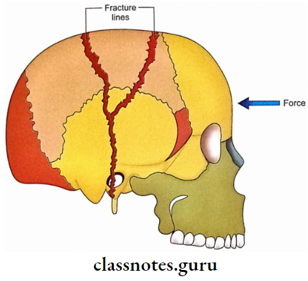
11. In depressed fractures, the inner table of calvaria is often more extensively fractured than the outer table.
12. Zygomatic fracture is very common in facial injuries due to its prominent position.
13. Fracture of the skull is usually due to direct blows. A forceful hit on the forehead causes linear fractures of both vertex and base.
14. Junctions of frontal and temporal processes of zygomatic bone form surgically important landmarks in the treatment of maxillofacial injuries.
15. The periosteum and attachment of strong temporal fascia limit the displacement of zygomatic bone following injuries.
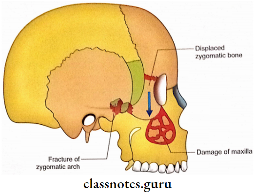
16. In a depressed fracture of the zygomatico-maxillary complex the maxilla is greatly damaged and there is displacement of the zygomatic bone without its fracture.
17. If a fracture of the zygomatic arch is associated with separation from the temporal fascia, then there occurs a downward displacement of the arch.
18. The weakest point of the zygomatic arch is its middle, just behind the temporo- zygomatic suture. This point is the most common fracture in cases of injuries to the arch.
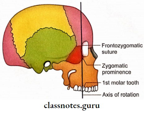
19. Frontozygomatic suture, the zygomatic prominence, the zygomatic buttress and 1st molar tooth lie in the same vertical line. In the majority of zygomatic-complex
(zygomatic bone + adjacent bones like maxilla and zygomatic process of temporal) fracture, the zygomatic bone rotates along this axis.