
Norma Frontalis Features
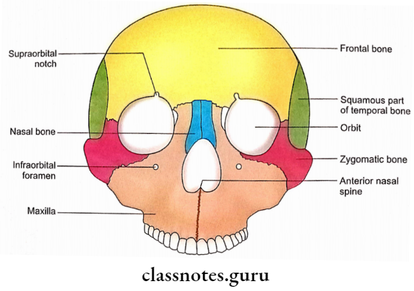
1. Three Large Apertures
One anterior nasal aperture and two orbital openings form the most striking feature of the norma frontalis.
- Anterior Nasal Aperture
- It is a midline aperture.
- It is piriform in shape and wider below than above.
- Its upper boundary is formed by the lower borders of nasal bones.
- Its lateral and inferior boundaries are contributed by nasal notches of two maxillae.
- The anterior nasal spine is a sharp projection at the lower margin of the nasal aperture, in the midline.
- Rhinion is the lower end of the internasal suture.
- A notch at the inferior border of the nasal bone is meant for passage of the external nasal nerve.
- Margins of the aperture give attachments to the nasal cartilages.
- Orbital Openings
- Each is present above and lateral to the anterior nasal aperture. It is quadrangular in shape and possesses four margins (supraorbital, infraorbital, lateral and medial).
- Supraorbital margin
- It is formed by the frontal bone.
- The supraorbital notch (or foramen) is situated at the junction of the medial 1/3rd (rounded) and lateral 2/3rds (sharp) of the supraorbital margin.
- The supraorbital notch transmits the supraorbital nerve and artery and a communicating vein between angular and superior ophthalmic veins.
- Infraorbital margin
- It is formed by maxilla medially and zygomatic bone laterally.
- Lateral orbital margin
- It is formed by the frontal process of the zygomatic bone below and the zygomatic process of the frontal bone above.
- Medial orbital margin
- It is formed by the frontal bone above and the lacrimal crest of the frontal process of the maxilla below.
2. Frontal Region
- Superciliary arch
- It is a curved elevation above the medial part of the supraorbital margin.
- Glabella
- It is the median elevation between two superciliary arches.
- Nasion
- It is the junction of internasal and frontonasal sutures.
- Frontal eminence (frontal tuber)
- It is a rounded elevation above each superciliary arch.
3. Maxillae
Each maxilla shows the following features:
- Infraorbital Foramen
- It is situated about 1 cm below the infraorbital margin.
- Infraorbital nerve and vessels pass through the infraorbital foramen.
- Incisive Fossa
- It is situated above the incisor teeth.
- Canine Eminence
- It is produced by the root of the canine tooth.
- Canine fossa
- It is situated just lateral to canine eminence.
- Frontal Process
- It is sandwiched between the nasal bone and the lacrimal bone.
- Zygomatic Process
- It articulates with the zygomatic bone.
- Alveolar Process
- It bears the sockets for the upper teeth.
4. Zygomatic Bones
- Each bone is situated below and lateral to the orbital opening.
- It is marked by a foramen called zygo- maticofacial foramen.
- Zygomaticofacial nerve traverses the zygomaticofacial foramen.
5. Mandible
- It forms the lower facial skeleton. For details of the features please consult the description of individual bone.
- It is situated just lateral to canine eminence.
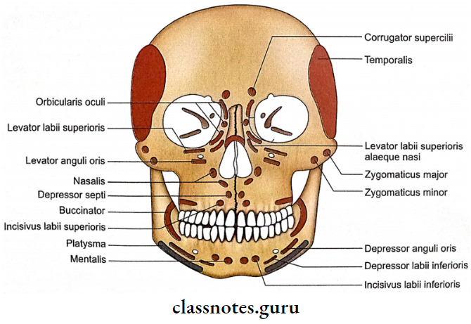
Norma Frontalis Attachments
- Nasal Bone: Procerus
- Superciliary Arch: Corrugator supercilii
- Frontal process of maxilla.
- The orbital part of orbicularis oculi (it is also attached to the nasal part of the frontal bone).
- Medial palpebral ligament.
- Levator babii superioris alaeque nasi.
- Between infraorbital margin and infra-orbital foramen: Levator labii superioris.
- Below the infraorbital foramen (to canine fossa): Levator anguli oris.
- Zygomatic bone just below the zygomaticofacial foramen: Zygomaticus minor.
- Lateral to zygomaticus minor: Zygomaticus major.
- Adjacent to nasal notch: Nasalis (transverse part above and alar part below).
- Incisive fossa.
- Medially: Depressor septi
- Laterally: Incisivus labii superioris.
- Alveolar process of maxilla opposite to molar teeth: Buccinator.
- Mandible: Please consult the description of individual bones.
Norma Frontalis Applied Anatomy
1. In about 8% of adult skulls a remnant of the lower part of the suture between two halves of the frontal bone (metopic suture) may persist. This suture is sometimes confused for a fracture in the radiograph.
2. Superciliary arches are elevated ridges from the surface of bone and any injury in this region will cause laceration of the skin and severe bleeding.
3. Compression of supraorbital nerves causes nerve pain. This fact may be used by anaesthetists to determine the depth of anaesthesia.
4. If the fracture of the frontal bone involves an inner table forming the roof of the frontal sinus, then the air may enter the cranial cavity (aerocele) causing meningitis or brain abscess.
5. An impact on the nose directed in the anteroposterior plane will cause a depression of the nasal bridge due to fracture of nasal bones, frontal processes of maxillae and septal cartilage.
6. A force on the nasal bridge directed from the lateral aspect will result in a deviation of the nasal bridge to the opposite side.
7. Traumatic alteration in the shape of the nose because of fractures of nasal bones is of great clinical importance due to cosmetic reasons, especially in young females.
8. A severe impact on the nasal bridge may involve frontal processes of maxillae.
9. Mid-facial skeleton is commonly involved in facial injuries. It includes maxillae, zygomatic bones, nasal bones and most of the bones which form a nasal cavity.
10. Mid-facial skeleton receives adequate blood supply from periosteal arteries and, therefore, all the fragments of fractured bone retain a periosteal blood supply.
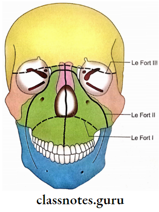
11. Fractures of maxillae and zygomatic bones show a constant pattern. Le Fort has classified such fractures into three types:
- Le Fort 1 fracture (Guerin’s fracture): It shows fractures of the lower 3rd of the nasal septum, maxillae and lower 3rd of pterygoid plates.
- Le Fort 2 fracture: It includes fractures of nasal bones, frontal processes of maxillae, lacrimal bones, ethmoid, vomer and pterygoid plates.
- Le Fort 3 fracture: In this fracture facial skeleton is separated from the skull base. It involves upper parts of nasal bones, frontal processes of maxillae, ethmoid, lesser wings of sphenoid and roots of pterygoid plates.
12. Zygomatic bone is very commonly involved in cases of fractures of the middle 3rd of the face. Fracture of the frontal process of the zygomatic bone may occur in conjunction with a comminuted fracture of the orbital rim and frontal bone.
13. Any of the four margins of the orbit may fracture as an isolated fracture or in combination.
Note: For applied anatomy of the mandible and other bones of norma frontalis, discussion on individual bones may be consulted.