Norma Basalis
Norma Basalis Definition
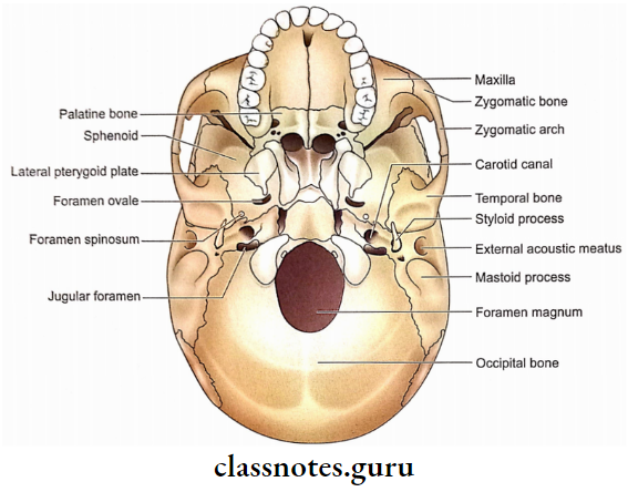
Observation of the cranium (skull without mandible) from the inferior aspect is called norma basalis.
Norma Basalis Boundaries
- Anterior: Incisor teeth.
- Posterior: Superior nuchal line.
- Lateral (side):
- Rest of teeth.
- Zygomatic arch.
- Posterior root of zygoma.
- Mastoid process.
Norma Basalis of Skull Notes
Norma Basalis Subdivisions
For the sake of convenience, norma basalis is divided into anterior, middle and posterior parts. The hard palate and alveolar arch are included in the anterior part.
An imaginary horizontal line passing through the anterior margin of the foramen magnum separates the posterior part from the middle part of the norma basalis.

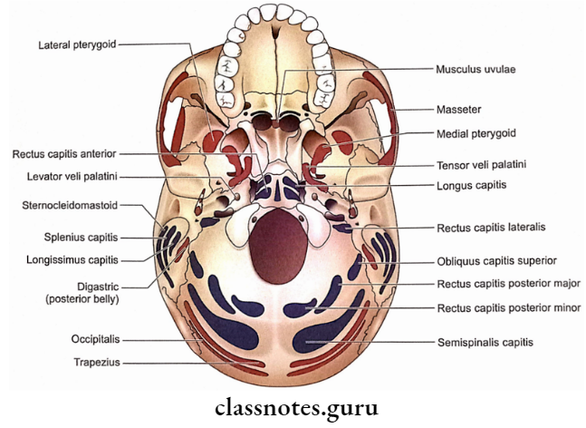
Norma Basalis Features And Attachments
1. Anterior Part Of Norma Basalis
- The posterior border of the hard palate
- It forms the junction of the anterior and middle parts of norma basalis.
- The posterior nasal spine is a spinous projection from its middle in the median plane.
- The musculus uvulae is attached to the posterior nasal spine.
- Alveolar arch
- It possesses sockets for the roots of the upper teeth.
- The number of sockets depends upon the number of roots. There is a single socket for each of the incisors, canines and premolars. There are three sockets for each of the upper molars.
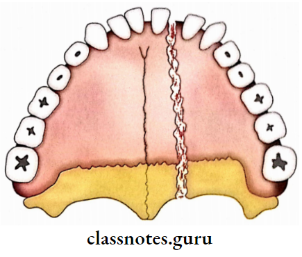
- Bones contributing to hard palate
- Palatine processes of two maxillae contribute to the anterior 2/3rd of the hard palate.
- The posterior 1/3rd of the hard palate is formed by the horizontal plates of palatine bones.
- The bony palate is marked by several depressions produced by palatine glands.
- Cruciform suture
- It is formed by the following three sutures.
- Intermaxillary suture.
- Interpalatine suture.
- Palatomaxillary sutures.
- Incisive fossa
- It is present anteriorly in the median plane of the hard palate.
- Incisive foramen (right and left) pierces its corresponding side.
- Each incisive foramen is transferred by the nasopalatine nerve and greater palatine vessels.
- Greater palatine foramen
- It is present behind the lateral part of the palatomaxillary suture.
- Greater palatine vessels and nerves pass through it.
- A groove observed between the greater palatine foramen and incisive fossa is meant for greater palatine vessels.
- Lesser palatine foramina
- These are 1-3 foramina in the pyramidal process of palatine bone and located just behind the greater palatine foramen on each side.
- Lesser palatine nerves and vessels pass through these foramina.
- Palatine crest
- It is a curved ridge observed in the hard palate near its posterior border.
- The palatine aponeurosis is attached to the palatine crest, the posterior border of the hard palate and the area between the two.
- Premaxilla
- It is a triangular piece of maxilla holding four incisor teeth.
- It is a separate bone in most vertebrates.
2. Middle part of norma basalis
For the sake of convenience, it is divided into a median area and two lateral areas (right and left).
Skull Norma Basalis Anatomy
1. Median area
- Posterior nasal apertures
- These are also known as choanae.
- The posterior border of the vomer
- It separates two choanae.
- Alae of vomer
- These are two bony plates formed by the splitting of vomer superiorly.
- It articulates with the rostrum of the sphenoid.
- Vomerovaginal canal
- It is formed between the latter! border of each ala of the vomer and the vaginal process of the medial pterygoid plate.
- It transmits branches of the pharyngeal nerve and vessels.
- Palatovaginal canal
- It is a canal between the vaginal process of the medial pterygoid plate and the sphenoidal process of the palatine bone.
- This canal leads anteriorly into the posterior wall of the pterygopalatine fossa.
- It transmits pharyngeal branches of the pterygopalatine ganglion and pharyngeal branches of 3rd part of the maxillary artery.
Note: Students are invariably confused as to which is the palatovaginal canal and which one is the vomerovaginal canal.
To differentiate keep in mind that the vaginal process of the medial pterygold plate is common to both but as the palatine bone is anterior to the medial pterygoid plate the palatovaginal canal is relatively anterior to the Domeroenginal canal.
- Broad bar of bone behind the alae
- It is formed by the continuation of the inferior surface of the body of the sphenoid and that of the basilar part of the occipital bone.
- It extends up to the foramen magnum.
- The pharyngeal tubercle is a median elevation just in front of the foramen magnum. It is better felt than seen.
- Pharyngeal tubercle gives attachments to:
- Highest fibres of superior constrictor.
- Pharyngeal raphe
- Longus capitis is inserted on the basilar part of the occipital bone just lateral to the pharyngeal tubercle.
- Rectus capitis anterior is inserted on each side just in front of the occipital condyle.
2. Lateral area
- Pterygoid processes
- Pterygoid processes are located just behind the posterior ends of the alveolar arch.
- Each pterygoid process descends vertically downwards from the junction of the body and the greater wing of the sphenoid.
- The pterygoid process consists of a lateral and a medial plate.
- Pterygoid plates unite anteriorly in the upper part to enclose a fossa called the pterygoid fossa.
- The lower ununited portions form a pterygoid fissure, which is filled by the pyramidal process of the palatine bone.
- The anterior surface of the pterygoid process forms the posterior boundary of the pterygopalatine fossa.
- The lateral surface of the lateral pterygoid plate forms the medial wall of the infra-temporal fossa and gives origin to the lower head of the lateral pterygoid muscle.
- The medial surface of the lateral pterygoid plate forms the lateral wall of the pterygoid fossa and gives origin to the deep head of the medial pterygoid muscle.
- The lateral surface of the medial pterygoid plate forms the medial wall of the pterygoid fossa and is related to the tensor palati muscle.
- The medial surface of the medial pterygoid plate forms the lateral wall of the corresponding posterior nasal aperture.
- The posterior border of the medial pterygoid plate shows the following features:
- At its upper end, it splits to enclose the scaphoid fossa, which gives origin to the tensor palati muscle.
- Its upper end shows a small projection called the pterygoid tubercle, which lies immediately below the posterior end of the pterygoid canal.
- The pharyngobasilar fascia is attached to its whole extent, while the superior constrictor arises from its lower part only.
- A hook-like process at its lower end is called pterygoid hamulus. Tendon of tensor palati winds around this process. Superior constrictor and pterygomandibular raphe are also. attached to it.
- An angular process projecting from the middle of this margin is called the processus tubarius. The posterior border above this process is called the notch of the auditory tube. This process and notch support the medial end of the auditory tube.
- The infratemporal surface of the greater wing of the sphenoid
- It is pentagonal.
- It forms roof of infratemporal fossa.
- It gives origin to the upper head of the lateral pterygoid muscle.
- It is crossed by deep temporal and masseteric nerves.
- The spine of the sphenoid is a projection from the posteriormost part of the infratemporal surface.
- The infratemporal crest is the lateral limit of the infratemporal surface.
- From the scaphoid fossa to the spine of the sphenoid, four foramina can be noticed, i.e. foramen of Vesalius, foramen ovale, canaliculus innominatus and foramen spinosum.
- Foramen ovale
- It is an oval foramen.
- It transmits the mandibular nerve, accessory meningeal artery, lesser petrosal nerve and emissary vein.
Note: For remembering the structures passing through the foramen ovale, remember MALE, in which M-Mandibular nerve, A-Accessory meningeal artery, L-Lesser petrosal nerve and E-Emissary vein.
- Foramen spinosum
- It is situated near the spine of the sphenoid, posterolateral to the foramen ovale.
- It transmits the middle meningeal artery, nervus spinosus (meningeal branch of the mandibular nerve) and parietal trunk of the middle meningeal vein.
- Foramen of Vesalius (sphenoidal emissary foramen)
- It is an infrequently seen foramen between the scaphoid fossa and the foramen ovale.
- It transmits an emissary vein connecting the cavernous sinus with the pterygoid venous plexus.
- Canaliculus innominatus (foramen innominatum)
- This is also an infrequently seen foramen between the foramen ovale and the foramen spinosum.
- It transmits the lesser petrosal nerve.
- Spine of the sphenoid
- It is related laterally to the auriculo-temporal nerve and medially to the chorda tympani nerve and the Eustachian tube.
- The sphenomandibular ligament is attached to its tip.
- Most posterior fibres of the tensor palatini originate from its anterior surface.
- Sulcus tube
- It is a groove between the posteromedial margin of the infratemporal surface of the greater wing of the sphenoid and the inferior surface of the petrous part of the temporal bone.
- The cartilaginous part of the Eustachian tube (also called the auditory tube or pharyngotympanic tube) occupies this sulcus.
- The inferior surface of the petrous part of the temporal bone
- It is located just behind the infratemporal surface of the greater wing of the sphenoid.
- Its anteromedial serrated end marks the apex of the petrous part.
- The quadrilateral area near the apex provides attachment to the levator palati muscle.
- The lower opening of the carotid canal is located just behind the quadrilateral area. It transmits the internal carotid artery with its sympathetic and venous plexuses.
- The Carotid canal runs forward and medially in the petrous part and perforates its apex as the upper opening of the carotid canal.
- Foramen lacerum
- It is located between the sphenoid and the apex of the petrous temporal.
- It is named ‘lacerum’ because of its irregular margins.
- The carotid canal and pterygoid canal open into it.
- Posterior part of norma basalis has irregular margins. For the sake of convenience, this part can be
- Only two structures pass through it, i.e. meningeal branch of the ascending pharyngeal artery and the emissary vein.
- The internal carotid artery traverses its upper part with its sympathetic and venous plexuses.
- The nerve of the pterygoid canal (Vidian nerve) is formed in its upper part by the union of the greater superficial petrosal and deep petrosal nerves.
- The tympanic part of the temporal bone
- It is a triangular bone which occupies the angle between the petrous and squamous parts of the temporal bone.
- Its anterior surface is related to the parotid gland.
- The squamous part of the temporal bone
Only a small part of the squamous part of the temporal bone is seen in norma basalis and shows the following features from posterior to anterior:
- Anterior (articular) part of mandibular fossa.
- Articular tubercle.
- Part of the roof of the infratemporal fossa.
- Squamotympanic fissure
- It marks the junction of the squamous and tympanic parts of the temporal bone.
- The downward edge of the tegmen tympani (a part of the petrous part of the temporal bone) divides the squamotympanic fissure into petrotympanic (posterior) and pterosquamous (anterior) fissures.
- The Chorda tympani nerve, the anterior tympanic artery and the anterior ligament of the malleus pass through the petrotympanic fissure.
Inferior View of Skull – Norma Basalis
3. Posterior Port Of Norma Basalis
For the sake of convenience, this part can be divided into a median area and two lateral areas (right and left).
- Median area
It consists of foramen magnum, external occipital crest and external occipital protuberance from anterior to posterior.
- Foramen magnum
- It is the largest foramen in the skull.
- It is a single foramen located in the lowest part of the posterior cranial fossa.
- It is oval.
- It is the communication between the cranial cavity and the vertebral canal.
- Anterior atlanto-occipital membrane is attached to its anterior margin.
- The posterior atlanto-occipital membrane is attached to its posterior margin.
- Lateral margins provide attachments to the alar ligaments.
- The following structures pass through its anterior part:
- Apical ligament of dens.
- Superior longitudinal band of the cruciform ligament.
- Membrana tectoria.
- The following structures pass through its posterior part:
- Medulla oblongata.
- Meninges.
- Spinal roots of accessory nerves.
- Meningeal branches of upper cervical nerves (C1-3).
- Vertebral arteries.
- Sympathetic plexuses around vertebral arteries.
- Anterior and posterior spinal arteries.
- External occipital crest
- It extends from the posterior margin of the foramen magnum to the external occipital protuberance.
- The upper margin of the ligamentum nuchae is attached to it.
- External occipital protuberance
- Foramen magnum
Norma Basalis Skull Diagram Explanation
Trapezius is attached to it superiorly and ligamentum nuchae inferiorly.
- Lateral area
- Occipital condyles
- These are located lateral to the anterior half of the foramen magnum.
- Each is oval and convex to articulate with a concave superior articular process of the atlas.
- Condylar fossa
- It is present just behind the occipital condyle.
- It may have a condylar canal for the emissary vein from the sigmoid sinus.
- Hypoglossal canal
- Lateral to the anterior part of the condyle is the outer opening of the hypoglossal canal.
- It transmits:
- Hypoglossal nerve.
- Meningeal branch of ascending pharyngeal artery.
- Emissary vein from basilar venous plexus.
- Squamous part of the occipital bone
- The superior nuchal line is a well-defined ridge which extends laterally from the external occipital protuberance on each side. Its medial 1/3rd provides origin to the trapezius while the lateral 1/3rd receives insertions of sternomastoid (above) and splenius capitis (below).
- Running laterally on each side from the middle of the external occipital crest is another ridge called the inferior nuchal line.
- A vertical line on each side along with the inferior nuchal line divides the region below the superior nuchal line into four areas, each meant for the attachment of a muscle as follows:
- Upper medial area for semispinalis capitis.
- Upper lateral area for obliquus capitis superior.
- Lower medial area for rectus capitis posterior minor.
- Lower lateral area for rectus capitis posterior major.
- Jugular foramen
- It is an interosseous foramen situated between the anterior margin of the jugular process of the occipital bone and the posterior margin of the petrous part of the temporal bone at the petro-occipital suture.
- It is divided into anterior, middle and posterior parts.
- 9th, 10th and 11th cranial nerves pass through its middle part.
- The inferior petrosal sinus and meningeal branch of the ascending pharyngeal artery pass through its anterior part.
- The sigmoid sinus and meningeal branch of the occipital artery traverse through its posterior part.
- At its posterior end, the anterior wall (petrous temporal) is hollowed out to form the jugular fossa which lodges the superior bulb of the internal jugular vein.
- The mastoid canaliculus is a minute canal in the lateral wall of the jugular fossa which transmits the auricular branch of the vagus.
- The glossopharyngeal notch is on the posterior border of the petrous temporal bone near the medial end of the jugular foramen.
- The cochlear canaliculus is located at the apex of the glossopharyngeal notch. The aqueduct of the cochlea opens into the cochlear canaliculus.
- Tympanic canaliculus is present in the ridge between the jugular fossa and the lower opening of the carotid canal. It transmits the tympanic branch of the glossopharyngeal nerve to the middle ear.
- Inferior surface of jugular process of occipital bone
- It is the area just lateral to the occipital condyle behind the jugular foramen.
- It provides attachment to rectus capitislateralis.
- Styloid process
- It is a conical projection just below the tympanic part of the temporal bone.
- It is directed downwards, forwards and slightly medially.
- It provides attachments to 3 muscles and 2 ligaments. Three muscles attached to it are the styloglossus (anteriorly), stylohyoid (posteriorly) and stylopharyngeus (medially). Two ligaments attached to it are stylomandibular (laterally) and stylohyoid (on the tip).
- It is interposed between the parotid gland (laterally) and the internal jugular vein (medially).
- Two structures cross it superficially, i.e. facial nerve (near the base) and external carotid artery (near the tip).
- Mastoid process
- It is a prominent projection from the temporal bone posterolateral to the styloid process.
- The medial aspect of this process shows a deep groove (digastric notch) which provides attachment to the posterior belly of the digastric.
- Medial to the digastric notch, there can be another groove for the occipital artery.
- Stylomastoid foramen
- It is present between the styloid and mastoid processes.
- The facial nerve and stylomastoid artery pass through this foramen.
- Occipital condyles
Anatomy Notes on Norma Basalis
Norma Basalis Applied Anatomy
A forceful hit on the forehead causes a linear fracture of both the vertex and base.
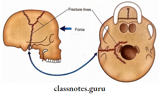
- Due to the presence of natural thick bony buttresses at the base of the skull, the fracture lines often converge towards the foramen magnum or sella turcica.
- In the case of basal fractures, the location of structures passing through the basal foramina may complicate the issue.
- A fracture line passing through the foramen lacerum may tear the internal carotid artery.
- If the fissure lines involve grooves having a resultant carotid-cavernous fistula. meningeal vessels and dural sinuses, then epidural haematoma might result.
- Fractures of the skull base are often zig-zag in appearance because the fracture lines avoid thickening and pass through the lines of least resistance.
- Tumours of the base of the skull
- Transitional cell carcinoma arising from the mucous membrane of paranasal sinuses or the fossa of the Rosenmuller of the nasopharynx usually erodes the skull base.
- Some of the very rare tumours which may arise from the base of the skull or adjacent tissue are as follows:
- Osteomas.
- Chondromas.
- Giant cell tumours of bone.
- Many malignant tumours metastasise to the base of skull bones from distant organs, for example. prostate, lung and breast.
- The bony palate may be fractured in an uncommon central split of the palate. It is paramedian because median sutures (intermaxillary and interpalatine) are relatively strong.
- Clinical signs and symptoms which support the involvement of the base of the skull are as follows:
- Discharge of CSF through the external acoustic meatus (C.S.F. otorrhoea)
- Tear of the tympanic membrane.
- Collection of blood in the middle ear.
- Facial paralysis due to damage to the 7th cranial nerve.
- Loss of hearing, vertigo and nystagmus due to the involvement of the 8th cranial nerve. The aforementioned signs are indicative of a fracture of the petrous part of the temporal bone.
- Discolouration and oedema of tissue over the mastoid process are an indication of sigmoid sinus damage.
- Cranial nerve damage is due to the involvement of foramina in the skull base by the fracture lines.