Hyoid
‘Hyoid’ is a Greek word that means ‘U’ shaped.
Hyoid Level
Hyoid lies at the level of the 3rd cervical vertebra.
Hyoid Location
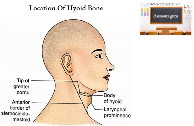
- The Hyoid is situated in the anterior midline of the neck above the thyroid cartilage.
- Its body (the bend of ‘U’) is the first resistant structure felt in the midline of the neck, inferior to the chin.
- The tip of the greater cornu (the limb of ‘U’) of the hyoid can be palpated in the relaxed neck near the anterior border of the sternocleidomastoid muscle midway between the laryngeal prominence and mastoid process.
Hyoid Features And Attachments
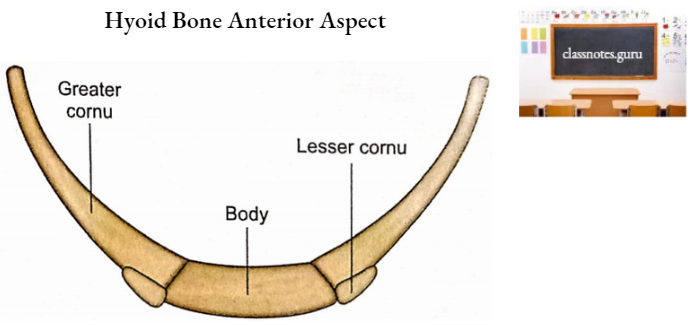
Hyoid bone consists of a central body, a pair of greater cornua, and a pair of lesser cornua.
Hyoid Body
It has two surfaces (anterior and posterior), two borders (upper and lower), and two lateral ends.
1. Surfaces
- Anterior Surface
- It is convex.
- A median ridge divides it into two lateral halves.
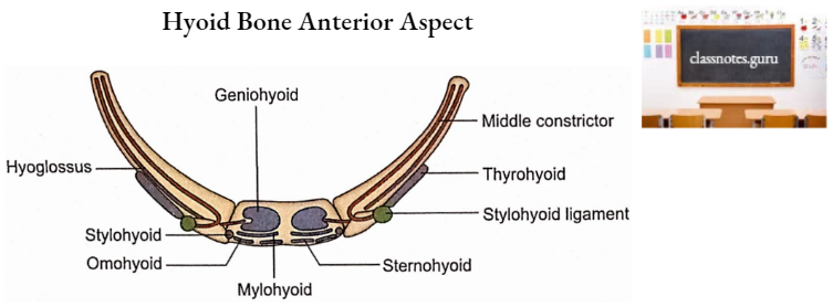
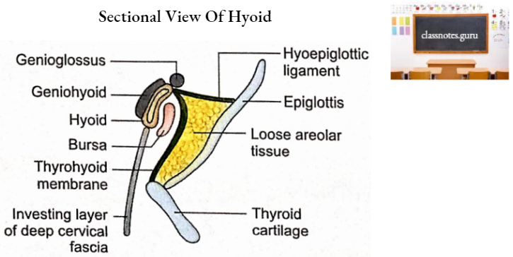
- Geniohyoid and mylohyoid muscles are inserted on this surface in its upper and lower parts respectively.
- Hyoglossus partly originates from anterior surface.
- The investing layer of the cervical fascia is attached below the insertion of the mylohyoid.
- Posterior Surface
- It is concave.
- It is related to the following structures.
- Bursa
- Thyrohyoid membrane
- Epiglottis
2. Borders
- Upper Border
- It provides attachment to 3 structures from anterior to posterior
- Genioglossus muscle
- Hyoepiglottic ligament
- Thyrohyoid membrane.
- It provides attachment to 3 structures from anterior to posterior
- Lower Border
- Two muscles are mainly attached to this border on each side of the midline from medial to lateral.
- Sternohyoid
- Omohyoid
- Two muscles are mainly attached to this border on each side of the midline from medial to lateral.
3. Ends
- Each end continues posteriorly as greater cornu.
- Lesser cornu projects upwards at the junction of the body and greater cornu.
Greater Cornua (Singular Greater Cornu)
Greater cornu has two surfaces (upper and lower), two borders (medial and lateral), and a tubercle (at the posterior end).
1. Surfaces
- Upper Surface
- It has the following attachments from medial to lateral.
- Middle constrictor along the whole length.
- Hyoglossus along the whole length.
- The stylohyoid muscle at the junction of lesser and greater cornua.
- Fibrous loop of digastric muscle-lateral to attachment of stylohyoid muscle.
- Lower Surface
- Fibroareolar tissue separates this surface from the thyrohyoid membrane.
2. Borders
- Medial Border
- It receives attachment of the thyrohyoid membrane.
- Lateral Border
- The thyrohyoid muscle is attached to this border anteriorly.
Lesser Cornua (Singular Lesser Cornu)
- It is a small conical projection attached to the bone at the junction of the body and greater cornu by fibrous tissue.
- It may form a synovial joint with the greater cornu.
- It has the following attachments:
- Stylohyoid ligament at the tip.
- Middle constrictor-posterolaterally.
Hyoid Ossification
- The hyoid ossifies from ventral portions of the cartilages of the 2nd and 3rd arches.
- Lesser cornua and the upper part of the body are developed from the 2nd arches.
- Greater cornua and the lower part of the body are developed from 3rd arches.
- The appearance of centres – 6 centers of ossification appears, 2 for the body and 1 for each cornu, as follows:
- Greater cornu just before birth Body-just after birth
- Lesser cornu-puberty
- The cartilage at the tip of each greater cornu persists up to the 3rd decade.
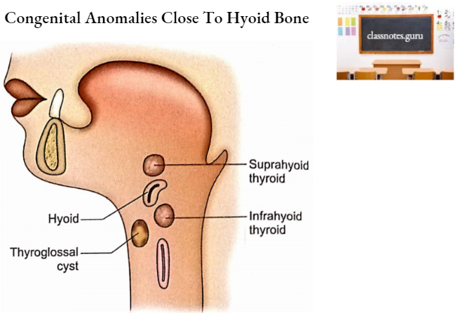
Hyoid Applied Anatomy
- Some congenital anomalies associated with developing thyroid are commonly observed adjacent to the hyoid, for example. suprahyoid thyroid, infrahyoid thyroid, and thyroglossal cyst.
- Sometimes a muscular band connects the body of the hyoid with the isthmus or pyramidal lobe of the thyroid gland. This is called levator glandular thyroid.
- The lingual artery arises from the external carotid artery posteroinferior to the tip of the greater cornu.
- The latter thus forms an important surgical landmark for locating the lingual artery which is ligated essentially in radical surgery of the tongue.
- The hyoid bone is of great medical importance. In suspected cases of death, a fracture of the hyoid bone suggests death by throttling or strangulation.