
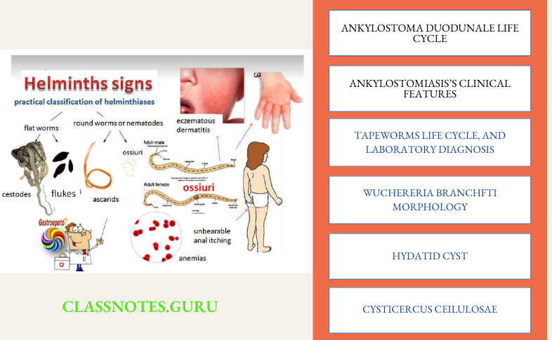
Helminths Long Question And Answers
Question 1. Describe the life cycle of Ankylostoma duodenale. Write a note on pathogenicity and laboratory diagnosis of Ankylostomiasis
Answer:
The common name of Ankylostoma duodenale is the Old World hookworm
Read And Learn More: Microbiology Question and Answers
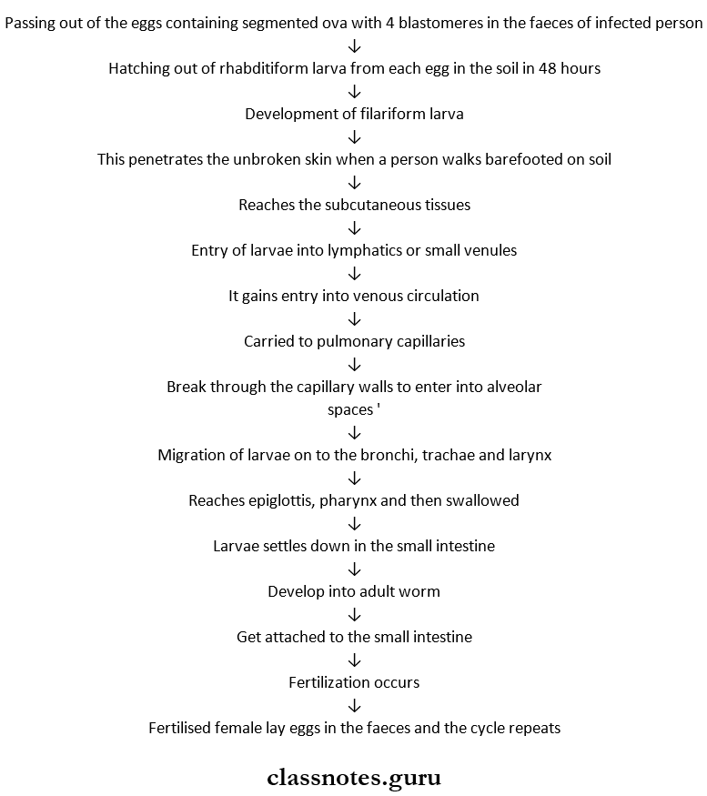
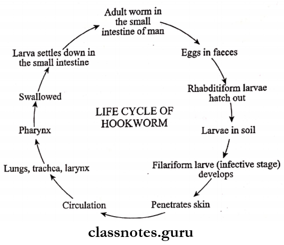
Ankylostoma Duodunale Life Cycle:
- Man is the only definitive host
- It involves following the steps
Use FAQ-style formatting or numbered lists for better readability.
Ankylostomiasis Pathogenicity:
- Migrating larvae may cause three types of lesions as follows
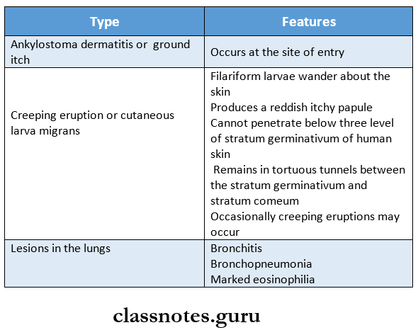
- The worm causes hookworm disease in man
Include diagrams or image descriptions
Ankylostomiasis’s Clinical Features Are:
- Microcytic, hypochromic anemia
- Epigastric pain
- Dyspepsia
- Vomiting
- Diarrhea
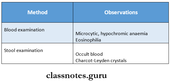
Ankylostomiasis Laboratory Diagnosis:
1. Direct Methods:
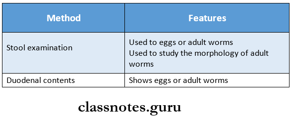
2. Indirect Methods:
helminths short questions for mbbs
Question 2. Enumerate the common tapeworms. Describe the morphology, life cycle, and laboratory diagnosis of taeniasolium
Answer:
Tapeworms:
- Taenia saginata
- Taeniasolium
- Echinococcus granulosus
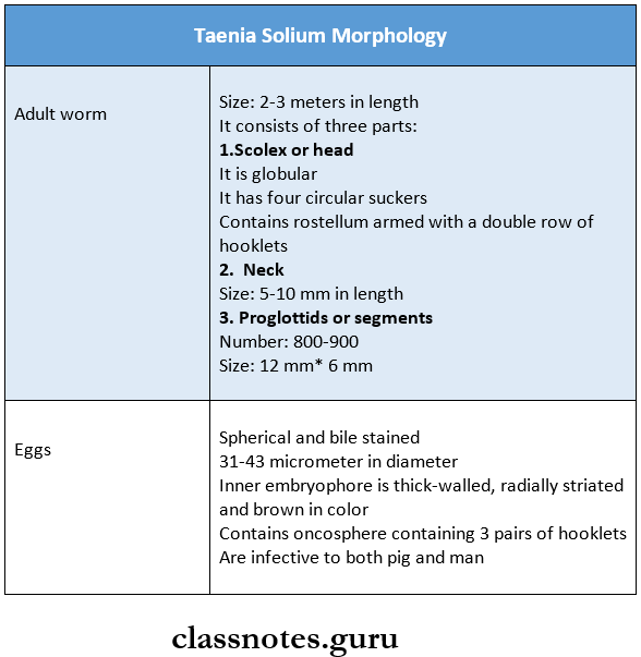
Taenia Solium Morphology:
Add schema markup (FAQ schema) to boost visibility in search results.
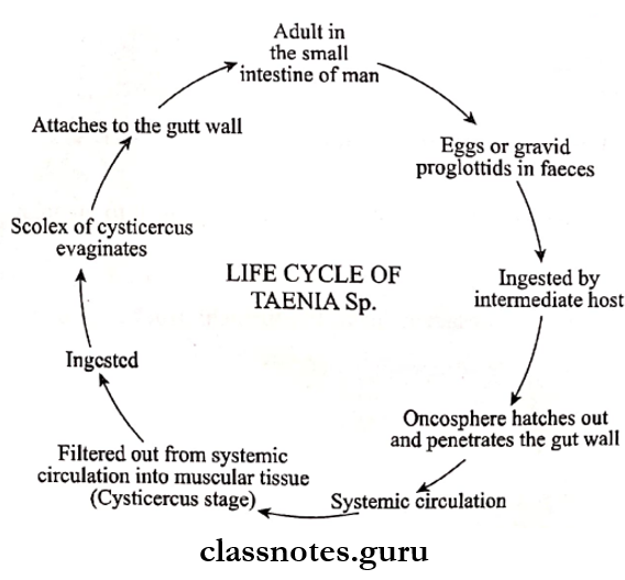
Taenia Solium Life Cycle:
- The worm passes its life cycle in two hosts: the definitive host, man, and an intermediate host, pig
- The adult worm lives in the small intestine of man
- Eggs are passed out with the feces
- The animal swallows these eggs and gets infected
- Oncosphere hatches out
- It penetrates the gut wall
- Carried in the systemic circulation
- The naked Oncospheres are transformed into cysticercus cellulose in the muscle
- By intake of uncooked or partially cooked pork, enters into the alimentary canal of man
- The scolex of cysticercus evaginates and attaches to the gut wall
- It develops into an adult worm
Taenia Solium Laboratory Diagnosis
1. Stool Examination
- It detects eggs of T. solium
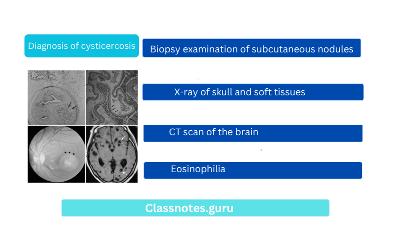
2. Diagnosis Of Cysticercosis
- It is done by
- Biopsy examination of subcutaneous nodules
- X-ray of skull and soft tissues
- CT scan of the brain
- Eosinophilia
Question 3. Describe the morphology, life cycle, and pathogenesis of the Wuchereria branch. Discuss the diagnosis of filariasis.
Answer:
Wuchereria Branchfti Morphology:
1. Adult Worms
- They are transparent, long hair-like structures
- Color-Creamy white
- Shape- Filiform with tapering ends
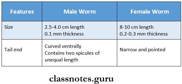
- Both remain coiled together
- Life span- 5-10 years
Write clear explanations using real-life examples suitable for undergraduates.
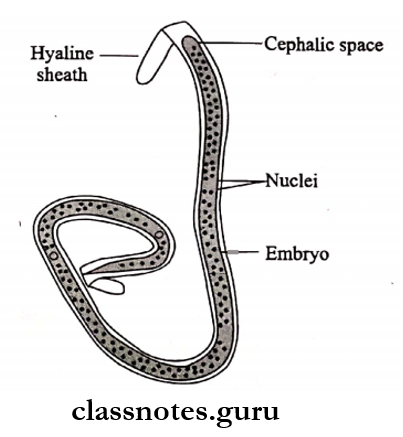
2. Embryos
- Color- colorless
- It is transparent
- Heads and tails- Heads are blunt while tails are pointed
- Size- 290 micrometer * 6-7 micrometer
- Covered by hyaline sheath
- Nuclei appear as granules
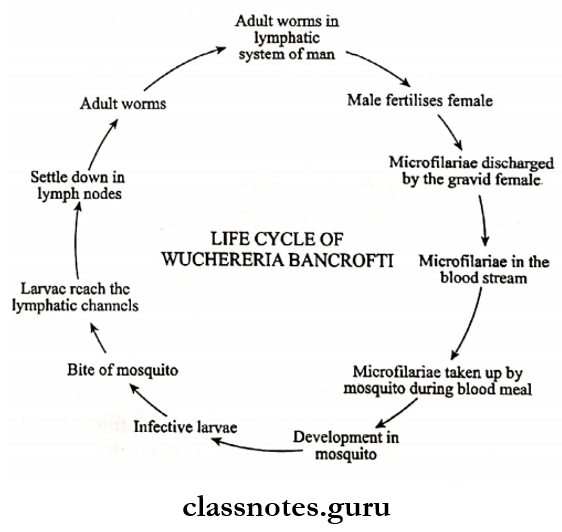
Wuchereria Branchfti Life Cycle:
- Wuchereria bancrofti passes its life cycle in man and mosquito
- Adult worms are present in the lymphatics system of man
- Male fertilises female
- Embryos or microfilariae are discharges
- These reach bloodstream
- Microfilariae are taken up by mosquitoes during a blood meal
- It develops in mosquito
- Through the bite of a mosquito, the infective larvae are deposited on the skin
- This penetrates the skin and reaches the lymphatic channels
- Gets settled in lymph nodes
- Develops into an adult worm
- Again fertilization occurs and the cycle is repeated
Wuchereria Branchfti Pathogenesis:
- W. bancrofti causes filariasis
Wuchereria Branchfti Types:
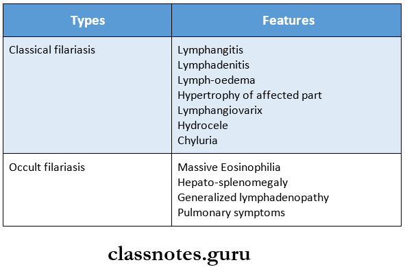
Make use of Hindi translations if targeting Indian audiences.
Diagnosis Of Filariasis:
1. Direct Methods
- Samples collected are
- Blood
- Chylous urine
- Exudate of lymph varix
- Hydrocele fluid
- Blood film should be made in night between 10 pm to 2 am as the number of microfilariae is higher at night
2. Indirect Methods
- It includes
- Blood examination- Detects Eosinophilia
- Serological tests like ELISA, indirect fluorescent antibody, and indirect haemagglutination assay.
helminths and virology short notes Q&A
Helminths Short Question And Answers
Question 1. Hydatid Cyst:
Answer:
- The cyst wall secreted by the embryo
- It consists of the following.
- Ectocyst
- Endocyst
1. Ectocyst:
- It is a hyaline membrane that forms a thick, tough outer circular layer and is elastic in nature.
- When incised it curls on itself
- As a result, it exposes the inner layer
2. Endocyst:
- It is an inner (or) germinal layer
- It forms an ectocyst on the outer side
- It gives rise to brood capsules and scolices on the inner side.
Hydatid Fluid:
- It is secreted by endocyst.
Hydatid Cyst Characteristics:
- It is clear, colorless (or) pale yellow fluid.
- It is a slightly acidic nature
- It is highly toxic, when absorbed it gives rise to anaphylactic symptoms.
- Due to its antigenic nature, it is used for Casoni’s test.
- It has a low specific gravity
- Composition- It contains
- Sodium chloride
- Sodium sulfate
- Sodium phosphate
- Sodium and calcium salts of succinic acid
Hydatid Sand:
- It is a granular deposit that consists of brood capsules, free scolices, and loose hooklets.
- It gets settled at the bottom of the hydatid cyst
Question 2. cassoni’s test
Answer:
Cassoni’s Test is an immediate hypersensitivity skin test introduced by Casoni in 1911.
Cassoni’s Test Method:
- Sterile hydatid fluid is used as an antigen.
- Hydatid fluid is obtained from hydatid cysts from humans or animals
- It is made of sterile
- Now 0.2 ml of it is injected intradermally in one arm
- While 0.2 ml of normal saline is injected intradermally in the other arm
Cassoni’s Test Result:
- It produces a large wheal measuring 5 cm in diameter or more within 30 minutes in all positive cases.
- Also shows multiple pseudopodia.
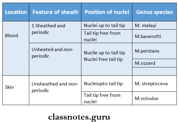
Question 3. Microfilaria
Answer:
- Microfilariae are the larvae of nematodes.
- The female nematodes are viviparous giving birth to larvae called microfilariae.
- It needs two hosts to complete its life cycle man and a blood-sucking insect
Microfilaria Features:
Question 5. Cysticercus Ceilulosae
Answer:
- Cysticercus cellulose is the larval stage of taenia solium
- It develops in the muscles of the pig which is an intermediate host
- A mature cyst is an opalescent ellipsoidal body and the long axis of the cyst is parallel to the muscle fiber.
- A dense milky white spot is present at the side where the scolex with its hooks and suckers remains invaginated.
- The cyst develops further when ingested by man which is the definitive host
- It may develop in any organ but is usually present in the subcutaneous tissues and muscles.
Various Features Of Cysticercosis.
- They cause palpable nodule in sub-cutaneous tissues and muscles
- In the brain leads to epileptic attacks.
- Neurocysticercosis involving the nervous system is the most serious form
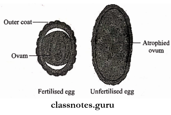
Question 6. Diagram of Fertilised egg of ascaris lumbricoides
Answer:
microbiology helminths Q&A pdf
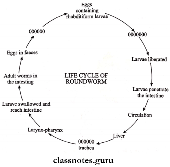
Question 7. Life cycle of Ascaris lumbricoides
Answer:
- Ascaris lumbricoides passes its life cycle in only one host i.e. Man
- Adult worms reside in the jejunum of man
- The passing of fertilised eggs containing the unsegmented ovale in the feces
- These develop in soil
- Rhabditiform larva develops from the unsegmented ovum
- This undergoes first molting
- Intake of food, drink or raw vegetables contaminated with eggs containing Rhabditiform larvae causes infection in man
- Liberation of Rhabditiform larvae occurs in the upper part of the small intestine
- They burrow through the mucous membrane of the small intestine
- It is carried to the liver and then enters pulmonary circulation
- They reach the lungs and enlarge
- They break through the capillary wall and reach alveoli
- The larvae reach the bronchi, then the trachea, and are swallowed after reaching the pharynx
- Pass down to the esophagus, and stomach and then gets localized in the upper part of the small intestine
- Here another molting occurs and the larvae grow into an adult worm
- Thus the cycle repeats
Question 8. Larva migrans
Answer:
It is a condition caused by the ingestion of embryonated eggs of some nematodes parasitizing animals
Larva Migrans Pathogenesis:
- Larvae are hatched in the small intestine
- These reach extraintestinal sites
- Gets settled in the liver, lungs, and other organs
- This leads to the formation of granulomatous lesions
Larva Migrans Clinical Features:
- Leucocytosis
- Eosinophilia
Larva Migrans Diagnosis:
- Serological tests detect antibodies in serum
Larva Migrans Prevention:
- Deworming animals prevent this disease
Use bullet points and tables for clarity and better SEO performance.
Question 9. Name four tapeworms.
Answer:
- Taeniasolium
- Taeniasaginata
- Echinococcusgranulosus
- Diphyllobothrium latum
- Hymenolepsis nana