Frontal Bone Terminology
The term ‘frontal’ is derived from the Latin word ‘frons’ which means ‘brow’ or ‘forehead’.
Frontal Bone Location
The frontal bone forms the forehead, the greater part of the roof of each orbit and most of the floor of the anterior cranial fossa.
Frontal Bone Features And Attachments
The frontal bone has a main part (frontal squama) and orbital parts.
Frontal Bone Squama (Main Part)
It has an external surface, right and left temporal surfaces, an internal surface, a nasal part and a margin (parietal or posterior).
1. Surfaces
- External Surface
- Supra-Orbital Margins.
- These are lower limits of the external surface on each side.
- They form the upper borders of the orbital opening.
- Supra-Orbital Notch Or Foramen
- The junction of the lateral two-thirds of the supra-orbital margin (sharp) with the medial one-third (rounded) is marked by a supra-orbital notch (sometimes foramen).
- This is meant for the passage of the supraorbital nerve, supraorbital artery and a communicating vein between angular and superior ophthalmic veins.
- Superciliary Arch
- This is an arched prominence just above the supra-orbital margin.
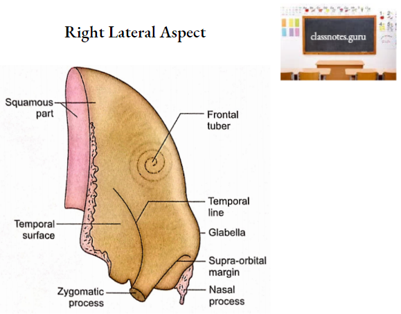
- Glabella
- It is the median prominence between superciliary arches.
- Frontal Eminence
- On each side, about 3 cm above the supra-orbital margin, there is an elevated area called frontal eminence or tuberosity.
- It is usually more marked in females.
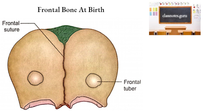
- Metopic Suture
- The frontal bone is bilateral in origin and the junction of the two halves is called frontal or metopic suture. Its remains can be seen even in adults in the region of glabella.
- Zygomatic Process
- Supra-orbital margin extends laterally on each side into a zygomatic process.
- The zygomatic process articulates with the frontal process of the zygomatic bone.
- A line curves upwards and backwards from the zygomatic process. The line soon divides into two lines called superior and inferior temporal lines.
- Supra-Orbital Margins.
- Temporal Surfaces
- An area on each side below and behind the temporal lines is called the temporal surface.
- It contributes to the anterior part of the temporal fossa on the lateral aspect of the skull (norma lateralis).
- The superior temporal line gives attachment to the temporal fascia.
- Inferior tempoRal Line And Temporal Surface Of the Frontal Bone Give Origin To the Temporalis Muscle.
- Internal Surface
- This surface shows depressions and elevations for cerebral gyri and sulci respectively.
- Sagittal Sulcus
- It is a midline sulcus in the upper part of the internal surface.
- Its margins provide attachments to falx cerebri.
- The sulcus itself lodges the superior sagittal sinus.
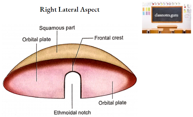
- Frontal crest
- Margins of the sagittal sulcus meet in the midline in the lower part and continue as a frontal crest.
- This also gives attachment to falx cerebri.
- A notch below the frontal crest is converted into foramen caecum by articulation with ethmoid bone. An emissary vein passing through it connects the vein of the nose with the superior sagittal sinus.
- Several depressions (granular foveolae) on each side of the sagittal sulcus are produced by arachnoid granulations.
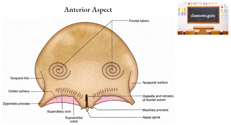
2. Nasal Part
- It is a downward projection of the frontal bone between two supra-orbital margins.
- Its lower serrated part is known as the nasal notch.
- Each half of the nasal notch articulates with the following three bones from anterior to posterior:
- Nasal bone.
- Frontal process of the maxilla.
- Lacrimal bone.
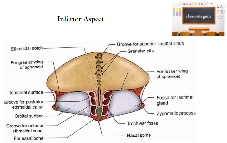
- The nasal spine is a midline downward continuation of the nasal part.
- On each side of the nasal spine, there is a grooved area which forms the roof of the nasal cavity.
- The nasal spine itself articulates with the crest of the nasal bone anteriorly and the perpendicular plate of the ethmoid posteriorly.
3. Posterior Margin
- This is also called the parietal margin because its major part articulates with parietal bones.
- The lower part of this margin is triangular and rough for articulation with the greater wing of the sphenoid.
Frontal Bone Orbital Parts
Orbital parts consist of two triangular laminae (orbital plates) separated by a gap called an ethmoidal notch.
1. Orbital Plate
It possesses two surfaces, orbital and internal.
- Orbital Surface
- It faces downwards.
- It forms the roof of the orbit.
- Its anterolateral part has a fossa for the lacrimal gland.
- Its anteromedial part (trochlear fovea) provides attachment to the fibrocartilaginous pulley for the tendon of the superior oblique muscle.
- Internal surface
- It faces upwards.
- It contributes to the anterior cranial fossa.
- It has impressions for the gyri of the frontal lobe of the cerebral hemisphere.
- It has grooves for meningeal vessels.
2. Ethmoidal notch
- It is a ‘U’ shaped gap occupied by a cribriform plate of ethmoid.
- Under surfaces of its lateral margins possess several incomplete air cells which complete the ethmoidal air cells when ethmoid bone is in position.
- Two grooves on the undersurface of each margin are converted into anterior and posterior ethmoidal canals by similar grooves on the superior surface of the ethmoidal labyrinth. These canals are meant for passages of anterior and posterior ethmoidal nerves and vessels.
- The undersurface of the anterior margin of the notch possesses openings for frontal sinuses (one on each side of the nasal spine).
Frontal Bone Frontal sinus
variable size. It is situated between the outer and Each frontal sinus is an irregular cavity of the inner tables of the frontal bone. They are separated from each other by a bony septum which is usually deviated to one side.
Frontal Bone Ossification
- The frontal bone ossifies in the membrane.
- Two primary centres appear one for each half of the frontal bone, in the region of the frontal tuberosity.
- Primary centres appear during the 8th week of intrauterine life.
- Ossification extends upwards to form the frontal squama, backwards to form the orbital part and downwards to form the nasal part.
- At birth frontal bone is made up of two halves separated by frontal or metopic suture.
- The union between two parts begins in 2nd year and completes at 8th year.
Frontal Bone Applied Anatomy
- Frontal squama is soft and pliable in neonates which can withstand a considerable amount of compression and moulding, a fact clinically important during childbirth.
- Frontal squama is prone to depressed or fissured fractures. In neonates and infants, the depressed fracture is often like a dimple in the bone. In adults, a depressed fracture is always associated with an irregular line of fracture.
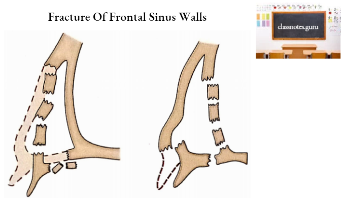
- A severe impact at the root of the nose leads to fractures of the frontal sinus walls. If the fracture involves the inner table forming the posterior wall of the frontal sinus, then the air may enter into the cranial cavity (aerocele) causing meningitis and brain. abscess.
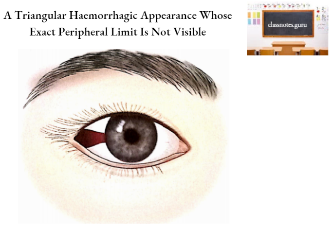
- Fracture of the orbital plate of the frontal bone causes haemorrhages in the orbit. The haemorrhage acquires a triangular shape under the conjunctiva whose apex is towards the corneoscleral junction and base towards the orbital margin.
- A crack in the inner table of the frontal squama may damage a large diploic vein and produce a small epidural haematoma.
- Almost invariably the fractures of frontal squama in children are associated with rupture of the dura mater.
- A gap in the frontal squama will not be regenerating and has to be filled with tantalum or titanium, a procedure called cranial prosthesis.