Ethmoid Bone
Ethmoid Bone Terminology
‘Ethmoid’ is a Greek word that means ‘sieve-like’. Ethmoid is so named because it possesses a perforated (sieve-like) plate called a cribriform plate.
Ethmoid Bone Location
- The single ethmoid bone is situated in the anterior part of the base of the cranium between the orbits.
- It forms part of the medial wall of the orbits and part of the bony septum, roofs, and lateral walls of the nasal cavities.
Ethmoid Bone Features And Attachments
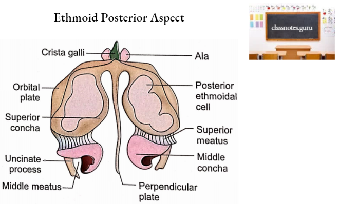
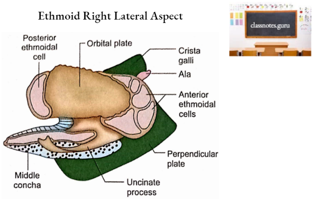
Ethmoid bone consists of a cribriform plate, a perpendicular plate, and two lateral masses called labyrinths.
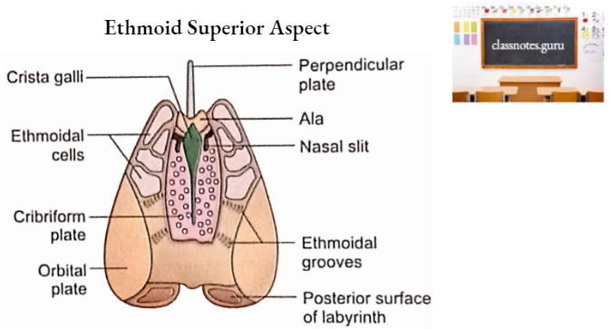
Ethmoid Bone Cribriform Plate
- It is the median part of the superior surface of the ethmoid.
- It contributes to the median portion of the anterior cranial fossa (anterior part of the interior of the base of the skull).
- It occupies the ethmoidal notch of the frontal bone.
- It possesses a median triangular upward projection called crista galli (named because it resembled the crown of a cock, the zoological name of which is Gallus domesticus).
- The posterior sloping border of the crista galli gives attachment to the falx cerebri.
- The anterior border of the crista galli has two alae which articulate with frontal bone to complete foramen caecum. An emissary vein passes through this foramen.
- On each side of the crista galli, the cribriform plate shows several perforations through which pass about 15-20 filaments of the olfactory nerve. This part is also related to the olfactory bulb superiorly.
- Just lateral to the anterior part of the crista galli there is a slit-like passage for a process of dura mater.
- Just lateral to the anterior end of the slit there is a foramen for the passage of the anterior ethmoidal nerve.
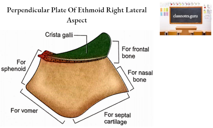
Ethmoid Bone Perpendicular Plate
- It is a quadrangular flat plate projecting downwards from the midline of the cribriform plate.
- Its anterior border articulates with the nasal process of the frontal bone and the crest of the nasal bones.
- Its posterior border articulates with a sphenoidal crest above and vomer below.
- Its superior border is attached to the cribriform plate.
- Its inferior border receives the attachment of septal cartilage.
- Its surfaces are mainly smooth except in the upper parts where there are grooves for filaments of olfactory nerves.
Ethmoid Bone Labyrinths
A large number of air-filled spaces (ethmoidal air cells) constitute the labyrinth. These air cells are divisible into anterior, middle, and posterior ethmoidal sinuses by bony plates.
Many of these air cells open on the surface and are completed only when articulating with the adjacent bones. Each labyrinth may be considered to have six surfaces.
1. Upper Surface
- It has several open-air cells which are completed only after articulation with the edges of the ethmoidal notch.
- It has two grooves which are converted into anterior and posterior ethmoidal canals by articulation with frontal bone.
2. Lower Surface
It articulates with the upper part of the nasal surface of the maxilla to complete the ethmoidal air cells from below.
3. Anterior Surface
It possesses half-cut air sinuses which are completed by the frontal process of maxilla and lacrimal bone.
4. Posterior Surface
It articulates with the sphenoidal concha and orbital process of palatine bone to complete the posterior ethmoidal sinus.
5. Lateral Surface
- It is a thin and smooth plate called an orbital plate.
- It covers the middle and posterior ethmoidal sinuses.
- It forms a large part of the medial wall of the orbit.
- It is quadrangular in shape and articulates as follows:
- Superiorly with the orbital plate of the frontal bone.
- Inferiorly with the maxilla and orbital process of the palatine bone.
- Anteriorly with lacrimal bone.
- Posteriorly with sphenoid bone.
6. Medial Surface
- It forms part of the lateral wall of the corresponding half of the nasal cavity.
- Its upper part is marked by numerous vertical grooves which lodge filaments of the olfactory nerve.
- Its posterior part is marked by an anteroposterior fissure called the superior meatus.
- The posterior ethmoidal sinus opens into the superior meatus.
- The superior meatus is bounded above by a curved plate called the superior nasal concha.
- Below and in front of the superior meatus is another curved plate of bone called the middle nasal concha.
- The lateral surface of the middle concha is concave and forms the medial wall of the middle meatus.
- The lateral wall of the middle meatus is marked by a swelling produced by the middle. ethmoidal air cells. This swelling is called bulla ethmoidalis.
- The middle ethmoidal sinus opens on the surface of the bulla or immediately above it.
- A thin bar of bone called an uncinate process projects downwards and backward from the anterior part of the labyrinth.
- The curved gap between the uncinate process and bulla is called hiatus semilunaris.
- The upper end of the hiatus semilunaris is continuous with a curved canal called the ethmoidal infundibulum.
- Anterior ethmoidal sinus opens into the infundibulum.
- In 50% of cases, the infundibulum continues superiorly as the frontonasal duct to reach the frontal sinus.
Ethmoid Bone Ossification
- At the age of 3rd month of intrauterine life, the walls of the nasal cavity are marked by a cartilaginous framework called a cartilaginous nasal capsule.
- The cartilaginous nasal capsule consists of two lateral regions and a median nasal part.
- A single center appears for each labyrinth in the lateral region of the nasal capsule at about 5th month of intrauterine life.
- Perpendicular plate and crista galli ossify from a single center which appears in the median septal part of the nasal capsule at the age of 1st year after birth.
- The labyrinths fuse with perpendicular plates in the region of the cribriform plate at about 2 years of age.
- The ethmoid air cells begin to develop during intrauterine life and are present in the form of narrow pouches at birth.
Ethmoid Bone Applied Anatomy
- A severe impact on the nasal bridge may involve frontal processes of maxillae and two orbital plates of ethmoid bones.
- In case of head injury, a discharge of CSF from the nose (CSF rhinorrhoea) is indicative of a fracture of the cribriform plate of ethmoid in the anterior cranial fossa.
- If the basilar fracture involves the cribriform plate, it may result in anosmia (loss of smell sensation) due to damage to olfactory nerve filaments.
- Fracture of cribriform plates may cause meningitis if it opens into the nasal cavity.
- The bony septum of the nose (perpendicular plate of ethmoid and vomer) is paper thin and does not resist much to forces responsible for the fracture.
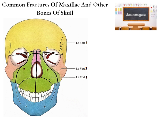
- The ethmoid is spared in Le Fort 1 fractures while involved in Le Fort 2 and 3 fractures.
- Since the ethmoid bone is clothed in mucosa over large areas of its surfaces, its fractures open into the nasal cavity or ethmoid air cells with the potential risk of infection.
- Nasal ethmoidectomy is performed in several operations involving the ethmoidal labyrinth.
- In this, an artificial opening is made in the ethmoidal labyrinth to drain the sinuses, for example. after removal of frontoethmoidal mucoceles or optic nerve decompression in the optic canal.