
What Happens in Reflex Actions
Define reflex action. Name the pathway undertaken by reflex action.
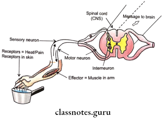
Reflex action is a type of very quick response to a stimulus in which delay can be harmful It eliminates the thinking and analytical component of the brain for its activity.
- Reflex action is found in most animals even where neuron networks have not come into existence. It persists in advanced animals because of its usefulness. Even here, the infants possess more reflex action than the adults. The total number is about 200.
- Reflex action was studied for the first time by Marshal Hall Cl TT U to central nervous system to effector organ.
Consultation by the brain is not carried out though the brain is often informed after the die reflex action is over. The short circuit pathway undertaken by reflex action is called reflex It consists of:
- Receptor. It is a sensory cell, tissue, or organ that is specialized to pick up the stimulus and get sensitized.
- Sensory or Afferent Neuron. The sensitized receptor activities cause the development of an impulse in the sensory or afferent neuron. The impulse travels along the sensory neuron and reaches the central nervous organ.
- Part of the Central Nervous System. It is a spinal cord for reflex action of the trunk and limbs. for the head region, the brain is used as part of the reflex arc, of course without involving its thinking part. In the central nervous system, the impulse brought by sensory neurons is transferred to a connector or interneuron. The interneuron passes the information to a motor neuron.
- Motor or Efferent Neuron. The sensitized motor neuron carries the impulse to a specific effector organ through its axon terminal.
- Effector Orean. It is a muscle, gland, organ, or tissue that on activation by the motor end plate provides a suitable response to the stimulus.
Importance of Reflex Action
- Control. Reflex action controls a number of activities of the body.
- Quick Response. Response to a stimulus is almost immediate.
- Accuracy. The response is accurate, useful, and purposeful.
- Survival Value. Reflex action has a survival value.
- Overtaxing of the Brain. Reflex action avoids overtaxing of the brain.
- Coordination. It coordinates many activities of the body.
- Conditioned Reflexes. They help us to perform many of our activities like reading, writing, cycling, typing, tying laces, etc.
Types of Reflex Actions
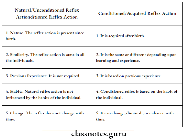
Based on origin, reflex action can be unconditioned or conditioned.
Unconditioned or Natural Reflex Action. It is a reflex action that is inborn or present right from birth. The response is the same in all the individuals. Some common examples are
- Closing of eyes when strong light is flashed on them or some moving object approaches them.
- Wide opening of pupil in dim light and narrowing of pupil in strong light
- Withdrawal of hand or foot when pricked,
- Jerking of the knee when it is hit below the knee cap.
Conditioned or Acquired Reflex Action. It is a reflex action that develops after birth due to learning, habit, or regular association of an indifferent stimulus with a natural stimulus. Conditioned reflexes can change or disappear with a change in habit or environment. Some common examples are
- Tying of shoe laces without looking
- Knitting without looking
- Pedaling
- Reading
- Writing.
Differences Between Unconditioned And Conditioned Reflex Actions
Functions Of Nervous System
- Awareness. It makes the individual aware of the surroundings.
- Sensations. With the help of receptors and sense organs, the nervous system has developed several sensations.
- Control. The functioning of different parts of the body is controlled by the nervous system.
- Coordination. It coordinates the functioning of interrelated parts.
- Memory. The nervous system retains the memory of past experiences and impressions.
- Interpretation. It interprets changes and happens based on memory and reasoning.
- Involuntary Functions. A section of the nervous system called the autonomous nervous system guides the different types of involuntary functions, for example., peristalsis.
- Reflex Actions. They are nerve-based immediate protective responses to certain stimuli.
Parts of the Human Nervous System
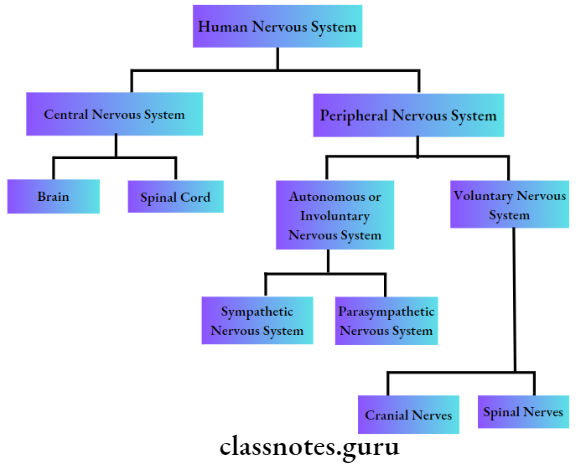
- The human nervous system consists of two parts, the central nervous system and the peripheral nervous system. The peripheral nervous system is a bridge between various body parts and the central nervous system.
- It picks up sensations and hands over the same to the central nervous system. On instructions from the central nervous system, through voluntary or involuntary control, it bring about activity in various parts of the body.
- The spinal cord is also a communication channel between the brain and the trunk part of the body. The brain is the major part of the nervous system that is involved in thinking, reasoning, memory, intelligence, emotions, and will.
Human Brain
Where is the brain located in the body? What are its three parts?
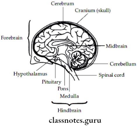
It is an anterior walnut-shaped large part of the nervous system that has a size of 1400 cc and a weight of about 1.4 kg. It is pinkish-grey in color.
- The brain is distinguished into three parts—fore-brain (thinking part), mid-brain (relay part), and hindbrain (involuntary part). Both mid brain and hind brain are also called reflex parts.
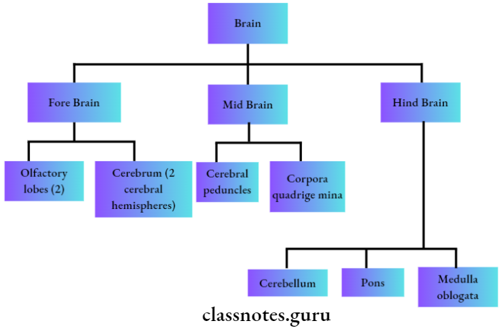
1. Olfactory Lobes. They are two club-shaped structures that lie on the inferior surface of the cerebrum. Olfactory lobes relay the sense of smell to the temporal lobes of the cerebrum.
2. Cerebrum. It is the largest part of the brain that occupies nearly 80% of its volume. The cerebrum is formed of two closely placed cerebral hemispheres. They are attached on the inferior surface by a thick band called the corpus callosum.
- Each cerebral hemisphere has a fluid-filled cavity called a lateral ventricle. The superior surface is convex and convoluted. The convolution are called gyri while the depressions are known as sulci.
- The left cerebral hemisphere controls the functioning of the right side of the body while the right cerebral hemisphere has control over the left part of the body. The cerebral hemispheres are divisible into 4 parts – frontal, parietal, temporal, and occipital.
- Frontal Lobes. They constitute the anterior or front part of the brain. Frontal lobes have several nerve centers like the motor area for controlling voluntary movements, the premotor area for the higher center of involuntary and autonomic functions, intelligence, the association area between sensation and movement, and motor speech or Broca’s area. Parietal Lobes.
- They are mid-dorsal lobes that control some components of speech (in the left lobe) but the major function is control of the sensation of pain, touch, pressure, temperature, and taste.
- Temporal Lobes. They are lateral lobes of the cerebrum. Temporal lobes have centers for smell, hearing, audiovisual memory, and some components of speech.
- Occipital Lobes. They lie on the posterior side of the cerebrum. They possess centers for a sensation of sight.
3. Diencephalon. It lies on the undersurface of the cerebrum. The diencephalon encloses a third ventricle which is connected with the lateral ventricles of the cerebral hemispheres. The roof of the diencephalon is called the epithalamus.
- It bears a pineal body. The sides of the diencephalon are called thalami. They function as relay stations of sensory impulses except that of smell.
- The floor of the diencephalon is called the hypothalamus. It has control centers for body temperature, hunger, thirst, sleep, fatigue, sweating, and emotional reactions. It secretes neurohormones for the pituitary gland which lies at the inferior surface.
4. Mid Brain. It has two tracts called cerebralpendimcles or crura cerebri, four swellings or corpora quadrigemina, and a narrow cavity called iter.
- Crura cerebri connects the forebrain with the hindbrain. The two superior corpora quadrigemina have centers for sight reflexes while inferior corpora quadrigemina have centers for auditory reflexes.
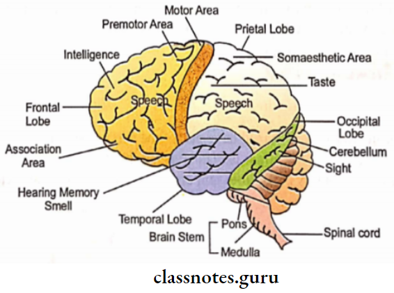
5. Cerebellum. It lies above the medulla oblongata, behind the cerebrum. The cerebellum is the second largest part of the brain, with 12.5% volume of the total.
- It has three parts, two large cerebellar hemispheres on the sides and a central small worm-like vermis.
- The cerebellum maintains the equilibrium of the body while walking, riding, dancing, picking up articles from the ground, etc. It also coordinates the muscular activities of the body.
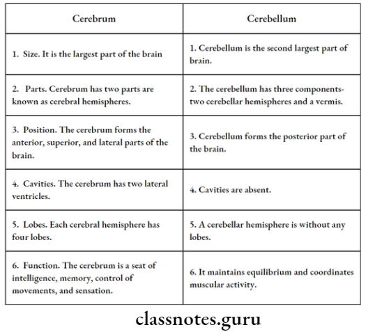
Differences Between Cerebrum and Cerebellum
6. Pons Varolii (= Pons). It is a crosswise band of nervous tissue that connects and relays information amongst the cerebrum, cerebellum, and medulla oblongata. It also possesses a pneumatic center of respiration.
7. Medulla Oblongata. It is a cylindrical conical that hinders most parts of the brain which lies below the cerebellum. The medulla oblongata encloses a fluid-filled cavity called the fourth ventricle. It controls many involuntary actions.
- For this, it has a respiratory center, cardiac center, blood pressure center, and reflex center for coughing, sneezing, vomiting, salivation, swallowing, and peristalsis.
- The medulla oblongata, pons, and midbrain are together called the brain stem. The midbrain and hindbrain do not have a thinking part. Instead, they control reflexes and involuntary actions connected with them.
How Are Nervous Organs Protected
Both the brain and spinal cord are delicate structures that are very efficiently protected. First of all, they lie in a fluid balloon. A bony covering occurs over the same.
- It is the cranium ( = brain box) of a skull in the case of the brain and the vertebral column or backbone in the case of the spinal cord.
- Fluid present inside and outside CNS is called cerebrospinal fluid. It filters out blood and performs all the functions of the blood. Additionally, it protects from shock.
- Three types of protective coverings or meninges occur around the brain and spinal cord—outer dura mater, middle arachnoid, and inner diameter.
- The Pia mater is in contact with the surface of the brain and spinal cord while the dura mater is in contact with the bony covering. Cerebrospinal fluid is present between the arachnoid and pia mater.
How Does The Nervous Tissue Cause Action
Nervous tissue collects the information from various receptors, processes the same, and decides the response. The response is conveyed to a particular muscle fiber through the axon terminal of a motor neuron.
- The knob or bouton of the axon terminal (motor-end plate) comes close to a depression or sole plate over the surface of the muscle fiber.
- As the excitation reaches the knob, the latter releases neurotransmitter. The neurotransmitter (for example., acetylcholine) is picked up by receptors of the sole plate.
- They become activated. They cause a release of calcium from the sarcoplasmic reticulum. It brings about the movement of myosin fibrils over actin fibrils. It causes shortening and changes in the shape of the muscle fiber. It is translated into movement.
- Nerve-controlled functioning of glands, other organs, and tissues also follow the same procedure.