Interior Of The Cranial Vault

Interior Of The Cranial Vault Definition

It is the internal surface of the skull cap.

Interior Of The Cranial Vault Shape

This is ovoid like norma verticalis.

Interior Of The Cranial Vault Bones

Same bones contribute to this part which were observed in norma verticalis, i.e.

  1. Frontal bone, anteriorly.
  2. Occipital bone, posteriorly
  3. Parietal bones, on each side.

Interior Of The Cranial Vault Sutures

Sutures correspond with those observed in norma verticalis which are as follows:

  1. Coronal suture, between frontal and parietal bones.
  2. Sagittal suture, between two parietal bones.
  3. Lambdoid suture, between parietal and occipital bones.

Interior Of The Cranial Vault Features

  • Frontal Crest
    • It is a midline crest seen at its anterior part.
    • Falx cerebri is attached to it.
  • Sagittal Sulcus
    • It is an anteroposterior groove in the median plane.
    • It is narrow anteriorly but widens posteriorly.
    • It contains superior sagittal sinus.
    • Falx cerebri is attached to its margins.
  • Bregma And Lambda
    • These mark the junctions of sagittal suture with coronal and lambdoid sutures respectively (see norma verticalis).
  • Parietal Foramen
    • It pierces the parietal bone on each side of midline about 3.5 cm in front of lambda. Emissary vein passes through it.
  • Granular Foveolae
    • These are irregular depressions on each side of sagittal sulcus.
    • These are produced by arachnoid granulations.
    • These are deep and more abundant in aged skull.
  • Grooves For Meningeal Vessels
    • Grooves for anterior (frontal) twigs of middle meningeal vessels are located just behind the coronal suture.
    • Grooves for parietal twigs of middle meningeal vessels are more posteriorly placed. These run backwards and upwards.
  • Impressions For Cerebral Gyri
    • These are less marked in cranial vault in contrast to the interior of the base of skull where cerebral impressions are well defined.

Interior Of The Cranial Vault Skull Cap Internal Surface

Ribs: Anatomy, Ligaments And Clinical Notes

Ribs General Considerations

  1. Ribs are bilateral bony arches forming the greater part of the thoracic wall.
  2. Normally there are 12 pairs of ribs which are numbered from above downwards.
  3. The length of the ribs increases from the 1st to the 7th rib and then decreases from the 7th to the 12th rib. Therefore, the 7th rib is the longest rib.
  4. The ribs are arranged obliquely, i.e. the anterior end is at a lower level than the posterior end. The obliquity is maximum in the 9th rib.
  5. The 8th rib is the most laterally projected rib.
  6. The width of the rib gradually reduces from above downwards.
  7. Intercostal spaces (gaps between adjacent ribs) are deeper in front than behind and deeper in the upper part than the lower part.

Ribs The Thoracic Cage Anterior And Posterior Views

First Ribs

Distinguishing Features

  1. It is the shortest.
  2. It is the broadest.
  3. It is most curved.
  4. It has no twisting.
  5. Angle coincides with tubercle.
  6. Head has got only a single facet.
  7. The costal groove is absent.
  8. The neck is rounded and elongated.
  9. It is flattened from above downwards and therefore has inner and outer borders and superior and inferior surfaces.

Side Determination

  1. Keep the larger end anteriorly and the smaller end posteriorly.
  2. Keep the surface of the shaft having two grooves separated by a ridge, superiorly.
  3. Keep the concave border towards the inner side and the convex border towards the outer side.

Note: Keep the rib on a flat surface considering its position in your own body.

The rib belongs to the side on which both ends touch the surface simultaneously. If the rib is placed on the wrong side then only the anterior end will be touching the table top.

Anatomical Position

  1. The posterior end is nearer the midline than the anterior end.
  2. The posterior end is 3.5 cm higher than the anterior end.
  3. The upper surface faces upwards as well as forwards.

Features And Attachments

Just like a typical rib, the first rib is comprised of two ends (anterior and posterior) and a shaft.

  1. Anterior End
    1. It is the larger end.
    2. It meets with 1st costal cartilage.
  2. Posterior End
    • It consists of the head, neck, and tubercle.
      1. Head
        1. It is small and rounded.
        2. It has a single rounded facet for articulation with the body of 1st thoracic vertebra to form a costovertebral joint.
        3. The capsular ligament of 1st costovertebral joint is attached to the margins of the facet.
        4. The radiate ligament is attached to the anterior margin of the head.
      2. Neck
        1. It is rounded.
        2. It is directed upwards, backward, and laterally.
        3. The inferior costotransverse ligament is attached to its posterior surface.
        4. The following structures form the anterior relations of the neck from medial to lateral:
          1. Sympathetic chain.
          2. First posterior intercostal vein.
          3. Superior intercostal artery.
          4. First thoracic root (T) of brachial plexus.
          5. Note: Remember SVAN for the relations of the anterior aspect of the neck from medial to lateral in which the S-sympathetic chain, V-Vein,- Artery, and N-Nerve.
      3. Tubercle
        1. It is large and prominent.
        2. It articulates with the transverse process of 1st thoracic vertebra.
        3. The lateral costotransverse ligament is attached laterally to the tubercle.
  3. Shaft
    • It consists of two borders (outer and inner) and two surfaces (upper and lower).
      1. Outer border
        1. It is convex.
        2. It is thick posteriorly and thin anteriorly.
        3. 1st digitation of the serratus anterior arises from its middle.
        4. It is related to the scalenus posterior muscle in its posterior part while clavipectoral fascia and pectoralis major muscle in its anterior part.
      2. Inner border
        1. It is concave.
        2. The scalene tubercle is situated near its middle.
        3. Sibson’s fascia (suprapleural membrane) is attached to it.
      3. Upper surface
        1. It is rough and irregular.
        2. It presents two shallow grooves separated by a ridge.
        3. The ridge continues medially with the scalene tubercle along the inner border.
        4. The scalenus anterior is inserted on the ridge and scalene tubercle.
        5. The subclavian vein lies in the groove anterior to the ridge.
        6. The subclavian artery along with the lower trunk of the brachial plexus occupies the posterior groove.
        7. Note: Remember ‘VAN’ is the sequence of structures occupying the grooves on the superior surface from anterior to posterior, i.e. Vein, Artery, and Nerve.
        8. The area anterior to the groove for the subclavian vein provides attachments to the subclavius muscle (anteriorly) and costoclavicular ligament (posteriorly). These attachments are located near the anterior end because they also extend over the costal cartilage.
        9. Scalenus medius is inserted on the rough area posterior to the groove for the subclavian artery.
      4. Lower surface
        1. It is smooth.
        2. It is related to costal pleura.
        3. Intercostal muscles are attached to this surface near its outer border.
        4. 1st intercostal nerve and vessels are related to this surface mainly in its posterior part.

Ribs First Rib Of Left Side Superior Aspect

Ribs First Rib Of Left Side Superior View

Ribs First Rib Of Left Side Inferior Aspect

Sternum: Anatomy, Parts, Pain And Diagram Notes

Sternum

Sternum Terminology

‘Sternum’ is derived from the Greek word ‘sternum’ which means chest. Sternum is also called ‘breast bone’.

It has three parts; manubrium, body, and xiphoid process. The manubrium is a Latin word that means ‘handle’. The term ‘xiphoid’ is borrowed from the Greek word ‘xiphos’ which means ‘sword’.

Sternum Location

It is a flat bone whose long axis is vertical. It lies in the median part of the anterior thoracic wall. Its surfaces are anterior and posterior. Its anterior surface also faces a little upwards.

Sternum Location Of Sternum

Sternum Length

It is about 7 inches (17 cm) long.

Sternum Structure

It is made up of mainly spongy bone and thus it is rich in red bone marrow.

Sternum Features

Sternum is made up of three pieces from above downwards:

  1. Manubrium.
  2. Body.
  3. Xiphoid process.

Sternum Manubrium

It is somewhat triangular in shape and is wider above than below. It has two surfaces (anterior and posterior) and four borders (superior, inferior, and two lateral).

Sternum Sternum Anterior Aspect

Sternum The Body

It has two surfaces (anterior and posterior), two borders (right lateral

Sternum Side Views Of Sternum And Thoracic Vertebrae

Sternum Xiphoid Process

It is the lowest and smallest part of the sternum and is of variable shape. It has two surfaces (anterior and posterior), two borders (right lateral and left lateral), and two ends (upper and lower).

Sternum Sternum Posterior Aspect

Sphenoid Bone: Anatomy, Function And Development Notes

Sphenoid Bone

Sphenoid Bone Terminology

‘Sphenoid’ is derived from the Greek word ‘sphen’ which means ‘a wedge’. The bone is so named because it is wedged between the frontal bone in front and the occipital bone behind.

Sphenoid Bone Anatomical Position

  1. Hypophyseal fossa faces upwards.
  2. Pterygoid processes descend vertically downwards.
  3. Openings of sphenoidal sinuses are directed forward.

Sphenoid Bone Articulations

The Sphenoid is a key bone in the cranial skeleton as it articulates with the following eight bones:

  1. Frontal
  2. Parietal
  3. Temporal
  4. Occipital
  5. Vomer
  6. Zygomatic
  7. Palatine
  8. Ethmoid

Sphenoid Bone Shape

Sphenoid resembles a ‘bat’ with its wings stretched out.

Sphenoid Bone Features And Attachments

Sphenoid consists of a central body, four wings (two greater and two lesser), and two pterygoid processes (right and left).

Sphenoid Bone Body

It has six surfaces (superior, inferior, anterior, posterior, and two lateral) and a pair of air-filled cavities (sphenoidal sinuses).

1. Surfaces

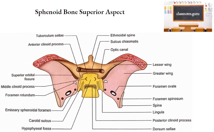
  1. Superior (cerebral) surface: It shows of following features from anterior to posterior.
    1. Jugum Sphenoidale
      1. It is smooth.
      2. It articulates with the posterior margin of the cribriform plate.
      3. It is related on each side to the gyrus rectus of the cerebral hemisphere and olfactor tract.
    2. Sulcus Chiasmatis
      1. It is a transverse groove behind the jugum sphenoidale
      2. Optic chiasma lies just above it.
      3. It leads laterally into the optic canal.
    3. Tuberculum Sellae
      • It is an elevation just behind the sulcus chiasmatis.
    4. Sella Turcica
      1. It is a depressed area behind the tuberculum sellae.
      2. The hypophyseal fossa is the deepest part of the Sella turcica. It lodges the pituitary gland.
      3. The anterior part of Sella turcica is bounded on each side by an elevation called the middle clinoid process.
    5. Dorsum Sellae
      1. It is a square plate of bone behind the sella turcica.
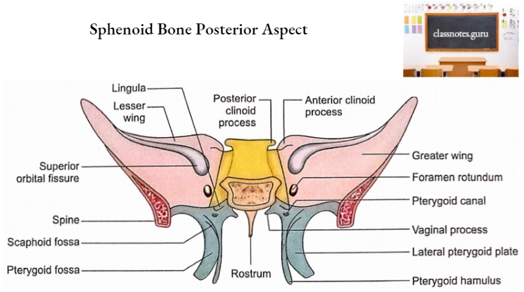
    6. Posterior Clinoid Process
      1. Superior angles of dorsum sellae project laterally into posterior clinoid processes.
      2. Attached margin of tentorium cerebelli is attached to this process on each side.
    7. The upper part of the clivus
      1. It is sloping behind the dorsum sellae.
      2. It is formed by the posterior parts of the body and dorsum sellae.
      3. It supports the pons.
  2. Posterior Surface 
    • It is rough.
    • It articulates with the basilar part of the occipital bone.
  3. Anterior And Inferior Surfaces 
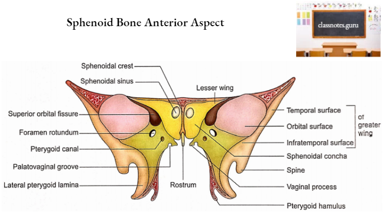
    1. The midline of the anterior surface is marked by a triangular crest called a sphenoidal crest.
    2. The sphenoidal crest articulates with the upper part of the posterior border of the perpendicular plate of the ethmoid.
    3. The midline of the inferior surface is marked by a triangular spine called a sphenoidal rostrum. It fits into the groove between the alae of the vomer.
    4. Both anterior and inferior surfaces of the body on either side of the midline, are occupied by a thin plate of bone called sphenoidal concha.
    5. Each sphenoidal concha consists of an anterior part which is vertical and quadrangular and a posterior part which is horizontal and triangular.
      1. Anterior Part
        1. It consists of an upper and lateral depressed area which completes the posterior ethmoidal sinus and articulates below with the orbital process of palatine bone.
        2. Its lower and medial part forms part of the roof of the nasal cavity and is perforated above by the round opening through which the sphenoidal sinus communicates with the sphenoethmoidal recess of the nasal cavity.
      2. Posterior Part
        • It forms part of the roof of the nasal cavity and completes the sphenopalatine foramen.
  4. Lateral Surface
    1. Its lower part unites with the greater wing and medial pterygoid plate.
    2. Its upper part is marked by a carotid sulcus which lodges the internal carotid artery and cavernous sinus.
    3. The lateral margin of the carotid sulcus at its posterior end projects backward into tongue-shaped lingula.
    4. The lingula lies just above the posterior opening of the pterygoid canal.

Sphenoid Bone Superior Aspect

Sphenoid Bone Posterior Aspect

Sphenoid Bone Anterior Aspect

2. Sphenoidal Sinuses

  1. These are two large air spaces present in the body of the sphenoid.
  2. The two sinuses are separated by a septum and are rarely symmetrical.
  3. Relations
    1. Superiorly:
      1. Optic chiasma.
      2. Pituitary gland.
    2. Laterally
      1. Internal carotid artery.
      2. Cavernous sinus.
  4. Size
    1. Vertical height: 2 cm (a little less than 2 cm)
    2. Transverse breadth: 1.8 cm
    3. Anteroposterior depth: 2.1 cm (little more than 2 cm).
      • Note: For simplification, students may consider all the measurements approximately as 2 cm.
  5. Each sinus communicates with the spheno-ethmoidal recess.
  6. Development
    1. Sphenoidal sinus starts developing as nasal mucosal evagination during intrauterine life.
    2. These are in the form of minute cavities at birth.
    3. It develops to its adult size in adolescence.

Sphenoid Bone Wings

1. Greater wings

There are two greater wings, a right and a left. Each has three surfaces (cerebral, lateral, and orbital) and several margins.

  • Surfaces
    1. Cerebral Surface
      • It is concave. It forms part of the middle cranial fossa. It is related to the temporal lobe of the cerebrum. It possesses the following foramina.
        1. Foramen rotundum
          • It is situated in the anteromedial part. The maxillary nerve passes through it.
        2. Foramen ovale
          • It is situated posterolateral to foramen rotundum. It transmits:
          • Mandibular nerve.
          • Accessory meningeal artery.
          • Lesser petrosal nerve.
          • Emissary vein.
        3. Emissary sphenoidal foramen
          • It is an inconstant foramen present medial to foramen ovale. It transmits the emissary vein.
        4. Foramen spinosum
          • It is lateral to foramen ovale. It transmits:
          • Middle meningeal artery.
          • Nervus spinosus.
        5. Canaliculus innominatus
          • It is occasionally present between foramen ovale and foramen spinosum. If present, it transmits lesser petrosal nerve.
    2. Lateral Surface
      1. It is convex from above downwards.
      2. The infratemporal crest is an anteroposterior ridge that divides the lateral surface into upper temporal and lower infratemporal parts.
      3. The temporal surface forms part of the temporal fossa and gives origin to the temporalis muscle.
      4. The infratemporal surface forms the roof of the infratemporal fossa and gives origin to the upper head of the lateral pterygoid muscle. This surface possesses openings of foramen ovale and foramen spinosum.
      5. The spine of the sphenoid is a projection at the posterior end of the lateral surface. It shows the following relations and attachments:
        1. Tip gives attachment to spheno-mandibular ligament.
        2. Medially it is related to the chorda tympani nerve and auditory tube.
        3. Laterally it is related to the auriculotemporal nerve.
    3. Orbital Surface
      1. It is quadrilateral in shape.
      2. It forms the posterior part of the lateral wall of the orbit.
      3. Its upper serrated edge articulates with the orbital plate of the frontal bone.
      4. Its lateral serrated margin articulates with the zygomatic bone.
      5. Its inferior smooth border forms the posterolateral boundary of the inferior orbital fissure.
      6. Its medial sharp margin constitutes the lower boundary of the superior orbital fissure. A projection from this border provides attachment to a common tendinous ring.
      7. Below the medial end of the superior orbital fissure is a depressed area pierced by foramen rotundum.
  • Margins
    1. The tip of the greater wing is called the parietal margin. It articulates with the sphenoidal angle of the parietal bone at the pterion.
    2. The posterior margin of the greater wing extends from the body of the sphenoid to its spine. Its medial half forms the anterior boundary of the foramen lacerum and receives the opening of the pterygoid canal. Its lateral half articulates with the petrous temporal.
    3. The lateral margin extends forward from the spine to the tip of the greater wing. This is also called the squamosal margin because it articulates with the squamous part of the temporal bone.
    4. Medial to the tip there is a triangular rough area for the frontal bone.
    5. The anterior angle of the triangular area continues with a serrated margin (lateral margin of the orbital surface) which articulates with the zygomatic bone.

2. Lesser Wings

It is a triangular bone extending laterally from the anterosuperior part of the body. It consists of a tip, two roots (anterior and posterior), two surfaces (superior and inferior), and two borders (anterior and posterior).

  • Тір
    1. It is the lateral end of the lesser wing.
    2. It is situated near the lateral end of the superior orbital fissure.
  • Roots
    1. The lesser wing is connected to the body by anterior and posterior roots.
    2. The two roots enclose the optic canal which transmits the optic nerve and ophthalmic artery.
  • Surfaces
    1. Superior Surface
      • It forms the posterior part of the floor of the anterior cranial fossa.
    2. Inferior Surface
      • It forms the superior boundary of the superior orbital fissure and the posterior part of the orbital roof.
  • Borders
    1. Anterior border
      • It articulates with the posterior border of the orbital plate of the frontal bone.
    2. Posterior Border
      • It is free.
      • Its medial end forms the anterior clinoid process to which is attached the free margin of tentorium cerebelli.

3. Superior Orbital Fissure

  1. It is a triangular slit-like communication between the orbit and the middle cranial fossa.
  2. Boundaries
    • Medial: Body of sphenoid Apex: Frontal bone
    • Superior: Lesser wing of sphenoid.
    • Inferior: Greater wing of sphenoid.
  3. It transmits the following structures:
    • Structures that enter the orbit
      1. Upper and lower divisions of the oculomotor nerve.
      2. Trochlear nerve.
      3. Three branches (lacrimal, frontal, and nasociliary) of the ophthalmic division of the trigeminal nerve.
      4. Abducent nerve.
      5. Orbital branch of the middle meningeal artery.
      6. Sympathetic filaments.
    • Structures that appear from the orbit
      1. Superior and inferior ophthalmic veins.
      2. Recurrent meningeal branch of the lacrimal artery.

Pterygoid Processes

  • The pterygoid process on each side descends vertically downwards from the junction of the body and the greater wing of the sphenoid.
  • Each consists of a lateral and a medial pterygoid plate.
  • The plates unite anteriorly in the upper part to enclose a fossa called pterygoid fossa.
  • The plates are not united in the lower portion to form a pterygoid fissure which is filled by the pyramidal process of palatine bone.
  • The anterior surface of the pterygoid process forms the posterior boundary of the pterygopalatine fossa.
  • The anterior opening of the pterygoid canal is located in this region. Some details of the two pterygoid plates are as follows:

1. Lateral pterygoid plate

It has two surfaces (lateral and medial) and two borders (anterior and posterior).

  • Surfaces
    1. Lateral Surface
      • It forms the medial wall of the infratemporal fossa and gives origin to the lower head of the lateral pterygoid muscle.
    2. Medial Surface
      • It forms the lateral wall of the pterygoid fossa which gives origin to the deep head of the medial pterygoid muscle.
  • Borders
    1. Anterior Border
      • It forms the posterior boundary of the pterygomaxillary fissure.
    2. Posterior Border
      • It is free.

2. Medial Pterygoid Plate

It has two surfaces (lateral and medial) and two borders (anterior and posterior).

  • Surfaces
    1. Lateral surface
      • It forms the medial wall of the pterygoid fossa and is related to the tensor palate muscle.
    2. Medial surface
      1. It forms the lateral wall of the corresponding posterior nasal aperture.
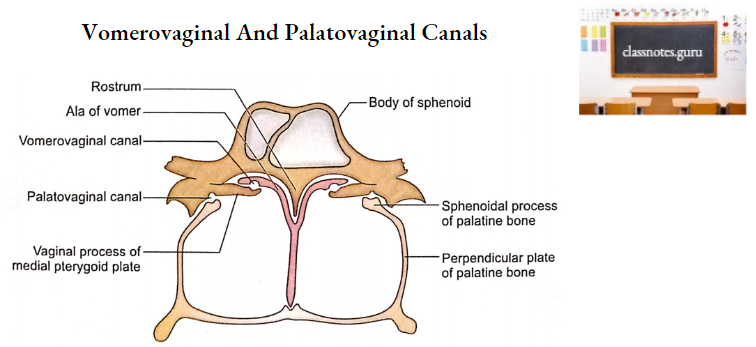
      2. Vaginal process is a thin lamina projecting medially from its upper part under the body of the sphenoid.
      3. A groove on the anterior part of the undersurface completes the palatovaginal canal with the sphenoidal process of the palatine bone.
      4. This canal transmits the pharyngeal branch of the maxillary artery and the pharyngeal branch of the pterygopalatine ganglion.
      5. The vaginal process articulates medially with ala of the vomer and forms vomero-vaginal canal between the two. This canal transmits branches of pharyngeal nerves and vessels.
  • Borders
    1. Anterior border
      • It articulates with the posterior border of the perpendicular plate of the palatine bone.
    2. Posterior border
      1. At its upper end, it splits to enclose the scaphoid fossa which gives origin to the tensor palati muscle.
      2. Its upper end shows a small projection called the pterygoid tubercle which lies immediately below the posterior end of the pterygoid canal.
      3. Pharyngobasilar fascia is attached to its whole extent while the superior constrictor arises from its lower part only.
      4. A hook-like process at its lower end is called pterygoid hamulus. Tondon of tensor palati winds around this process. Superior constrictor and pterygomandibular raphe are also attached to it.
      5. An angular process projecting from the middle of this margin is called processus tubarius.
      6. The posterior border above this process is called the notch of the auditory tube. This process and notch support the medial end of the auditory tube.

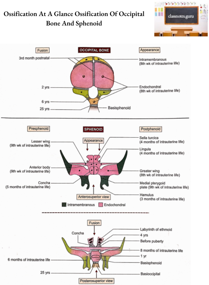
Sphenoid Bone Ossification

  1. Sphenoid ossifies partly in the membrane and partly in cartilage.
  2. Parts ossifying in the membrane are as follows:
    • Greater wings except for their roots.
    • Pterygoid processes except pterygoid hamuli.
  3. Parts ossifying in cartilage are as follows:
    • Body of sphenoid.
    • Lesser wings.
    • Sphenoidal conchae.
    • Roots of greater wings.
    • Pterygoid hamuli.
  4. From an ossification point of view, the sphenoid is divided into presphenoidal and post-sphenoidal parts.
    1. The presphenoidal part is comprised of parts lying in front of tuberculum sellae, i.e. anterior part of the body, lesser wings, and sphenoidal conchae.
      1. Two centers appear for each of these components as follows:
      2. Anterior body-9th week of intrauterine life.
      3. Lesser wings-9th week of intrauterine life.
      4. Conchae-5th month of intrauterine life.
    2. Rest of the sphenoid is included in post sphenoidal part. Two centers appear for each of the following components of the post-sphenoidal part.
      • Sella turcica-4th month of intrauterine life.
      • Lingulae 4th month of intrauterine life. Greater wings (including lateral pterygoid plates)-8th week of intrauterine life.
      • Medial pterygoid plates-9th week of intrauterine life.
      • Hamuli-3rd month of intrauterine life.
  5. Fusions of different components of sphenoid take place as follows:
    1. Medial and lateral pterygoid plates fuse with each other at about 6th month of intrauterine life.
    2. The presphenoidal part of the body fuses with the postsphenoidal part of the body at about the 8th month of intrauterine life.
    3. At birth, the sphenoid is in three parts, a central part consisting of the body and lesser wings and two lateral parts, each consisting of the greater wing and the pterygoid process.
    4. Greater wing fuses with the body at about 1st year.
    5. Concha fuses with the ethmoidal labyrinth at about 4th year.
    6. Concha fuses with the body of the sphenoid before puberty.
    7. The body of the sphenoid fuses with the basilar part of the occipital bone at about the 25th year.

Sphenoid Bone Applied Anatomy

  1. In the anterior part of the hypophyseal fossa, there is occasionally a vascular foramen termed as craniopharyngeal canal. The canal sometimes extends inferiorly to the exterior of the skull and is said to mark the original position of Rathke’s pouch.
  2. Premature ossification of sutures between pre and post-sphenoidal parts and sphenoid and occipital bones is often observed in achondroplasia.
  3. Anomalous development of the pre-sphenoidal elements may lead to excessive separation of the two orbits (hypertelorism).
  4. Observation of the sella turcica and the hypophyseal fossa in radiographs is important clinically because they may reflect pathological changes such as a pituitary tumor or aneurysm of the internal carotid artery.
  5. Decalcification of the dorsum sellae is one of the signs of a generalized increase in intracranial pressure.
  6. The lateral wall of the optic canal is fractured during optic nerve decompression in the optic canal.
  7. A fracture of the sphenoid bone may lacerate the optic nerve resulting in blindness.
  8. Basilar fracture of the skull through the sphenoid bone may lacerate the internal carotid artery resulting in the carotid-cavernous fistula. This leads to pulsating exophthalmos.
  9. Collection of air in the cranial cavity (aerocele) may occur if the basilar fracture of the skull involves a sphenoidal sinus.
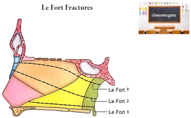
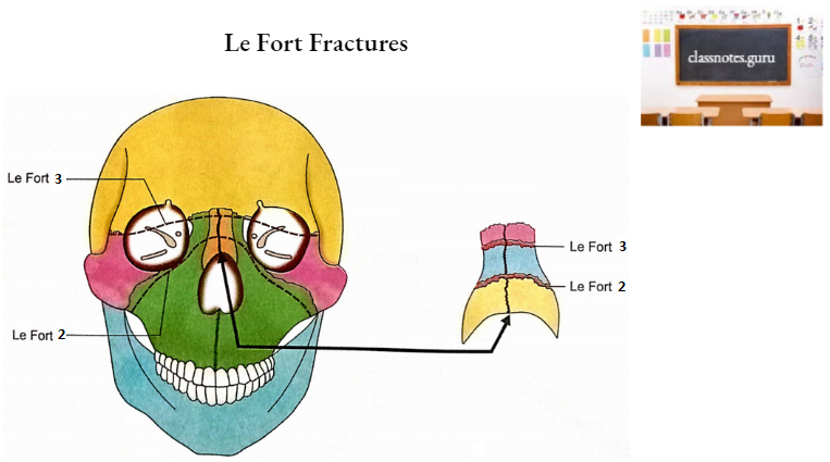
  10. Involvement of the pterygoid processes of the sphenoid is a constant feature in cases of Le Fort fractures of the mid-facial skeleton but the location of a fracture depends upon its type.
  11. In Le Fort 1 fracture, the lower 3rd of the pterygoid plates is involved while in Le Fort 3 fracture the roots of pterygoid plates are fractured.
  12. Large areas of the body and medial pterygoid plates are clothed in mucosa and, therefore, fractures of these parts of the sphenoid may open into the nasal cavity or sphenoidal sinus with potential risk of infection.

Vomer: Anatomy, Location, And Function Notes

Vomer

Vomer Terminology

‘Vomer’ is a Latin word. The term is used for the thin plate of bone between the nostrils.

Vomer Location

Vomer forms the posteroinferior part of the septum of the nose.

Vomer Left View

Vomer Features And Attachments

Vomer has got two surfaces (right and left) and four borders (superior, inferior, anterior, and posterior).

Vomer Surfaces

  1. It has small grooves for vessels.
  2. A large groove runs downwards and forwards. This is meant for nasopalatine nerve and vessels.

Vomer Borders

  • Superior Border
    1. It is thick.
    2. Two lateral projections (alae) enclose a deep furrow that fits over the rostrum of the sphenoid.
    3. The margin of ala intervenes between the body of the sphenoid and the vaginal process of the medial pterygoid plate. Under the surface of the ala forms a vomer-ovaginal canal with the vaginal process.
  • Inferior Border
    • It articulates with the nasal crest formed by the maxillae and palatine bones.
  • Anterior Border
    1. It is the longest border.
    2. Its upper half articulates with the posterior border of the perpendicular plate of the ethmoid bone.
    3. Its lower half is attached to septal cartilage.
  • Posterior Border
    1. It is free.
    2. It is situated between two posterior nasal apertures (choanae).

Vomer Le Fort Fractures

Vomer Ossification

  1. Vomer develops by ossification of the membrane covering the median septal part of the cartilaginous nasal capsule.
  2. One center of ossification appears on each side of the cartilage at about the 8th week of intrauterine life. giving rise to two bony plates separated by a cartilage.
  3. Two bony plates fuse with each other in the lower part at about the 12th week of intrauterine life.
  4. The cartilaginous plate is gradually absorbed allowing the fusion of two bony plates which proceed upwards from below. Fusion is completed at puberty.

Vomer Applied Anatomy

  1. The vomer is paper thin and does not resist much force responsible for the fracture.
  2. The vomer is involved in all three types of Le Fort fractures of the mid-facial skeleton.
  3. Vomer receives adequate blood supply from periosteal arteries and therefore all the fragments of fractured bone retain a periosteal blood supply.
  4. A transverse fracture of the vomer due to a direct blow on the nose can lead to deviation of the nasal septum (DNS).
  5. The vomer is clothed in mucosa over large areas of its surfaces, and therefore, its fracture opens into the nasal cavity with a potential risk of infection.
  6. Vomer may be deviated from the median plane as a result of birth injury or a congenital malformation.
  7. In case of severe deviation, the nasal septum comes into contact with the lateral wall of the nasal cavity. Surgical repair (submucosal resection-SMR) is usually necessary to correct the deviation.

Vomer Vomerovaginal And Palatovaginal Canals

Nasal Conchae: Anatomy, Structure And Function Notes

Inferior Nasal Conchae

Inferior Nasal Conchae Terminology

‘Concha’ is a Latin word that means ‘shell’. Conchae (superior, middle, and inferior) are bracket-like projections of thin (like eggshell) bones from the lateral wall of the nose.

Inferior Nasal Conchae Location

An inferior concha is an independent bone whose long axis occupies the whole length of the lower part of the lateral wall of each half of the nasal cavity.

Inferior Nasal Conchae Features And Attachments

Each inferior concha has two ends (anterior and posterior), two surfaces (medial and lateral), and two borders (superior and inferior).

Inferior Nasal Conchae Ends

  1. Anterior End
    • It is pointed and directed forward.
  2. Posterior End
    • It is directed backward and is more pointed and tapering.

Inferior Nasal Conchae Surfaces

  • Medial Surface
    1. It is convex.
    2. It has numerous apertures and grooves for vessels.
  • Lateral Surface
    1. It is concave.
    2. It forms the medial wall of the inferior meatus of the nose.

Inferior Nasal Conchae Right Inferior Concha Medial Aspect

Inferior Nasal Conchae Right Inferior Concha Lateral Aspect

Inferior Nasal Conchae Borders

1. Superior Border

    1. It is thin and irregular.
    2. It is divided into three parts:
  • Anterior Part: This articulates with the conchal crest of the maxilla.
  • Posterior Part: This articulates with the conchal crest of the palatine bone.
  • Middle Region: This part possesses three processes which are as follows from anterior to posterior:
    1. Lacrimal Process: It is an upward projection to articulate the descending process of the lacrimal bone.
    2. Maxillary Process: It is a curved downward projection that articulates with the nasal surface of the maxilla and the lower part of the anterior border of the perpendicular plate of the palatine bone.
    3. Ethmoidal process: It is an upward projection to articulate with the uncinate process of the ethmoid.

2. Inferior Border

  • It is free.
  • It is thick.

Inferior Nasal Conchae Ossification

1. It develops from the lowest part of the lateral region of the cartilaginous nasal capsule.

2. The center of ossification appears during the 5th month of intrauterine life.

Inferior Nasal Conchae Applied Anatomy

Inferior nasal concha is at great risk in cases of mid-facial injuries.

  1. Inferior concha receives adequate blood supply from periosteal arteries and, therefore, all the fragments of the fractured bone retain a periosteal blood supply.
  2. Inferior concha is clothed in nasal mucosa over large areas of its surfaces and, therefore, the fractures usually open to the nasal cavity with potential risk of infection.
  3. Infracture of the inferior concha is sometimes needed during the management of congenital lacrimal defects.

Inferior Nasal Conchae Infracture Of Inferior Concha

Lacrimal Bone: Anatomy, Borders And Function Notes

Lacrimal Bones

Lacrimal Bones Terminology

‘Lacrimal’ is a Latin word which means ‘tear’. The bone is so named because of its relation with the tear sac.

Lacrimal Bones Peculiarities

  1. It is most fragile among the cranial bones.
  2. It is the smallest of the cranial bones.

Lacrimal Bones Location

  1. There are two lacrimal bones.
  2. Each lacrimal bone is located in the anterior part of the medial wall of the orbit.
  3. It also contributes to the middle meatus of the nose.

Lacrimal Bones Features And Attachments

Lacrimal bone is rectangular in shape. It has two surfaces (medial and lateral) and four borders (anterior, posterior, superior, and inferior).

Lacrimal Bones Surfaces

1. Medial Surface

  • It is also called the nasal surface.
  • Its anteroinferior part contributes partly to the middle meatus of the nose.
  • Its posterosuperior part articulates with the ethmoid and completes a few anterior ethmoidal air cells.

Lacrimal Bones Right Lacrimal Bone Medial Surface

2. Lateral Surface

  • It is also known as the orbital surface.
  • It is divided into anterior and posterior parts by a vertical crest called the posterior lacrimal crest.
  • The anterior part is grooved and forms the posterior half of the floor of the lacrimal groove.
  • The anterior half of the lacrimal groove is formed by the frontal process of the maxilla. The groove lodges the lacrimal sac.
  • The portion behind the posterior lacrimal crest is smooth and forms part of the medial wall of the orbit.
  • The lower end of the posterior lacrimal crest projects forward as the lacrimal hamulus.
  • It articulates with the maxilla to complete the upper end of the nasolacrimal canal.
  • The posterior lacrimal crest provides attachment to the lacrimal fascia.
  • The crest and small area of the lateral surface immediately behind it give origin to the lacrimal part of the orbicularis oculi muscle.
  • The medial wall of the groove projects downwards in a descending process.
  • This process articulates with the lips of the nasolacrimal groove of the maxilla and the lacrimal process of the inferior concha to complete the bony canal for the nasolacrimal duct.

Lacrimal Bones Right Lacrimal Bone Lateral Surface

Lacrimal Bones Borders

  1. Anterior Border
    • It articulates with the frontal process of the maxilla.
  2. Posterior Border
    • It articulates with the orbital plate of the ethmoid.
  3. Superior Border
    • It articulates with the nasal notch of the frontal bone.
  4. Inferior Border
    • It articulates with the orbital surface of the maxilla.

Lacrimal Bones Ossification

Lacrimal bone ossifies in membrane.

2. A single center of ossification appears in the mesenchyme around the cartilaginous nasal capsule.

3. The center appears at about the 12th week of intrauterine life.

Lacrimal Bones Applied Anatomy

  1. A severe impact on the nasal bridge may involve the lacrimal bone and damage the lacrimal passage.
  2. Lacrimal bone is included by clinicians in the central portion of the middle 3rd facial skeleton.
  3. All the bones of the middle 3rd facial skeleton receive adequate blood supply from periosteal arteries and, therefore, all the fragments of fractured bone retain a periosteal blood supply.
  4. Lacrimal bone is involved in Le Fort 3 fracture.
  5. Since the anteroinferior part of the nasal surface of the lacrimal bone is covered with nasal mucosa, the fracture of the bone may open into the nasal cavity with the potential risk of infection.
  6. To reach the medial wall of the optic canal during a surgical procedure called optic nerve decompression, most of the bones of the medial wall of the orbit (including the lacrimal bone) are fractured.
  7. Lacrimal bone is very fragile, therefore extra precautions should be taken to avoid trauma during surgery of the lacrimal system.
  8. In some of the cases of obstruction of the lacrimal sac or nasolacrimal duct, dacryocystorhinostomy is performed.
  9. In this operation, an artificial passage is made for drainage into the nasal cavity, by breaking the lacrimal bone.

Parietal bone: Anatomy, Borders and Surfaces

Parietal Bones Terminology

The word parietal is derived from the Latin word ‘paries’ which means ‘wall’, because two parietal bones form a large part of the walls of the calvaria.

Parietal Bones Side Determination

  1. Keep the bone by the side of your own cranial vault in such a way that the outer surface is convex and the inner surface is concave.
  2. Inferior (squamosal) border is concave.
  3. The anteroinferior angle is prominent and has a vascular and narrow groove on its inner aspect.
  4. The posteroinferior angle has a shallow and wide groove for the sigmoid sinus on its inner aspect.

Parietal Bones Features And Attachments

1. Parietal Bones Surfaces

It has two surfaces, external and internal.

  • External Surface
    • It is relatively smooth.
    • The most prominent part of this surface is called parietal tuberosity or eminence.
    • There are two curved lines running anteroposteriorly. These are called superior and inferior temporal lines.
    • The superior temporal line gives attachment to temporal fascia while the area below the inferior temporal line gives attachment to the temporalis muscle.
    • The area above the superior temporal line is covered by galea aponeurotica.
    • A foramen may be present near the posterior part of the sagittal border. This is called parietal foramen. It transmits the emissary vein.
  • Internal Surface
    • It is concave and exhibits elevations and depressions for cerebral sulci and gyri respectively.
    • Near the sagittal border, there is a longitudinal half groove (to be completed with that of the opposite side) for the superior sagittal sinus. The margins of the groove provide attachment to falx cerebri
    • Grooves for the branches of middle meningeal vessels are present at the anteroinferior angle and at the middle of the lower border of the bone.
    • Adjacent to the groove for the superior sagittal sinus there are deep irregular pits (granular foveolae) produced by arachnoid granulations.
    • The bone is grooved near the posteroinferior angle by the sigmoid sinus.

Parietal Bones Right Parietal Bone External Surface

Parietal Bones Right Parietal Bone Internal Surface

2. Parietal Bones Borders

It has four borders, superior, inferior, anterior, and posterior.

  • Superior Border
    • This is also called the sagittal border.
    • It articulates with a similar border of opposite sides to form a sagittal suture.
  • Inferior Border
    • This is also called the squamosal border.
    • It articulates with the following three bones from anterior to posterior:
      • Greater wing of the sphenoid.
      • Squamous part of temporal.
      • Mastoid portion of temporal.
  • Anterior Border
    • This is also called the frontal border.
    • It articulates with the frontal bone to form a coronal suture.
  • Posterior Border
    • This is also called the occipital border.
    • It articulates with the squamous part of the occipital bone to form a lambdoid suture.

3. Parietal Bones Angles

The parietal bone has four angles (frontal, sphenoidal, occipital, and mastoid).

  • Frontal Angle
    • This is also called the anterosuperior angle.
    • It corresponds to bregma, i.e. the junction of coronal and sagittal sutures.
  • Sphenoidal Angle
    • This is also called an anteroinferior angle.
    • It corresponds to pterion, i.e. a small area enclosing four bones (frontal, temporal, parietal, and greater wing of sphenoid).
  • Occipital Angle
    • This is also called the posterosuperior angle.
    • It corresponds to lambda, i.e. junction of sagittal and lambdoid sutures.
  • Mastoid Angle
    • This is also called posteroinferior angle.
    • It corresponds to asterion, i.e. small area enclosing three bones, parietal, temporal, and occipital.

Parietal Bones Ossification

  1. Parietal bones ossify in the membrane.
  2. Each ossifies from two centers which appear at parietal tuberosity at about the 7th week of intrauterine life.
  3. The centers soon fuse with each other and then the ossification spreads radially.
  4. Angles are the parts last to be ossified explaining the existence of a fontanelle at each angle before the ossification is completed.

Parietal Bones Age Changes

  • At birth
    • Temporal lines are present at quite a lower level.
  • Adult
    • Higher and permanent positions of temporal lines are reached only after the eruption of permanent molar teeth.

Parietal Bones Applied Anatomy

  1. Occasionally the parietal bone is divided into upper and lower parts by an anteroposterior suture. The condition may be confused with fracture radiologically.
  2. The latter can be ruled out easily because the anomalous parietal suture is usually bilateral.
  3. Parietal bones are loosely attached to the adjacent bones at sutures during the intrauterine period allowing moulding (change in shape of calvaria) at the time of childbirth.
  4. Calvaria returns to normal shape within a few days after birth.
  5. Parietal bones undergo remodeling to allow enlargement of calvaria during childhood. This is only possible because of their loose attachments to the adjacent bones.
  6. Granular foveolae are more numerous and marked in aged parietal bones. This fact is of great medical importance.
  7. The regenerating capacity of the parietal bone is negligible due to the lack of a cambium layer in the periosteum.
  8. In neonates, the parietal bone is pliable and soft, and, therefore, a depressed fracture (pond fracture) is like a dimple.
  9. In adults such fractures are produced by direct blows and always show an irregular line of fracture at the periphery of the depressed area. The depression of the inner table forms the lowest limit of the depressed area also
  10. Almost invariably all fractures the known as apex.
  11. A crack in the inner table of the parietal bone may damage a large diplomatic vein and produce a small epidural hematoma. parietal bone in children is associated with rupture of the dura mater.
  12. In adults, the parietal bone shows a fissured or linear fracture if the force is transmitted to this bone from frontal or occipital blows.

Parietal Bones A Depressed Fracture Of Parietal Bone In Adult

Nasal Bones Osteology Notes

Nasal Bones

Nasal Bones Terminology

The nasal bone is so named because of its location. It forms the bridge of the nose.

Nasal Bones Location

Two nasal bones meet with each other in the midline in the upper part of the external nose. They are located below the nasal part of the frontal bone and between the frontal processes of the maxillae.

Nasal Bones Features And Attachments

Each nasal bone has two surfaces (external and internal) and four borders (superior, inferior, lateral, and medial).

Nasal Bones Surfaces

1. External Surface

  • It is convex from side to side.
  • It is covered by the procerus and nasalis muscles.
  • A foramen in the center (vascular foramen) allows the transmission of a small vein.

Nasal Bones Right Nasal Bone External Surface

2. Internal Surface

  • It is concave from side to side
  • It presents a vertical groove for the anterior ethmoidal nerve.

Nasal Bones Right Nasal Bone Internal Surface

Nasal Bones Borders

1. Superior Border

  • It is serrated.
  • It articulates with the nasal part of the frontal bone.

2. Inferior Border

  • It is notched for the passage of the external nasal nerve.
  • It is continuous with the lateral nasal cartilage.

3. Lateral Border

It articulates with the frontal process of the maxilla.

4. Medial Border

  • It is thicker above than below.
  • It articulates with the opposite nasal bone (to form an internasal suture) and is prolonged behind as a nasal crest.
  • The nasal crest articulates with the following structures from above downwards:
    1. The nasal spine of the frontal bone.
    2. Perpendicular plate of ethmoid.
    3. Septal cartilage.

Nasal Bones Ossification

Nasal bone ossifies in the membrane overlying the anterior part of the cartilaginous nasal capsule

The Centre of ossification appears in its middle during 3rd month of intrauterine life.

Nasal Bones Applied Anatomy

  1. Nasal bone is usually fractured due to a direct hard blow.
  2. The fractures of the nasal bone are transverse in nature.
  3. The common site for the fracture of the nasal bone is half (2) inches above its inferior border.
  4. The slight mobility of the anteroinferior part of the nasal bone protects the nose against mild injuries.
  5. An impact directed in the anteroposterior plane will cause a depression of the nasal bridge due to a fracture of the nasal bone, frontal process of the maxilla, and septal cartilage.
  6. A force directed from the lateral aspect will result in a deviation of the nasal bridge to the opposite side.
  7. Traumatic alteration in the shape of the nose because of fracture of nasal bones is of great clinical importance due to cosmetic reasons, especially in young females.
  8. In cases of Le Fort 2 and Le Fort 3 fractures of maxillae, nasal bones are also involved. Le Fort 1 fracture of maxillae spares the nasal bone.

Nasal Bones Le Fort Fractures

Ossification Osteology Notes

Ossification At A Glance

Ossification At A Glance Definition

Ossification is defined as the deposition of calcium salts in the membranous or cartilaginous background of a bone. The former is called intramembranous, while the latter endochondral ossification.

Ossification At A Glance Centre Of Ossification

The site in the developing bone where calcium salts start depositing is called the centre of ossification. The centre of ossification is first microscopic but soon becomes macroscopic.

Ossification At A Glance Examples

  • Bones ossifying in the membrane are most of the mandible, the upper part of the squamous part of the occipital bone, the frontal bone, parietal bones, and squamous and tympanic parts of temporal bones.
  • Upper parts of greater wings and pterygoid processes of sphenoid; palatine, lacrimal and zygomatic bones; maxillae, vomer and nasal bones. The rest of the bones in the body are endochondral in origin.

Primary And Secondary Centres

  • Though many bones (For example. lacrimal, nasal, zygomatic bones; inferior nasal conchae and auditory ossicles) ossify from a single centre, the majority of them ossify from several foci.
  • One centre in these bones appears first in late embryonic and early foetal life (7th week to the primary centre of ossification.
  • Remaining 4th month of intrauterine life). These are called centres, called secondary centres appear later during the period from birth to 12 years.

Fusion Of Ossification Centres

  • The secondary centres fuse with each other and then with the bone derived from the primary centre.
  • The process of fusion begins as early as 3 months of intrauterine life (Example. fusion between 4 centres of squamous part of occipital bone) or may be observed as late as 40 years of age (Example. fusion between body and xiphoid process of sternum).

Fusion Between Adjacent Bones

Some adjacent bones may fuse to develop continuity, for Example. basilar parts of both sphenoid and occipital bones fuse at the age of 25 years.

Ossification At A Glance Clinical Significance

Xiphoid process fuses with the body of the sternum at the age of 40 years, therefore, enhancing the scope of age determination at a later age.

Cartilaginous Nasal Capsule

  • It is the cartilage which forms medial and lateral walls as well as the roof of the nasal cavity. It plays an important role in the development of the nasal framework.
  • Structures derived from this capsule are sphenoidal conchae, ethmoidal bone, inferior nasal conchae, vomer, nasal bones and septal, lateral nasal and alar cartilages.
  • Nasal bones and vomer develop in the membrane while the rest of the above bones are endochondral in origin.

Appearance And Fusion Of Ossification Centres

The times of appearance of the ossification centre (in case of single focus) or appearance, as well as a fusion of primary and secondary centres (in case of multiple foci), are represented diagrammatically.

Ossification At A Glance Ossification Of Mandible, Hyoid, Parietal Bone And Frontal Bone

Ossification At A Glance Ossification Of Occipital Bone And Sphenoid

Ossification At A Glance Ossification Of Temporal Bone, Lacrimal Bone, Zygomatic Bone And Maxilla

Ossification At A Glance Fate Of Cartilaginous Nasal Capsule