General Considerations Of Bone Definition
Bone is the hard part of the body providing a dynamic framework for it.
Bone Properties

Bone Functions
- Bones provide a framework for the body.
- Bones accord shape to the body.
- Bones act as levers for muscles and, therefore, help in the movements of the body.
- Bones provide protection to a number of viscera, for example. brain, lungs and heart.
- Bone is the site of blood formation.
- Bone plays an important role in the immune responses of the body by producing cells of the reticuloendothelial system.
- Bones are storehouses of calcium and phosphorus
Bone Chemical Composition
Bone is one-third organic and two-thirds inorganic. Inorganic calcium salts [calcium phosphate, calcium carbonate and crystals of hydroxyapatite, i.e. Ca10 {P04}4 (OH)2] make it hard and rigid. the organic connective tissue(collagen fibre) makes it tough and resilient. The collagen protein of collagen fibres is characterised by hydroxyproline amino acid.
Structure Of Bone
1. Macroscopically
There are two types of bones, spongy or cancellous bone and compact or dense bone. The outer part of all bones is made up of compact bone. Cancellous bone fills up the interior of the except the following.
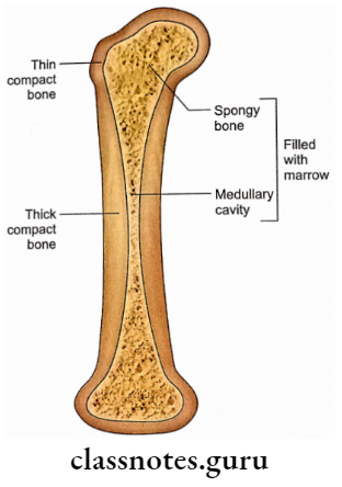
In the shaft of a long bone, it is replaced by a medullary cavity. This is filled with red marrow in newborns but replaced by yellow or fatty marrow in adults.
- In the maxilla, sphenoid, ethmoid and frontal bones, it is replaced by large air spaces called sinuses.
- In many places, the cancellous bone is replaced by marrow. The red marrow is active in hematopoiesis. Yellow marrow is mainly inert and fatty.
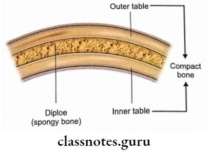
- The flat bones of the skull cap (calva) have a spongy bone (diploe) sandwiched between two compact bones called outer and inner tables. Red marrow persists in spongy bone throughout life.
The compact bone is more radio-opaque, while the spongy bone is relatively more radiolucent. In the radiograph, therefore, the compact bone looks whiter than the spongy bone which appears relatively darker.
2. Microscopically
Microscopically the bones can be classified into four types:
- Lamellar Bone: Most of the mature human bones, both compact and spongy, are of this type.
- Fibrous Bone: It is found in early foetuses.
- Dentine: It is found in teeth.
- Enamel: It is found in teeth.
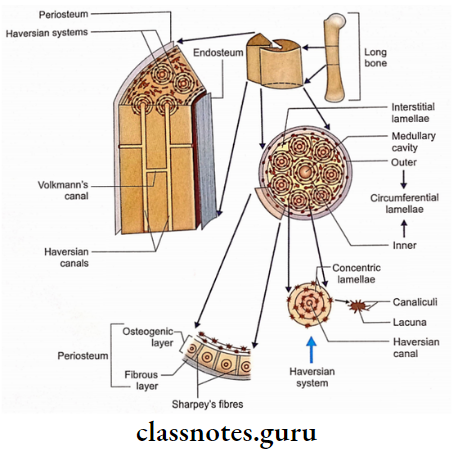
The compact bone shows typical Haversian systems each of which is comprised of a central canal along the long axis of bone surrounded by concentric lamellae.
- Volkmann’s canals connect the adjacent Haversian canals. Osteocytes are located in the small spaces (lacunae) between adjacent lamellae.
- The cytoplasmic processes of osteocytes extend into canaliculi diverging from lacunae. Circumferential lamellae adjoin the surface or medullary cavity of long bones. Interstitial lamellae fill the spaces between Haversian systems.
- Spongy bone differs from ct fa ‘lacking Haversian systems’ and ‘having Spongy bone differs from compact bone in irregularly arranged bony lamellae’.
- Periosteum, the outer covering of bone, consists of an external collagen fibrous layer and an inner osteogenic cellular layer.
Collagen fibres from the periosteum piercing the bone are called Sharpey’s fibres. The periosteum has a rich nerve supply which makes it the most sensitive part of the bone.
Classification Of Bones
Bones may be classified according to their development, shape or location.
1. Phylogenetic classification
From a comparative anatomy point of view skeleton may be classified as:
Exoskeleton
Nails, hairs and enamel of teeth are the only remnants of exoskeleton observed in human beings.
Endoskeleton
It includes most of the bones.
2. Developmental Classification
Developmentally bones may be classified as:
- Cartilaginous bones.
- Membranous bones
3. Morphological classification
According to shape, the bones may be classified as:
- Long Bones – Femur, humerus
- Short Bones – Carpal and tarsal bones
- Miniature Long– Metacarpals and bones metatarsals
- Flat Bones – Parietal bone
- Irregular Bones – Hip bone
- Pneumatic Bones – Maxilla, ethmoid, sphenoid and frontal bone.
4. Regional classification
Bones may be classified regionally as:
Axial Bones
It includes 80 bones as shown below.
- Skull bones – 22
- Vertebrae – 26
- Ribs – 24
- Sternum – 1
- Auditory ossicles – 6
- Hyoid – 1
Appendicular Bones
It includes 126 bones which are further subgrouped as:
- Upper limb bones – 64
- Lower limb bones – 62
The total number of bones is 206
5. Miscellaneous classification
Accessory Bones
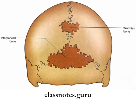
An accessory bone is a small piece of bone which develops from a separate centre of ossification but fails to unite with the main mass of bone, for example. sutural (Wormian) bones and interparietal bones.
Sesamoid Bones
A sesamoid bone is a bone usually small, developing in the tendon of a muscle, ligament or joint capsule. they ossify after birth and are devoid of periosteum. sesamoid bones possibly resist pressure, they alter the direction of pull of muscle and minimize the friction.
Blood Supply Of Bones
- Short Bones: These are supplied by numerous periosteal vessels.
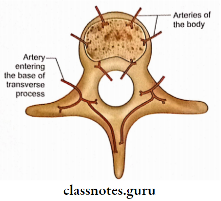
- Vertebrae: The body of the vertebra is supplied by the anterior and posterior vessels. The vertebral arch is supplied by large vessels entering through the bases of transverse processes.
- Ribs: These are supplied by nutrient and periosteal vessels.
- Flat Bones: These are supplied by nutrient and periosteal vessels.
Nerve Supply Of Bones
Nerves accompany the blood vessels of bone. Periosteal nerves are sensory (carry pain) while others are vasomotor.
Lymphatic Drainage Of Bones
Lymphatics have not been demonstrated within bone but these are very much present in the periosteum which drains into regional lymph nodes.
Ossification Of Bones
Bones ossify from centres of ossification from where the laying down of long lamellae starts by osteoblasts.
- Centres of ossification may be primary or secondary. The primary centre appears before birth, usually during the 8th week of intra-uterine life and gives rise to the diaphysis. The secondary centre appears at or after birth and gives rise to epiphysis.
- Most of the long bones have epiphysis at each end but the growth in length occurs mainly at one end. This end is called the growing end.
- Here, the epiphysis usually appears earlier and fuses with the body later than that at the non-growing end.
Applied Anatomy Of Bones
- Organic matter in the bone is greatest in childhood making it more flexible.
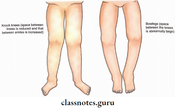
- In rickets and osteomalacia, there is inadequate calcium in bone leading to knocked knees and bowlegs.
- Metaphysis is the most common site of infection due to rich vascular anastomosis which has relatively fewer lymphocytes and has a hairpin loop arrangement of blood vessels.
- Capsular relations of metaphysis are clinically important. The inflammation of intra-articular metaphysis may result in septic arthritis, e.g. upper end of the femur.
- Injury of the growing ends of long bones. is more dangerous in young children because it will directly affect their growth.
- In certain conditions example. pernicious anaemia) the yellow marrow is replaced by red marrow to enhance the formation of red blood cells.
- Some interesting facts regarding fractures in young children are as follows:
- It is more common due to carefree activities.
- Green-stick fracture (incomplete fracture with bending) is common in children due to excessive elasticity in bone.
In old age, there is generalized skeletal atrophy called osteoporosis which makes the bone very weak. Osteoporosis is relatively more common in females, therefore, fracture of the femoral neck is more common in elderly ladies.
- In sternal puncture, the needle pierces the compact bone to reach the central spongy bone from where red marrow is aspirated for haematological examination. The same procedure is used for bone marrow transplantation.
- For perfect healing, the fractured ends of a bone should be properly aligned. This is called reduction. Healing is difficult and defective if the bony ends are mobile. To make them immobile, a hard cast is made around the fractured site and adjacent joints. This is called plaster immobilization.
- The age of a person can be determined by observing the ossification centres of the bones and their fusion in the radiographs. This is of medicolegal importance.
Part of a bone may be deprived of blood supply after fracture. This leads to avascular necrosis. The best example of avascular necrosis is the head of the femur after a fracture of the neck.
- A fibrous capsule is the most sensitive structure in a joint.
- A bone cyst is the most common cause of pathological fracture in children.
- Increased density in metaphysis is seen in hypervitaminosis.
- Senile osteoporosis is radiologically manifested only when 30% of the skeleton has been lost.
- Multiple bone fracture in a newborn is seen in osteogenesis imperfecta.
- Two interesting facts regarding Ewing’s tumour are:
- It arises from diaphysis.
- It is very sensitive to radiotherapy.