
Auditory Ossicles Terminology
There are three ossicles malleus, incus and stapes.
These names are Latin in origin, the meanings of which are as follows:
- Malleus: Hammer
- Incus: An anvil
- Stapes: A stirrup
Note: Remember that MIS is situated between the tympanic membrane and the oval window where M-Malleus, 1-Incus and S-Stapes.
Auditory Ossicles Features And Attachments
Features And Attachments Of Malleus
It consists of a head, neck and handle.
- Malleus Head
- It is the large upper end of the bone.
- It is located within the epitympanic recess.
- Its posterior surface articulates with the body of the incus.
- Malleus Neck
- It is the constricted part below the head.
- Its medial surface is crossed by chorda tympani nerve.
- Malleus Handle
- It is the lower elongated part of the malleus.
- It is embedded in the tympanic membrane and moves with it.
- Its upper end (root) shows the following features:
- A slight projection on the medial aspect provides attachment to the tendon of the tensor tympani.
- Anterior process projects forward. Anterior ligament of the malleus is attached to it. This ligament extends into the petrotympanic fissure.
- The lateral process projects laterally from where extend anterior and posterior malleolar folds to the ends of the tympanic sulcus.
Auditory Ossicles
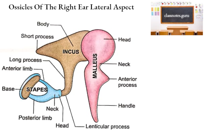
Features And Attachments Of Incus
It has a large body and two processes (long and short).
- Incus Body
- It is cubical in shape.
- Its anterior surface is concave and articulates with the head of the malleus.
- Incus Processes
- Long Process
- It projects downwards parallel to the handle of the malleus.
- Its lower end (lenticular process) bears an articular surface on the medial aspect for articulation with the head of stapes.
- Short Process
- It is directed backwards.
- It is attached by a ligament to the fossa incudis just below the aditus.
- Long Process
Features And Attachments Of Stapes
It has a head, a neck, two limbs (anterior and posterior) and a foot plate (base).
- Stapes Head
- It is rounded
- It articulates with the long process of incus.
- Stapes Neck
- It is a constricted part adjacent to the head.
- The Tendon of the stapedius is attached to its posterior surface.
- Stapes Limbs (Crura)
- Anterior and posterior limbs diverge from the neck.
- These two limbs are attached to the footplate.
- Stapes Foot Plate (Base)
- It is oval in shape.
- It fits into the fenestra vestibuli.
Auditory Ossicles
Auditory Ossicles Ossification
- Malleus and incus develop from the dorsal end of Meckel’s cartilage.
- Stapes develop from the dorsal end of the hyoid arch cartilage.
- Malleus ossifies by two centres:
- One endochondral centre near the neck.
- One centre for the anterior process appears in dense connective tissue.
- Appearance
- 4th month of intrauterine life.
- Fusion
- 6th month of intrauterine life.
- Appearance
- Incus ossifies by the single endochondral centre in the upper part of the long process. This centre appears in the 4th month of intrauterine life.
- Stapes ossify by a single endochondral centre which appears in the base at 4th month of intrauterine life.
- At birth, the auditory ossicles are of almost adult size.
Auditory Ossicles Functions
The malleus functions as a lever as it is attached to the tympanic membrane.
- The base of stapes is considerably smaller than the tympanic membrane.
- Due to this fact, the vibratory force of the stapes is about 10 times that of the tympanic membrane.
- Thus the auditory ossicles increase the force but decrease the amplitude of vibrations transmitted from the tympanic membrane.
Auditory Ossicles Applied Anatomy
- Treacher-Collins syndrome is a condition in which there are abnormalities of ossicles and craniofacial skeleton.
- This may be one of the causes of congenital conductive deafness.
- Damage to ossicles in cases of head injury with fracture of temporal bone leads to very severe and permanent conductive deafness.
- Late conductive deafness due to aseptic necrosis of the long process of incus can occur some years after head injury.
- Ankylosis of stapes is a common occurrence in cases of otosclerosis.